American Journal of Botany 89(3): 383–392. 2002.
COMPARATIVE
ANATOMICAL ANALYSIS OF THE
MEDITERRANEAN (FAGACEAE)1
COTYLEDONARY REGION IN THREE
BASIN QUERCUS
GEMMA PASCUAL,2 MARISA MOLINAS,3
AND
DOLORS VERDAGUER2,4
Unitat de Biologia Vegetal, Departament de Cie`ncies Ambientals, Universitat de Girona, Campus de Montilivi s/n 17071 Girona, Spain; and 3 Unitat de Biologia Cel.lular, Departament de Biologia, Universitat de Girona, Campus de Montilivi s/n 17071 Girona, Spain 2
Anatomical changes at the cotyledonary node from the embryo to the seedling stage in Quercus coccifera, Q. ilex, and Q. humilis were investigated by light and scanning electron microscopy techniques. Mature embryos of Q. humilis possess 2–3 pairs of leaf primordia and a pair of cotyledonary buds, whereas in Q. coccifera and Q. ilex there are two incipient primordia, and cotyledonary buds are not observed until 1 wk after germination. In all three species the cotyledonary buds multiply, forming bud clusters, and a vascular connection is well established within 5–6 wk after germination. As development proceeds, the cotyledonary region becomes woody, but buds, which are exogenous in origin, never become embedded in the periderm. In comparison with Q. suber, another native Mediterranean Basin oak, the cotyledonary node is short and axillary buds are not present below the insertion of cotyledons. In addition, starch accumulation in the cotyledonary region is not observed from histological analysis in the three oaks. Therefore, in Q. coccifera, Q. ilex, and Q. humilis seedlings the cotyledonary node can be considered to be an important regenerative structure enabling them to resprout after the elimination of the shoot above the cotyledons, despite the absence of a lignotuberous structure. Key words: sprout.
cotyledonary bud; cotyledonary node; lignotuber; Quercus coccifera; Quercus humilis; Quercus ilex; Quercus suber;
In the northeast of the Iberian Peninsula four oak species are dominant in the native forest: the evergreen sclerophyllous Quercus coccifera L. (kermes oak, subgenus Sclerophyllodrys O. Schwarz); Q. ilex L. (holm oak, subgenus Sclerophyllodrys O. Schwarz); and Q. suber L., (cork oak, subgenus Cerris ¨ rsted); and the winter deciduous Q. humilis Miller (Spach) O (pubescens oak, subgenus Quercus). Mature forms of these species are well adapted to the common disturbances in the Mediterranean ecosystems (wildfire, clear-cutting, and herbivory), showing a high capacity for resprouting from the hypogeal system (see Malanson and Trabaud, 1988, for Q. coccifera; Gratini and Amadori, 1991; Ducrey and Turrel, 1992; Lo´pez-Soria and Castell, 1992; Retana et al., 1992; and Fuente, Sardans, and Roda`, 1997, for Q. ilex; Pausas, 1997, for Q. suber; Trabaud, 1987, for Q. ilex and Q. suber; and Di Pasquale and Garfi, 1998, for Q. humilis; and others). The above-mentioned oaks species are commonly used in the reforestation of Mediterranean type forests. Although all of them are successful resprouters in their adult stage, in disturbance-prone ecosystems seedling establishment is one of the main limitations of reforestation and natural recruitment (Kozlowski, 1971; Weltzin, Archer, and Heitschmidt, 1998; Retana et al., 1999). It has been reported that in holm oak forests in northeast Spain the two major disturbances affecting seedling recruitment are thinning and fire (Retana et al., 1999). Understanding early stages of the life cycle in oaks may help to develop sylviculture strategies to improve reforestation.
Thus, given that little has been written about the anatomy and morphology of young forms of the above-mentioned Mediterranean oaks, our studies focus on the analysis of morphological traits that will permit these species to resprout at the seedling stage and hence to cope with some adverse conditions. Moreover, the processes that operate during the seed, seedling, and juvenile phases are important for understanding patterns, dynamics, and succession in plant communities (Grime and Hillier, 1992). For cork oak we have already described the ontogeny of the cotyledonary or root-shoot transition region (Molinas and Verdaguer, 1993a, b). In the cork oak seedlings, all the transition region is considered a true lignotuber ontogenically produced and ready for vegetative regeneration. In this species, the presence in the cotyledonary region of numerous buds placed underground and the accumulation of starch allow resprouting after damage to aboveground tissues. Furthermore, we have also studied the role of the cotyledonary node in the production of new shoots after disturbance in Q. humilis, Q. ilex, and Q. suber seedlings (Verdaguer et al., 2001). After eliminating the shoot above the cotyledons, all three species resprouted, but when the shoot was removed from below the cotyledon level, only Q. suber seedlings survived. Based on these observations and considering the importance attributed to the cotyledonary node in the formation of anatomical structures related to sprouting (Kauppi, Rinne, and Ferm, 1987; Del Tredici, 1992; Graham, Walwork, and Sedgley, 1998; Mibus and Sedgley, 2000), we investigated the development of the cotyledonary node in Q. coccifera, Q. humilis, and Q. ilex and identified the site of lateral shoot origin in these species. This paper focuses on the anatomy of the cotyledonary node from the mature embryo to 6-mo-old seedlings with special emphasis on the development of the buds in the axils of the cotyledons and on the bud vascular connection. The results will be discussed in comparison to embryo and seedling anat-
Manuscript received 15 June 2001; revision accepted 25 September 2001. The authors thank Dr. Tina Bell for valuable help in preparing the original manuscript; Mr. Blavia and Mrs. Carme Carulla from the Electron Microscopy Service of the University of Girona for technical advice; and Mrs. Nuri Niell for technical assistance. This research was supported by the UdG grant SUdG97-17 and UdG research grant for GP to obtain the Ph.D. degree in Biology. 4 Author for reprint requests (e-mail:
[email protected]). 1
383
384
AMERICAN JOURNAL
OF
BOTANY
[Vol. 89
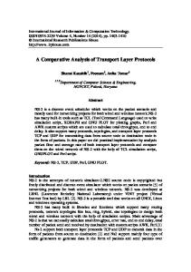
Figs. 1–9. Mature embryos. Quercus humilis: Figs. 1–3. Quercus coccifera: Figs. 4, 6, 8. Quercus ilex: Figs. 5, 7, 9. In Figs. 2, 6, 7 longitudinal sections were cut near to the cotyledonary plane. In Figs. 8–9 longitudinal sections were cut near to the intercotyledonary plane. 1. SEM of the epicotyl with three leaf primordia. Note a bud primordium in the axil of the cotyledonary petiole that begins to protude (arrow). 2. Longitudinal near-median section showing a developing bud (arrowheads) in each axil of the cotyledonary petioles. Observe the provascular cotyledonary strand continuous from the root meristem to the
March 2002]
PASCUAL
ET AL.—COTYLEDONARY REGION IN
QUERCUS
SPECIES
385
RESULTS
Embryos from mature Q. coccifera and Q. ilex acorns, ;3.5 mm long, were less developed, and both species had a flat or dome-shaped shoot apical meristem with two buttresses in a decussate position to the cotyledons, the first leaf primordia (Figs. 4–8). The cotyledonary axillary buds were not yet developed (Figs. 4–7). With regard to the embryo provascular tissue, the three oak species showed a similar pattern of development. In longitudinal cotyledonary sections, procambial strands could be traced from the root meristem to the petioles of the cotyledons (Figs. 2, 6, 7). In longitudinal sections passing between the cotyledons (intercotyledonary plane) procambial traces joined the two meristems (Figs. 8, 9). Despite the vascular pattern of the embryo being similar in all three oaks, serial transverse sections revealed that the number of cotyledonary vascular traces, both lateral and median, varied in the three species and even in the same species. The lateral traces arose from two bundles lying in the intercotyledonary plane, and the median traces arose from one bundle lying in the cotyledonary plane ramifying each bundle only once or not at all (Figs. 10–21). Cotyledonary traces matured earlier than bundles in the axis tissue of the provascular embryo. Just above the point where the cotyledons were attached to the embryo axis, the two cotyledonary petioles were almost fused, leaving at the center the epicotyl in which a hollow vascular cylinder was observed. Traces of the cotyledonary petioles also began to join with the vascular cylinder (Fig. 13). Below the cotyledonary insertion the tissue of the cotyledonary petioles fused to the embryo axis tissue and the vascular cylinder was interrupted by the exit of the lateral and median cotyledonary traces that enter the cotyledons (Figs. 16–18). At 0.3–0.5 mm below the cotyledonary insertion, the hypocotyl-root vascular tissue formed a hollow vascular cylinder (Figs. 19–21) that became solid at the root pole.
Mature embryo stage—Quercus humilis embryos were more developed than those of Q. coccifera and Q. ilex. Quercus humilis embryos were ;4.5 mm long and showed an epicotyl with two to three pairs of leaf primordia overlapping each other and arching up over the shoot apex (Fig. 1). There were several trichomes at the margins and the abaxial surfaces (Fig. 1). At this developmental stage, a pair of rudimentary buds were present in the axils of the cotyledons (Figs. 1, 2). Longitudinal sections passing medially through both cotyledons (cotyledonary plane) also showed the shoot apex with one to two pairs of well-developed leaves and the exogenous origin, from superficial embryo tissues at the base of the epicotyl, of the first pair of cotyledonary axillary buds (Fig. 2). In transverse sections, these cotyledonary axillary buds formed oval-shaped protuberances (Fig. 3).
Germinated and 1-wk-old seedlings—With acorn germination the radicle was pushed out of the seed as a result of elongation of the cotyledonary petioles. Then it grew and penetrated into the soil. In the three oak species, cotyledonary petioles of newly germinated acorns measured ;8.5 mm in length and the radicle ;8 mm. The hypocotyl elongated slightly or not at all and the epicotyl remained protected by the cotyledonary petioles. The epicotyl showed different stages of development of leaf primordia in the three species of Quercus (Figs. 22–24). For Q. coccifera and Q. ilex, the cotyledonary buds were not visible in any of the germinated acorns examined (Figs. 23, 24). One week after germination seedlings had developed a root ;3 cm long and the epicotyl was still protected by the cotyledonary petioles. In Q. humilis the epicotyl measured ;2.5
omy of Q. suber and to the presence of a lignotuber. This study will establish a base for further comparative studies on vegetative reproduction of oaks. MATERIALS AND METHODS Mature acorns of Quercus coccifera, Q. humilis, and Q. ilex were collected from parental trees in experimental plots of the ‘‘Laboratori del Suro’’ of the University of Girona (Spain) during the 1996–1997 fall-winter period and at about the time of maximum acorn production for each species. Acorns were sown in 30 cm deep, 23.5 cm diameter pots in a mixture of 25% vermiculite and 75% peat and were kept moist by controlled irrigation in laboratory conditions for germination. A total of 24 seedlings per species from germination to 6-mo-old were taken at different stages to obtain a developmental sequence. For optical microscopy, we cut small fragments containing the embryo, root fragments, and fragments including tissue of the cotyledonary region of seedlings at different stage of development. Tissue fragments were fixed with 4% formaldehyde in phosphate-buffered saline (pH 7.5) in a vacuum at room temperature for 48 h minimum. The fragments were dehydrated through an isopropyl alcohol series and embedded in glycol-methacrylate (GMA). Sections ranging in thickness from 3 to 5 mm were cut on a rotatory microtome Autocut 1150 (Reichert-Jung, Wien, Austria) and mounted on glass slides. Routine staining was performed with toluidine blue, and to enhance the presence of carbohydrates, some sections were stained with periodic acid-Schiff (PAS) (Verdaguer and Molinas, 1997). Specimens were photographed using an Olympus-Vanox light photomicroscope (Olympus Optical, London, UK). For scanning electron microscopy (SEM), mature embryos carefully prepared under a dissecting microscope removing one of the cotyledonary petioles and seedling fragments containing the cotyledonary node were fixed as for optical microscopy. Fragments were dehydrated in a graded ethanol series, exchanged through amyl-acetate, and critical-point dried. The pieces were mounted on copper stubs and coated with gold. Specimens were observed using a Zeiss DSM 960A scanning electron microscope (Zeiss, Oberkochen, Germany) of the Electron Microscopy Service of the University of Girona.
← cotyledonary petioles. 3. Transverse section through a cotyledonary bud. Bud primordium is visible as an oval-shaped group of densely cytoplasmic cells (arrowheads). The cotyledonary petiole is seen above, with four cotyledonary lateral traces. 4. SEM showing the epicotyl with a dome-shaped shoot apex and two buttresses corresponding to the first pair of leaf primordia (arrowheads). 5. SEM of the first leaf primordia arching over the apex. 6. The epicotyl shows a leaf primordium. The procambial tissue is continued from the root meristem to the cotyledonary petioles. 7. The epicotyl shows a dome-shaped shoot apex. Starch granules are observed in the cotyledonary petioles and in parenchyma cells of the embryo axis near the root meristem (arrowheads). 8. The first leaf primordia arch over the apex. The provascular tissue forms a cylinder connecting both apical meristems. 9. The shoot apical meristem is nearly a flat surface. Figs. 2, 3, 6, 8, 9 stained with PAS with toluidine blue. Fig. 7 stained with PAS. Bars 5 100 mm in Figs. 3–5, 200 mm in Figs. 1, 2, and 250 mm in Figs. 6–9. Figure Abbreviations: AB, accessory bud; Ad, adventitious bud; CB, cotyledonary bud; CP, cotyledonary petiole; CT, cotyledonary tissue; E, epicotyl; EA, embryo axis; FN, first internode; LT, lateral cotyledonary trace; MT, median cotyledonary trace; P, leaf primordium; PAS, periodic acid-schiff; Pi, pith; PV, provascular tissue; R, root; RM, root meristem; S, shoot; Sc, scale; SEM, scanning electron micrograph; SM, shoot meristem; T, trichome; V, vascular tissue.
386
AMERICAN JOURNAL
OF
BOTANY
[Vol. 89
Figs. 10–21. PAS and toluidine blue-stained transverse embryo sections of the three Quercus taken at different levels proceeding acropetally. Quercus humilis: Figs. 10, 13, 16, 19. Quercus coccifera: Figs. 11, 14, 17, 20. Quercus ilex: Figs. 12, 15, 18, 21. Figs. 10–12. Sections through the leaf primordia. Figs. 13–15. Sections at the epicotyl level. Figs. 16–18. Sections at mid-embryo axis level. Figs. 19–21. Sections proximal to the root meristem. 10. The two cotyledonary petioles are almost fused. Each petiole shows four lateral traces grouped two by two and no median cotyledonary traces (arrowheads). 11. Two or three lateral traces (arrowheads) and two median cotyledonary traces (arrows) are viewed in each petiole. 12. The two-cotyledonary petioles are almost fused (arrows). Note the three median cotyledonary traces in one of the petioles, whereas in the other only two are seen (arrowheads). 13. Cotyledonary traces begin
March 2002]
PASCUAL
ET AL.—COTYLEDONARY REGION IN
mm in length and was made up of three or more foliar primordia, a shoot apex, and a short subjacent stem with some trichomes (Figs. 25, 26). Cotyledonary buds had 1-2 pairs of leaf primordia and several trichomes (Figs. 25, 27). Serial transverse sections revealed an incipient connection between the cotyledonary buds and the provascular cylinder (Fig. 28). In Q. coccifera and Q. ilex the epicotyl was ;1 mm long and showed two incipient leaf lamina with dorsiventral symmetry. At the base of the epicotyl, the cotyledonary buds were readily visible for the first time (Figs. 29–31). Cotyledonary buds were discernible as a protuberance of undifferentiated and somewhat densely stained cells and originated exogenously as in Q. humilis. In several Q. coccifera and Q. ilex seedlings, buds located in the axils of the cotyledons developed from the outer layers of the cotyledonary tissue instead of embryo axis tissues (Fig. 30). We called these buds adventitious buds in order to differentiate them from cotyledonary buds originated from embryo axis tissue. Five- to six-week-old seedlings—Within 5–6 wk of germination, the average lengths of the root and shoot for the three oaks were 19 and 5 cm, respectively. The cotyledonary node region measured ;1 mm in length in Q. ilex and Q.coccifera and ;4 mm in Q. humilis. The cotyledonary region in Q. humilis was readily distinguishable as the two petioles formed a whitish sheath of tissue surrounding the base of the stem (Fig. 32). The cotyledonary buds of the three oak species gave rise to a relatively large first bud with 2–3 leaf primordia and laminar scales rich in tannins that developed marginal trichomes (Figs. 33–36). Beside these structures, one or more accessory buds developed from the outer tissues at the base of the first cotyledonary buds on each side (Figs. 33, 35). Adventitious buds arising from cotyledonary tissue of Q.ilex and Q. coccifera resembled those formed from the embryo tissue showing fully developed meristems and foliar primordia (Fig. 37). As development proceeded, the cotyledonary region that eventually formed the root–shoot transition region of the seedling became woody showing secondary thickening with a well-developed cambial zone (Figs. 33, 35–37) and, eventually, a phellogen. Six-month-old seedlings—Seedlings of the three oaks possessed a root between 40 and 70 cm long and a shoot of 15– 30 cm. The cotyledonary region of Q. humilis seedlings appeared to be slightly thicker than the shoot and the root. In each species, the exogenous proliferation of cotyledonary buds produced small clusters of 3–5 buds formed by the first cotyledonary bud and some accessory buds at different stages of development (Figs. 38–42). These clusters were often protected by the cotyledonary petioles (Fig. 41); parenchymatic tissue and/or the periderm of the rapidly expanding seedling axis never engulfed them (Figs. 38–42). Accessory buds had 2–3 pairs of leaf primordia and some hypertrophied scales (Figs. 41–42). The scales were rich in tannins and develop
QUERCUS
SPECIES
387
some trichomes. At this stage, the connection of vascular tissue of the buds with the central vascular cylinder was complete (Fig. 40), and it was possible to distinguish some mature xylem and phloem elements in the connection. Secondary vascular connection was established later due to the activity of cambial cells. In each species, the cells of the pith and the xylem parenchymatic cells and the uniseriated rays contained some starch grains. Wild-grown and greenhouse-grown 2- to 3-yr-old trees of Q. ilex and Q. coccifera did not show any swelling at the shoot base or at the upper root, nor the production of abundant bud clusters. Only in Q. humilis, the first internode above the cotyledons was slightly thicker than the stem, and the formation of few epicotyledonary buds and some adventitious roots were observed (Fig. 43). DISCUSSION Embryo anatomy and early seedling developmental processes differ in Q. humilis, Q. ilex, and Q. coccifera from those previously described for Q. suber (Molinas and Verdaguer, 1993a, b). This morphological study confirms the presence of clusters of dormant buds at the cotyledonary node of Q. humilis, Q. ilex, and Q. coccifera seedlings, enabling them to resprout from the stem base. However, no distinct organ of clonal regeneration or lignotuber comparable to those of Q. suber has been observed in these species. The presence of buds in the axil of the cotyledons at the embryo stage may be related to the number of leaf primordia and its stage of development. The axillary bud originates a bit later than the leaf primordia, and in general, during the second plastochron (Esau, 1985). Thus, we suggest that in embryos of Q. coccifera and Q.ilex, cotyledonary buds are not formed as leaf primordia at the second node have not yet developed (we consider cotyledons the first pair of leaves); whereas in Q. humilis and Q. suber (Molinas and Verdaguer, 1993a), cotyledonary buds would be present as more than two pairs of leaf primordia are developed in the shoot apex of mature embryos. In other species, such as Ginkgo biloba (Del Tredici, 1992), Betula pubescens (Kauppi, Rinne, and Ferm, 1987), and Eucalyptus cinerea (Graham, Walwork, and Sedgley, 1998), cotyledonary buds develop after germination, but additional information about the leaf primordia development at the embryo stage of these species is not available. The anatomy of the cotyledonary node is similar for Q. coccifera, Q. ilex, and Q. humilis, but it differs from that described for Q. suber (Molinas and Verdaguer, 1993a). In embryos of Q. coccifera, Q. ilex, and Q. humilis, the cotyledonary petioles fuse with the tissue of the embryo axis and only one pair of cotyledonary buds is shown. In these oak species the cotyledonary node does not elongate or elongates only slightly. In contrast, in Q. suber embryos, the cotyledonary petioles are not completely fused to the embryo axis tissue, and in sections passing through the cotyledonary plane, an entire epidermal surface layer extending from the apical dome
← to fuse with the vascular tissue of the embryo axis (arrows). Note the presence of a group of cells with dense cytoplasm that correspond to the cotyledonary bud. 14. The unfused epicotyl is seen between the cotyledonary petioles. 15. Note the continuous vascular cylinder of the epicotyl. 16. Cotyledonary tissue is completely fused with the embryo axis tissue. Lateral cotyledonary traces (arrowheads) are semifused with the vascular tissue of the embryo axis (arrows). 17. Cotyledonary traces are still not joined with the procambium of the embryo axis. The vascular cylinder of the embryo axis is interrupted at the cotyledonary plane (arrows). 18. As in Fig. 17. 19. Section showing the provascular cylinder. 20. As in Fig. 19. 21. As in Fig. 19. Bars 5 300 mm.
388
AMERICAN JOURNAL
OF
BOTANY
[Vol. 89
Figs. 22–31. Epicotyl and cotyledonary bud development of germinating seedlings. Figs. 22–24. Epicotyls from germinated acorns. Figs. 25–31. One-weekold seedlings. Quercus humilis: Figs. 22, 25–28. Quercus coccifera: Figs. 24, 29, 31. Quercus ilex: Figs. 23, 30. 22. SEM showing the cotyledonary bud with two smooth protuberances corresponding to the first leaf primordia at the base of the epicotyl (arrows). 23. SEM showing the first leaf primordia arching over the apex. Some marginal trichomes can be seen (arrows). 24. SEM showing the shoot apex with two leaf primordia with bilateral symmetry and their corresponding stipules (stars). Note the presence of some developing trichomes. 25. In the cotyledonary bud the first leaf primordia are developed. Abundant trichomes are seen on the leaf primordia and the epicotyl surface. 26. Longitudinal cotyledonary section showing the cotyledonary bud at the base of the epicotyl and axillary to the cotyledon. Note the abundant starch grains in the parenchyma cells of the epicotyl and cotyledonary petiole (arrowheads). 27. Detail of a
March 2002]
PASCUAL
ET AL.—COTYLEDONARY REGION IN
to the lower insertion point of the cotyledons can be seen (Molinas and Verdaguer, 1993a). Furthermore, in Q. suber embryos ;5–7 pairs of lateral buds develop along the length of the cotyledonary node which, after germination, elongates to ;12 cm and remains buried, leaving most of the dormant buds underground. The presence of buds clusters in the axils of the cotyledons in Q. coccifera, Q. ilex, and Q. humilis seedlings confers the high capacity for regeneration shown by these species when the shoot is cut above the cotyledons, whereas the absence of dormant buds below the soil causes the lack of sprouting when the aerial shoot is eliminated below the cotyledons (Verdaguer et al., 2001). The present histological analysis shows that dormant buds are strictly limited to the axils of the cotyledons and that no differences in starch accumulation can be observed between the shoot, the upper part of the root, and the rest of the root of these oak species. However, the quantification of starch reserves in above- and underground organs should be undertaken in order to gain a better understanding of starch distribution and its use in response to sprouting. The proliferation of cotyledonary buds forming clusters of three or more buds in young seedlings is a common characteristic of the Quercus species investigated in this study and in Q. suber (Molinas and Verdaguer, 1993b). Such accessory buds are exogenous in origin and arise from cells at the base of the cotyledonary buds that are already partially or completely differentiated. However, some buds in Q. coccifera and Q. ilex were determined to arise from cells of the cotyledonary petioles, indicating that this tissue is also capable of generating adventitious buds. The high meristematic potentiality of the cotyledonary node has already been described in resprouting species, such as the burl in Betula pubescens (Kauppi, Rinne, and Ferm, 1987), the lignotuber in some species of Eucalyptus (Kerr, 1925; Bamber and Mullette, 1978; Graham, Walwork, and Sedgley, 1998) and Banksia (Mibus and Sedgley, 2000), or the basal chichi in Gingko biloba (Del Tredici, 1992). Furthermore, cotyledonary nodal explants are commonly used in plant clonal regeneration programs, including woody species (Distabanjong and Geneve, 1996–1997; Tyagi and Kothari, 1997; Pradhan, Pattnaik, and Chand, 1998). The origin of accessory buds in the species cited above is exogenous, although some buds in Eucalyptus (Chattaway, 1958) and Banksia (Mibus and Sedgley, 2000) can also originate endogenously. With the exception of Quercus and Betula pubescens, cotyledonary buds are generally embedded in periderm, and their growth and development are under the surface. Hidden cotyledonary buds should favor survival of the species in comparison to that have non engulfed dormant buds. In Q. coccifera, for example, Malanson and Trabaud (1988) showed that increasing fire intensity significantly reduced sprout emission, suggesting a negative effect of fire on bud viability. In Mediterranean-type ecosystems, where fire is an important part
QUERCUS
SPECIES
389
of the functioning of the system, it would be interesting to analyze the anatomical and morphological characteristics that are involved in the resprouting behavior of key species and determine how this might influence stand structure and composition. Lignotubers are broadly described as woody organs of storage with a supply of protected buds responsible for sprouting. Lignotubers and burls are found in trees and shrubs of the Mediterranean vegetation including different Quercus species. The term lignotuber was originally applied to the swellings found in the axils of the cotyledons of eucalyptus (Carrodus and Blake, 1970). These swellings are ontogenetically programmed and not the result of physical or environmental stresses (Carr, Jahnke, and Carr, 1984; Graham, Walwork, and Sedgley, 1998). Moreover, in eucalyptus the lignotuber is a transitory organ characteristic of the young forms (Chattaway, 1958). Ontogenetically programmed lignotubers have also been documented in other species including Banksia (Mibus and Sedgley, 2000), Cryptocarya alba, Colliguaya odorifera, and Lithraea caustica (Montenegro and Avila, 1982), and Quercus suber (Molinas and Verdaguer, 1993a, b). According to James (1984), only ontogenetically produced structures rich in carbohydrates and dormant buds located at the stem base should be defined as lignotuber. In mature forms of Q. ilex (Canadell and Roda`, 1991) and Q. coccifera (Kummerow, Kummerow, and Trabaud, 1990), thick woody swellings are seldom found at the upper part of the root or root crown. These structures are often referred to as lignotubers because of their high nutrients and carbohydrates content. However, our histological analysis do not reveal any underground structure showing bud and starch accumulation at the seedling stage nor in 2- to 3-yr-old seedlings, as was the case in Q. suber (Molinas and Verdaguer, 1993b). It is likely that Q. ilex and Q. coccifera seedlings produce new shoots from axillary buds and, as the plant matures, the continuous, secondary growth of the stem base results in the fusion of the main stem and the sprouts. Additionally, axillary buds at the base of sprouts can become buried beneath the developing periderm. This may explain the presence of a thick woody stem base but, to date, no information is available about the accumulation of dormant axillary buds. So, for these structures the term lignotuber should not be used in order to avoid possible misunderstandings. In 6-mo-old and older seedlings of Q. humilis, the first internode above the cotyledons has a high meristematic capacity as it develops adventitious buds and roots. This characteristic could act in favor of the seedling, allowing it to cope with adverse conditions and possibly leading to the formation of a specialized sprouting structure in mature plants. However, data about the presence in Q. humilis mature trees of swellings, burls, or lignotubers are not available. Further research on older plants is needed to clarify the fate of the cotyledonary buds and the development of the shoot–root transition region in Q. humilis.
← cotyledonary bud with three leaf primordia. Note the disrupted cotyledonary tissue. 28. Transverse section through the cotyledonary node. The vascular connection is initiate between the bud and the provascular cylinder (arrows). 29. SEM of a shoot apex showing dorsiventral symmetry and abundant trichomes. Note at the base of the epicotyl a bump that corresponds to the cotyledonary bud. 30. Transverse section close to the cotyledonary node showing two adventitious cotyledonary buds originating from the cotyledonary tissue. 31. Transverse section close to the cotyledonary node. Note the developing cotyledonary bud formed by small and densely stained cells. Figs. 26, 28, 30, 31 stained with PAS and toluidine blue. Bars 5 100 mm in Figs. 22, 26–28, 31 and 200 mm in Figs. 23– 25, 29, 30.
390
AMERICAN JOURNAL
OF
BOTANY
[Vol. 89
Figs. 32–37. Detail of the cotyledonary region of 5- to 6-wk-old seedlings. Figs. 33, 35–37. Transverse sections through the cotyledonary node Quercus humilis: Figs. 32–33. Quercus coccifera: Figs. 34–35. Quercus ilex: Figs. 36–37. 32. The two cotyledonary petioles are fused, giving rise to a whitish tissue that surrounds the upper part of the hypocotyl-root axis, ;4 mm in length (30.75). 33. An accessory bud is seen at the left side of the first cotyledonary bud. Note the hypertrophied scales rich in tannins of the cotyledonary and accessory bud (short arrows). A secondary vascular connection occurs between the bud and the axial vascular cylinder (long arrows). 34. Cotyledonary node with two cotyledonary buds. The cotyledonary node measures ;0.5–1.5 mm (39). 35.
March 2002]
PASCUAL
ET AL.—COTYLEDONARY REGION IN
QUERCUS
SPECIES
391
Figs. 38–43. Cotyledonary region of 6-mo-old Q. coccifera, Q. humilis, and Q. ilex seedlings. Quercus coccifera: Figs. 38, 40, 42. Quercus ilex: Fig. 39. Quercus humilis: Figs. 41, 43. 38. SEM of bud proliferation to form a cluster. Some hypertrophied scales protecting the cotyledonary bud can be seen. 39. SEM showing an accessory bud on each side of the first cotyledonary bud with some protective flat laminar scales. 40. Transverse section showing the vascular connection between the axial vascular tissue and an accessory bud (arrows). Scales rich in tannins protect cotyledonary buds (arrowheads). 41. Transverse section showing a cluster of buds still protected by the cotyledonary tissue. The secondary vascular connection between the main cotyledonary bud and the axial vascular tissue is shown (arrows). 42. Transverse section showing a cluster of buds with three accessory buds in various developmental stages originating from superficial meristems (arrows). 43. Note at the first internode, just above the cotyledonary node, the presence of several buds (arrowheads) and an adventitious root (arrow) (31). Figs. 40–42 stained with PAS and toluidine blue. Bars 5 200 mm.
LITERATURE CITED BAMBER, R. K., AND K. J. MULLETTE. 1978. Studies of the lignotubers of Eucalyptus gummifera (Gaertn. and Hochr.). II. Anatomy. Australian Journal of Botany 26: 15–22. CANADELL, J., AND F. RODA`. 1991. Root biomass of Quercus ilex in a montane Mediterranean forest. Canadian Journal of Forestry Research 21: 1771–1778.
CARR, D. J., R. JAHNKE, AND M. G. S. CARR. 1984. Initiation, development and anatomy of lignotubers in some species of Eucalyptus. Australian Journal of Botany 32: 415–437. CARRODUS, B. B., AND T. J. BLAKE. 1970. Studies on the lignotubers of Eucalyptus obliqua l’Heri. I. The nature of the lignotuber. New Phytologist 69: 1069–1072. CHATTAWAY, M. M. 1958. Bud development and lignotuber formation in Eucalypts. Australian Journal of Botany 6: 103–115.
← Accessory bud at the right side resembling the first cotyledonary bud. 36. The first cotyledonary bud with some scales arches over the shoot meristem. The cotyledonary petiole covers a cotyledonary bud. 37. Adventitious bud arising from the superficial tissue of the cotyledonary petiole. A vascular connection between the bud and the cotyledonary traces is absent. Figs. 33, 35–37 stained with PAS and toluidine blue. Bars 5 100 mm in Figs. 33, 35, 36 and 50 mm in Fig. 37.
392
AMERICAN JOURNAL
DEL TREDICI, P. 1992. Natural regeneration of Ginkgo biloba from downward growing cotyledonary buds (basal chichi). American Journal of Botany 79: 525–530. DI PASQUALE, G., AND G. GARFI. 1998. Comparative analysis of the regeneration patterns of Quercus suber and Q. pubescens after grazing exclusion in the forest. Ecologia-Mediterranea 24: 15–25. DISTABANJONG, K., AND R. L. GENEVE. 1996–1997. Multiple shoot formation from cotyledonary node segments of eastern redbud. Plant, Cell, Tissue, and Organ Culture 47: 247–254. DUCREY, M., AND M. TURREL. 1992. Influence of cutting methods and dates on stump sprouting in holm oak (Quercus ilex L.) coppice. Annals Science Forestry 49: 449–464. ESAU, K. 1985. Anatomia vegetal, 3rd ed. Omega Edition, Barcelona, Spain. FUENTE DE LA, J., J. SARDANS, AND F. RODA`. 1997. Creixement de rebrots d’alzina en una brolla postincendi en el perı´ode entre tres i sis anys despre´s de fertilitzar-los. Orsis 12: 117–125. GRAHAM, A. W., M. A. WALWORK, AND M. SEDGLEY. 1998. Lignotuber bud development in Eucalyptus cinerea (F. Muell. Ex. Benth). International Journal of Plant Sciences 159: 979–988. GRATINI, L., AND M. AMADORI. 1991. Post-fire resprouting of shrubby species in Mediterranean maquis. Vegetatio 96: 137–143. GRIME, J. P., AND S. H. HILLIER. 1992. The contribution of seedling regeneration to the structure and dynamics of plant communities and larger units of landscape. In M. Ferrer [ed.], Seeds: the ecology of regeneration in plant communities, 349–364. CAB International, Wallingford, UK. JAMES, S. 1984. Lignotubers and burls: their structure function and ecological significance in mediterranean ecosystems. Botanical Review 50: 225– 266. KAUPPI, A., P. RINNE, AND A. FERM. 1987. Initiation, structure and sprouting of dormant basal buds in Betula pubescens. Flora 179: 55–83. KERR, L. R. 1925. The lignotubers of eucalypt seedling. Proceedings of the Royal Society of Victoria 37: 70–97. KOZLOWSKI, T. T. 1971. Growth and development of trees, vol. I. Academic Press, New York, New York, USA. KUMMEROW, J., M. KUMMEROW, AND L. TRABAUD. 1990. Root biomass, root distribution and the fine-root growth dynamics of Quercus coccifera L. in the garrigue of southern France. Vegetatio 87: 37–44. LO´PEZ-SORIA, L., AND C. CASTELL. 1992. Comparative genet survival after fire in woody Mediterranean species. Oecologia 91: 493–499. MALANSON, G. P., AND L. TRABAUD. 1988. Vigour of post-fire resprouting by Quercus coccifera L. Journal of Ecology 76: 351–365. MIBUS, R., AND M. SEDGLEY. 2000. Early lignotuber formation in Banksia—
OF
BOTANY
[Vol. 89
investigations into the anatomy of the cotyledonary node of two Banksia (Proteaceae) species. Annals of Botany 86: 575–587. MOLINAS, M. L., AND D. VERDAGUER. 1993a. Lignotuber ontogeny in the cork-oak (Quercus suber, Fagaceae) I. Late embryo. American Journal of Botany 80: 172–181. MOLINAS, M. L., AND D. VERDAGUER. 1993b. Lignotuber ontogeny in the cork-oak (Quercus suber; Fagaceae) II. Germination and young seedling. American Journal of Botany 80: 182–191. MONTENEGRO, G., AND G. AVILA. 1982. Presence and development of lignotubers in shrubs of the Chilean matorral. Canadian Journal of Botany 61: 1804–1808. PAUSAS, J. G. 1997. Resprouting of Quercus suber in NE Spain after fire. Journal of Vegetation Science 8: 703–706. PRADHAN, C., S. PATTNAIK, AND P. K. CHAND. 1998. Rapid in vitro propagation of East Indian Rosewood (Dalbergia latifolia Roxb.) through high frequency shoot proliferation from cotyledonary nodes. Journal of Plant Biochemistry and Biotechnology 7: 61–64. RETANA, J., J. M. ESPELTA, M. GRACIA, AND M. RIBA. 1999. Seedling recruitment. In F. Roda`, J. Retana, C. A. Gracia and J. Bellot [eds.], Ecology of Mediterranean evergreen oak forests, Ecological studies, vol. 137, 89–103. Springer-Verlag, Berlin, Germany. RETANA, J., M. RIBA, C. CASTELL, AND J. M. ESPELTA. 1992. Regeneration by sprouting of holm-oak (Quercus ilex) stands exploited by selection thinning. Vegetatio 99–100: 355–364. TRABAUD, L. 1987. Natural and prescribed fire: survival strategies of plants and equilibrium in mediterranean ecosystems. In J. D. Tenhunen [ed.], Plant response to stress, vol. 15, 607–621. Springer-Verlag, Berlin, Germany. TYAGI, P., AND S. L. KOTHARI. 1997. Micropopagation of Capparis decidua through in vitro shoot proliferation on nodal explants of mature tree and seedling explants. Journal of Plant Biochemistry and Biotechnology 6: 19–23. VERDAGUER, D., E. GARC´ıA-BERTHOU, G. PASCUAL, AND P. PUIGDERRAJOLS. 2001. Sprouting of seedlings of three Quercus species (Q. humilis Miller, Q. ilex L., and Q. suber L.) in relation to repeated pruning and the cotyledonary node. Australian Journal of Botany 49: 67–74. VERDAGUER, D., AND M. MOLINAS. 1997. Development and ultrastructure of the endodermis in the primary root of cork oak (Quercus suber). Canadian Journal of Botany 75: 769–780. WELTZIN, J. F., S. R. ARCHER, AND R. K. HEITSCHMIDT. 1998. Defoliation and woody plant (Prosopis glandulosa) seedling regeneration: potential vs realized herbivory tolerance. Plant Ecology 138: 127–135.