Chapter 3
Soft Tissue Techniques
Ted A. Cook Robert A. Guida
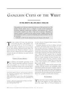
Expertise in facial plastic and reconstructive surgery requires knowledge and skills of basic soft tissue surgery. The face, with its excellent blood supply, variety of relaxed skin tension lines, and numerous anatomic boundary markers, offers great flexibility and challenge for rewarding cosmetic and reconstructive procedures. Superb results can only be obtained if certain rudimentary principles are practiced. It is the purpose of this chapter to discuss equipment, techniques, and surgical principles that we feel are necessary to achieve optimal results. EQUIPMENT Soft tissue instrument set To obtain good soft tissue handling, proper instrumentation is mandatory. The basic soft tissue instrument set should include the ten items listed in the box and depicted in Figure 3-1. Brown-Adson forceps are toothed to provide a firm grasp of deep soft tissue. For finer, less traumatic manipulation, as in skin closure, the delicate, single-toothed Bishop-Harman forceps are used. Skin hooks inflict less tissue crushing than forceps and are essential for skin edge eversion and retraction needed in skin
undermining or placement of subcuticular sutures. Two varieties of needle holders are used to effectively manage the smaller needles used for facial surgery. The Webster needle holder easily grasps smaller needles and is used for subcuticular or deep suturing of the head and neck. The delicate Castroviejo needle holder allows accurate placement of finer sutures for skin closure, nerve grafting, or other precise work. A variety of Stevens tenotomy scissors are used for undermining or cutting through Basic soft tissue instrument set 1. Brown-Adson tissue forceps 2. Bishop-Harman tissue forceps 3. Single and doublt skin hooks 4. Webster needle holder 5. Castroviejo needle holder 6. Stevens tenotomy scissors 7. Castroviejo calipers 8. Small suture scissors 9. No. 15 and No. 10 knife blades 10. Moist sponge
Figure 3-1. Soft tissue instrument set.
Figure 3-2. Local anesthesia set.
soft tissues or skin. The tips are curved and blunted and should be used with the points directed upward when undermining and in the direction of the incision when cutting through skin. Smaller suture scissors are necessary to achieve more accurate cutting of the fine sutures used in facial work. Precision work and flap design frequently require measurement with Castroviejo calipers because of its easy maneuverability, 0 to 20 mm scale, and sharp tips that can be used for skin markings. Knife handles on the set include no. 3, no. 9, and the Beaver handle. The no. 15 and no. 11 knife blades are the most frequently used in facial plastic surgery. The larger no. 10 blade on facial skin is rarely required. Finally, moist sponges are always used in soft tissue work on the face. A wet sponge causes less pulling and trauma to the soft tissue and blood vessels than a dry sponge. Local anesthesia set The majority of work done on the face can be accomplished with local anesthesia only (Figure 3-2). The local set should consist of a 10 ml three-ringed, Luer-Lok syringe. The three-ringed holder offers control for injecting small amounts of anesthesia. A 27-gauge 1.5-inch needle minimizes the pain of injection. A sealed packaged anesthetic agent, most commonly 1% lidocaine with 1: 100,000 epinephrine, is used in conjunction with sodium bicarbonate in a 9:1 mixture. Preservatives in the packaged anesthetic bottle lowers the pH, causing increased pain on injection, which can be minimized by the buffering effect of the sodium bicarbonate.
EXCISIONS Planning of excisions and incisions Proper planning is necessary for achieving good results when excising lesions on the head and neck.2 The face has several unique features compared with the rest of the body that must be recognized before planning any surgical maneuvers. An abundance of anatomic boundary markers between aesthetic units can be found on the face (e.g., the sublabial crease, the nasolabial fold, and the preauricular crease) that are ideal for incision placement (Figure 3-3). Care must be taken not to advance tissue from one anatomic unit to another because the quality of skin will often change, resulting in a disfiguring scar. The relaxed skin tension lines (RSTL) and a general lack of tension movement in the face enables one to hide and minimize most facial incisions (Figure 3-4). All incisions, especially in the face, must be made so that the long axis of the incision is in one of the
Photographic equipment Finally, a camera is an absolute necessity in the practice of facial plastic surgery. Not only is it helpful for insurance documentation and liability coverage, but it is most significant in maintaining good patient rapport. Important features include a 35-mm camera body, transparency film, a macro lens with a focal length of 105 mm that will allow for good portrait shots, an off-centered metered flash, and a sturdy carrying case capable of carrying extra miscellaneous equipment.
Figure 3-3. Variety of anatomic units of the face.
en the final length of the wound.8 However, the trade-off for shortening the overall incision length is to create an additional small incision on each side and a wound that is a bit more complex to close. This technique is used when large defects may be closed as an ellipse and when avoiding extension of an ellipse across a normal anatomic border such as the vermilion. Serial excisions at 2 to 3 month intervals will allow excision of larger defects with a final straight-line closure. Reexcision may be prevented in some areas like the scalp or forehead by use of tissue expanders at the sides of the defect before final closure. Execution of incisions
Figure 3-4. Multiple relaxed skin tension lines of the face. (From Thomas JR and Holt GR, editors: Facial Scars: incision, revision, and camouflage, ed 1, St Louis, 1989, The CV Mosby Co.).
A
B Figure 3-5. A, Planning and creating a fusiform excision. B, Mplasty for a shorter scar.
RSTL even if it curves or, in some instances, double curves. It is essential that these lines be learned and used. Incisions made in the RSTL will close with minimal tension and without widening the scar; however, incisions made across the lines will close under significant tension and the scar will spread as it heals. Another unique feature of the face is its excellent blood supply, which results in a greater resistance to infection and higher rates of wound healing. The simplest type of excision is a fusiform design commonly referred to as an ellipse. The fusiform excision involves wide undermining of all sides and bilateral advancement of tissue for closure. As mentioned previously, the fusiform must always be designed in a RSTL and with a length-to-width ratio of 2.5: 1 to 3.0: 1, thus helping to avoid the standing cone or dog-ear deformity at the ends of the wound on closure (Figure 3-5). There are methods of altering the size of the fusiform excision. An M-plasty created at the ends of the fusiform will short-
Most incisions in the facial region can be made either with a no. 15 or no. 11 blade using the no. 9 knife handle. The no. 11 blade is most useful for straight cuts made perpendicular to the skin in a sawing-type motion (e.g., creating the small cuts in a running W-plasty incision). The initial knife position is perpendicular to the skin edge, and the tip of the blade should be used to begin the incision. Then, the handle of the knife should be arched downward so that the midportion of the incision is made with the belly of the blade. At the end of the incision, the handle should be arched upward again so that the end of the incision is made with the tip of the blade. The blade should always be perpendicular to the skin surface, and the incision should be made completely through skin and subcutaneous tissue for the entire length of the cut. When making an incision to excise a lesion, instead of creating the cut at right angles to the skin surface, the incision should be beveled outward (Figure 3-6). This accomplishes the following three things: it aids in skin edge eversion, which is helpful in preventing depressed scars; it helps to create the plane for undermining; and it gives better assurance of excising completely around the lesion. A skin hook should then be placed into the starting end of the first-side incision providing traction and stabilization for the second-side incision. Without this stabilization of the end of the wound, it is difficult to make the second incision precisely. The final step in executing an excision before closure is undermining. This should be done in the immediate subdermal plane (Figure 3-7). Undermining should be used in virtually all surgical
Figure 3-6. Beveling outward from lesion when creating skin incision.
Figure 3-7. Undermining skin with skin hook and No. 15 blade VS. forceps and scissors.
Figure 3-8. Principle of halving.
Figure 3-9. Equalizing length of edges with a Burow’s triangle.
procedures in the skin of the head and neck and should be done completely around the incision, the ends as well as the sides, for a distance of about 2 cm from the wound edges. Recent studies have shown that undermining beyond 2 cm is probably counterproductive in removing tension from the wound edges.4 Although undermining has been traditionally performed with curved scissors and tissue forceps, the use of a skin hook and a no. 15 scalpel blade is more advantageous. The skin edge should be everted outward with the skin hook and middle finger and the belly of the no. 15 blade beveled upward to scrape the skin’s undersurface to undermine. This technique provides more effective skin eversion, less tissue crushing of the skin edges, and more accurate and less traumatic undermining. Undermining allows for movement of the wound edges toward the center of the defect, decreases tension on the wound edges, and, with differential undermining, one can move the center of the excision to a more desirable spot.
ter wound healing: minimize trauma to the wound edges, obtain minimal tension on the wound edges, and provide eversion of the skin closure. When closing a defect, especially a fusiform excision, it is important to equalize the sides of the incision to avoid bunching at the ends of the wound. The best way to avoid this problem is to use the principle of halving (Figure 3-8). This is done by placing the first suture in the center of the defect. The next suture should bisect the two remaining halves, and by continuing this halving, the wound will be closed with equal tension and eversion throughout, even if one side of the incision is initially longer than the other. Another technique to equalize the length of both sides of an excision is to remove a triangle of skin (Burow’s triangle) from the longer side of the wound (Figure 39). Often, if adequate undermining is performed, this step can be avoided. There are several methods of closing a wound (Figure 3-10). The standard simple suture should be inserted with the needle slightly angled outward to achieve slight eversion of the wound edge with the knot tied as far as possible from the wound edge (Figure 3-10, A). The simple suture provides little wound-edge eversion and hardly any relief of wound edge tension and therefore, is not the best technique for closing facial skin. The vertical
Closure of excisions Depending on the size and location of the defect, closure of an excision can be done primarily (e.g., in a fusiform defect), with a flap, or using a graft. Regardless of the technique chosen, the following three principles should be remembered to achieve bet-
A
B
C
D
Figure 3-10. Excision closure techniques. A, Standard simple suture B, Vertical mattress suture C, Subcuticular suture. D, Stainless steel staple.
mattress suture involves placing the small intercuticular portion of the suture first, holding up the wound edges with the suture, and causing eversion of the wound for placement of the wide portion of the suture (Figure 3-10, B). This suture provides both eversion and tension relief but has little use in facial plastic surgery because of its tendency for unnecessary needle marks and suture tracks. The most useful suture technique for accomplishing both eversion of wound edges and relief of tension of the edges is the subcuticular buried everting suture (Figure 3-10, C). In placing the suture in the head and neck region a small, one-half curved, reverse cutting needle such as the Ethicon P-2 needle is most helpful. The subcuticular suture is placed while everting the wound edge outward between a skin hook and the middle finger. The surgeon should reach back with the needle under the dermis and insert the suture 2 to 3 mm back from the wound edge, tying the knot in a buried fashion. A monofilament absorbable synthetic material polydioxinone (PDS) is preferred for the buried subcuticular suture. The skin is then closed with fine nylon loops or fast absorbing gut to align the depth of the two sides of the wound. Stainless steel staples provide for wound eversion and ride above the level of the skin surface when in place (Figure 3-10, D). Because of this and the low tissue reaction from stainless steel there is very little incidence of skin marking from staples themselves. They are particularly used for enclosing neck and scalp defects providing full-thickness closure. Simple interrupted sutures are most commonly used to reapproximate skin edges once the subcuticular layer has been placed. Because they can be time consuming, some prefer using simple running sutures or a running locking suture technique. The advantage of the running suture is that it is quicker to place
and provides even tension throughout the length of the wound. However, if pulled too tightly, the running suture may compromise vascularity to the skin edge. The running intracuticular suture provides a nice closure of straight wounds, especially in children, but does not relieve tension from the wound edges. Although more time consuming and difficult to use, it can provide an excellent result once the technique is mastered. When proper subcuticular closure of a facial wound has been done, there should be eversion of the skin edges with the edges approximating each other under minimal tension. The skin can then be properly closed using a 6-0 fast absorbing gut that has minimal tissue reaction and will dissolve spontaneously in 3 to 5 days. The repair is then reinforced with Steri-strips to relieve wound edge tension further. Steristrips are almost always placed across the wound for 10 days after suture removal. The final aspect of closing and excision involves an adequate dressing to provide protection and pressure to the site. This will prevent collection of fluid or blood underneath the wound edges. The dressing usually involves antibiotic ointment, a nonadhesive gauze, and cotton or gauze sponge followed by tape. A tissue adhesive on the surrounding skin is used to prevent the tape from losing its firm attachment to the skin. LOCAL FLAPS The simplest local flap is the fusiform excision that was previously discussed. As a general rule, one should favor the simplest repair and avoid more complex closures. Because the skin of the head and neck has a superb subdermal vasculature, almost all local flaps in this area can be designed as random-patterned flaps, rather than axial-pattern flaps based on specific arteries (Figure 3-11).7 The flaps are almost always elevated in the immediate
A
B Figure 3-12. Unilateral advancement flap.
Figure 3-11. A, Random pattern flap. B, Axial pattern flap.
subdermal plane and sharp undermining is necessary in all sides of the flap and receptor site in this same plane. The principles of soft tissue surgery previously mentioned should be strictly adhered to when executing these flaps. Unilateral advancement flap The classic, single-pedicled advancement flap moves entirely in one direction, advancing over the defect (Figure 3-12). It has limited value in the head and neck because of its somewhat restricted flexibility. The forehead and lip, especially the upper lip, are the two areas where this flap may occasionally be useful. In the face, these flaps are rarely designed in a straight line but commonly follow the natural skin lines. The length-to-width ratio may be up to 4:1 and seldom requires excision of Burow’s triangles for closure, if adequate undermining is performed. For smaller defects, a variation of the single advancement flap is the V to Y flap. This design recedes the flap away from the defect and can be used to bulk up an area slightly widened and lengthen an axis of an area (Figure 3-13).
Figure 3-13. V-Y advancement flap.
Bilateral advancement flap The bilateral advancement flap is more flexible in planning though the same principles of design utilized for the single advancement flap also apply here. A flap is advanced from both sides of a defect that is too large to be closed with a single advancement flap (Figure 3-14). The two flaps do not need to be of equal length. The flap should be designed so that closure of the final line where the two flaps meet is placed in an optimal location such as the filtrum of the upper lip. The bilateral advancement flap requires extensive undermining and long inci-
Figure 3-14. Bilateral advancement flap.
Figure 3-15. Classic rotation flap.
Figure 3-16. Bilobed interposition flap.
sion lines. With adequate undermining, closure without Burow’s triangles can be accomplished. Although limited in its use in the head and neck, it can close some forehead defects and, less often, dorsal nasal defects quite nicely. Rotation flap The classic rotation flap covers a triangular defect by rotating a semicircular flap around the pivotal point (Figure 3-15). It is a versatile, broad-based flap capable of closing large defects in the head and neck. The length of the perimeter of the flap should be at least 4 times the width of the defect itself to allow easy closure with no donor site defect. The relaxing-side incision should be placed in a RSTL or an anatomic borderline (i.e., nasolabial groove or preauricular crease). Rarely is there a need for an equalizing Burow’s triangle at the distal end of the flap if the surrounding tissue is undermined appropriately. Interposition flaps Interposition flaps, sometimes called transposition flaps, are flaps that are raised from their donor site and rotated over adjacent tissue to be placed in the defect site.1 This usually requires a combination of rotation and advancement of tissue. The interposition of flaps in such a manner provides great flexibility for closure of a variety of large and small defects in the head and neck. The bilobed flap is actually two transposition flaps. A smaller flap is used to close a defect left by a larger flap when that defect is too large to close primarily (Figure 3-16). As a general rule, the first flap (A) should be slightly smaller than the original defect, and the second flap (B) should be half the diameter of the first. The length of both flaps, however, must equal the length of the original defect. The flaps should be created as half ellipses with the direction of the final closure planned along the orientation of the RSTL. Flap A should be 90 degrees to the long axis of the original defect and flap B 180 degrees to that axis. Care must be taken so that the base of flap A, which becomes the common base of both flaps at final closure, is not
Figure 3-17. Rhombie interposition flap.
made too narrow by extending the second side of flap B too far back for the defect. The rhombic (Limberg) interposition flap is a reliable alternative for closure of many smaller defects of the head and neck (Figure 3-17).3 This flap takes advantage of the elasticity of the skin adjacent to the defect by rotating and advancing that skin over the recipient site. The tension across the secondary defect is perpendicular to that of the original defect. Once the flap is interposed into the defect the secondary defect can be easily closed primarily. The flap can be designed from virtually any angle around a given defect so that the final closure can almost always be in the direction of the RSTL. All four sides of the basic defect and both legs of the flap must be of equal length. The outer edge of the flap must be parallel to the sides of the rhombic defect. The Webster 30-degree angle flap and the Duformental flap are other frequently used variations of the original flap design by Limberg.
Figure 3-18. Single Z-plasty.
Figure 3-20. W-plasty scar revision.
from 45 degrees to 70 degrees adjusting to the individual case. For longer scars, a multiple Z-plasty can be used (Figure 3-19). All Z-plasties are planned, marked out with Castroviejo calipers, scratched out with the tip of an 18-gauge needle, and created with no. 11 scalpel. Wide undermining is necessary in all sides for tension-free rotation of the interposing flaps. Closure depends on meticulously placed, interrupted subcuticular sutures of monofilament synthetic absorbable suture. Leveling of the sides of the wound edges is done with fine nylon or fastabsorbing gut cutaneous sutures. The closure is then reinforced with Steri-strips. W-plasty and geometric broken line
Figure 3-19. Multiple Z-plasty.
Z-plasty Z-plasty is basically two flaps that are raised and interposed over each other such that the tissue is borrowed from areas of excess and interposed to areas of deficiency (Figure 3-18).3 Z-plasty has the following three basic functions: to rotate the long axis of a scar from an unfavorable to a favorable position; to lengthen a contracted scar line; and to align anatomic lines that have been misaligned. By achieving the desired result when doing a Z-plasty, one must accept a new scar that is 3 times the length of the original. The amount of lengthening achieved by Z-plasty is related to the length of the central and lateral limbs, as well as the angle of the Z-plasty. In general, a 30-degree Z-plasty achieves a 25% increase in length of the axis to the central limb, a 45 degree Z-plasty achieves a 50% increase, and a 60 degree Z-plasty achieves a 75% increase.6 Our general rule is to use no Z-plasty with arms greater than 1 cm in the face or 1.5 cm in the neck. When a greater length gain is needed, the scar is broken up with two or more nonadjoining Z-plasties. Most often, we use a 60-degree, angled Z-plasty, although we will work with angles
The running W-plasty and the geometric broken line (GBL) closure of Webster are two techniques used to camouflage scars in the face that are not parallel to RSTL, are not contracted, do not cross anatomic boundaries, and are greater than 2 cm in length.9 The objective is to excise the scar and close the defect to disguise a straight line, which is easy for the eye to follow, into a pattern that is more difficult for the eye to follow. A GBL closure is the more effective of the two because of its random pattern but is more complex in design and more of a challenge to execute properly. The W-plasty scar revision procedure was first described by Borges in 1959 (Figure 3-20). It is important to realize that the running W-plasty is not a multiple Z-plasty and does not cause any increase in scar length. Scar revision of this type should be delayed as long as possible in children under 21 years of age because delayed scar maturation will exaggerate the resultant defect. There are some general guidelines to aid in designing a W-plasty scar revision. The entire width of the original scar must be excised when designing the W-plasty. No part of the pattern should cross into the scar. No leg of the scar should be over 6 mm in length and the angles of the Ws should be about 60 degrees with one set of legs in the direction of the RSTL. Avoid designing a repair that disrupts a normal anatomic boundary line such as the eyebrow or philtrum ridge.
REFERENCES 1. Bernstein L: Transposed an interposed flaps in head and neck surgery, Otolaryngol Clin North Am 5(3):531, 1972. 2. Borges AF: Elective review of Z-plastic techniques, Clin Plast Surg 4:207, 1977. 3. Borges AF: Historical review of Z-plastic techniques, Clin Plast Surg 4:207, 1977. 4. Cox KW and Larabee WF: A study of skin flap advancement as a function of undermining, Arch Otolaryngol 108:151, 1982. 5. Gunter JP: Rhombic flaps, Facial Plast Surg 1:69, 1983. 6. Gustafson J, Larabee WF, and Borges AF: Experimental analysis of the adjunctive Z-plasty in the closure of fusiform defects, Arch Otolaryngol 110:41, 1984.
Figure 3-21. GBL scar revision.
After one side is marked out, the mirror image on the other side is drawn. The design should be scratched in with the tip of an 18-gauge needle. Using a no. 11 blade each cut should be made toward the scar itself. The scar in the W-plasty should be excised as one piece. The techniques of skin-hook stabilization, wound edge handling, and wide, sharp undermining on all sides must be strictly adhered to. The sides are then brought together using interrupted, subcuticular, synthetic, and absorbable sutures at the tips of each point along one side only. Skin closure is accomplished with a running locked suture using 6-0 mild chromic cat gut at each tip, up one side and then back down the other side. The wound is then reinforced with steri-strips. Although the design of the GBL closure is somewhat more complex, the same principles apply to its excision and closure (Figure 3-21). Patients undergoing this type of scar camouflage must be warned that the wound will be somewhat erythematous for several months after surgery before becoming pale. They should also plan for dermabrasion of the scar at 6 to 12 months after surgery. SUMMARY The basics of soft tissue surgery are not difficult concepts to comprehend, yet their execution and daily practice require patience and discipline. By maintain a working knowledge of these basic principles, a creative approach to facial soft tissue surgery, and good patient rapport, a surgeon can hope to achieve further improvement in surgical results.
7. Tardy ME: Regional flaps: principles and applications, Otolaryngol Clin North Am 5:551, 1972. 8. Webster RC, Davidson TM, Smith RC et al: M-plasty techniques J Dermatol Surg 2:393, 1973. 9. Webster RD, Davidson TM, and Smith RC: Broken line scar revision, Clin Plast Surg 4:263, 1977.