GANGLION CYSTS
OF THE
WRIST
BY PHIL MINOTTI, MD, AND JOHN S. TARAS, MD
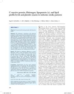
The ganglion cyst is the most common soft-tissue mass of the hand and wrist. The etiology remains unclear, but it is thought to involve mucoid degeneration. Presenting complaints include pain, tenderness, and deforming mass. Treated nonsurgically, ganglion cysts have a high rate of recurrence. Knowledge of regional anatomy is essential for safe surgical excision. Complete excision of the cyst and pedicle as well as a swath of surrounding joint capsule minimizes the risk for recurrence. Copyright © 2002 by the American Society for Surgery of the Hand he most common soft-tissue mass presenting in the hand and wrist is the ganglion cyst, accounting for 50% to 70% of all masses in this anatomic region.1 Ganglion cysts occur at all ages but are most prevalent during the second, third, and fourth decades of life. Women are affected 3 times as often as are men.2 Surgical excision is often required to eliminate the pain and deformity associated with the ganglion cyst.
T
CLINICAL CHARACTERISTICS ain, weakness, and unsightly appearance are the most common presenting complaints of patients with wrist ganglion cysts. At least 10% of patients associate a preceding traumatic event with the appearance of a ganglion cyst, and most investigators theo-
P
From Thomas Jefferson University; and the Division of Hand Surgery, Department of Orthopaedic Surgery, MCP/Hahnemann University, Philadelphia, PA. Address reprint requests to John S. Taras, MD, The Philadelphia Hand Center, PC, 834 Chestnut St, Ste G-114, Philadelphia, PA 19107. Copyright © 2002 by the American Society for Surgery of the Hand 1531-0914/02/0202-0008$35.00/0 doi:10.1053/jssh.2002.33318
102
rize that a history of repeated minor trauma is a factor in their development.1-4 In general, ganglions are firm or rubbery, not fixed to the skin, and usually range in size from 1 to 3 cm.5 They are often nontender to palpation. Wrist motion, particularly at the extremes, exacerbates pain, which is usually dull and persistent. Several investigators have noted smaller ganglions to be more painful than larger ones.3,6 Dellon and Seif7 postulate that an emerging ganglion cyst may compress the posterior interosseous nerve as it passes through the fourth extensor compartment. Less typical presenting symptoms include carpal tunnel syndrome or trigger digit resulting from a volar carpal ganglion cyst’s interference with the flexor tendon sheaths. Office diagnostic procedures include aspiration of the mucinous, jelly-like material, and radiographs, which will reveal any related interosseous component. The differential diagnoses include solid tumors and proliferative tenosynovitis. The examiner will note that a proliferative tenosynovitis will move along with the long extensors or flexors, but a ganglion cyst will remain stationary.
PATHOGENESIS here have been numerous theories about the etiology of the ganglion cyst, and confusion exists about their origin. Eller in 1746 and Volkmann in
T
JOURNAL OF THE AMERICAN SOCIETY FOR SURGERY OF THE HAND 䡠 VOL. 2, NO. 2, MAY 2002
GANGLION CYSTS 䡠 MINOTTI & TARAS
1882 believed that ganglion cysts were herniations of synovial tissue from joints. The belief that ganglion cysts arise de novo from within the connective tissue forms the basis of most modern theories. This idea was first postulated by Ledderhose in 1893. In 1928, Carp and Stout8 furthered this premise by theorizing that ganglion cysts resulted from mucinous degeneration of connective tissue because of chronic damage. The accumulation of collagen fibers, intra- and extracellular mucin, and decreased collagen fibers and stroma cells supported this theory.1 Carp and Stout8 felt that a ganglion cyst’s communication with the joint capsule was the result of later degeneration of the capsule. Soren5 in 1966 reinforced this theory and postulated that a constitutional factor may contribute to the development of ganglion cysts because some patients display multiple ganglion cysts on their wrists and ankles. At present, most investigators agree that ganglion cysts arise from modified synovial or mesenchymal cells at the synovial-capsular interface in response to repetitive minor injury.1 Repetitive stretching of the capsular and ligamentous supporting joint structures appears to stimulate the production of the tissue lubricant hyaluronic acid by fibroblasts at the synovial-capsule interface. The resultant mucin accumulates in small channels, eventually pooling in the ganglion cyst. These observations, however, do not explain why the cyst fluid reaccumulates after aspiration or incomplete excision. There is currently no single theory that fully explains the pathogenesis of ganglion cysts.
103
DORSAL WRIST GANGLION CYSTS he dorsum of the wrist is the most common location of ganglion formation, accounting for 60% to 70% of all hand and wrist ganglion cysts.1,3 Ganglions in this region usually are directly over the scapholunate ligament, though they appear anywhere between the long thumb extensor laterally and the common finger extensors medially (Fig 1). The main body of the cyst is tethered to the wrist capsule by a pedicle. This pedicle often penetrates the capsule and
T
MICROSCOPIC ANATOMY ight microscopy reveals ganglion cysts to be single or multiloculated, having a smooth, shiny lining. Extensive studies by Psaila and Mansel4 by using scanning electron microscopy showed that the walls of ganglion cysts consist mainly of sheets of collagen fibers arranged in multidirectional strata. The walls show sparse, flattened cells resembling fibroblasts, but an epithelial or synovial lining is distinctly absent. Most cysts contain a clear, highly viscous, jelly-like fluid, significantly thicker than synovial fluid. This viscosity is attributed to its high concentration of hyaluronic acid and other mucopolysaccharides.5
L
FIGURE 1. Common locations of dorsal wrist ganglion cysts.
104
GANGLION CYSTS 䡠 MINOTTI & TARAS
FIGURE 2. A representation of a ganglion cyst with its pedicle attachment to the scapholunate ligament.
enters the scapholunate ligament (Fig 2). Angelides and Wallace9 found this to be true in all of 500 (100%) ganglion cysts excised over a 25-year period. Clay and Clement10 showed this in 47 of 62 (76%) cases and found most of the remainder to have arisen from the region of the capitate. In 1985, Gunther6 presented a series of patients in which he showed that an occult scapholunate ganglion cyst could be the cause of wrist pain and tenderness. He postulated that shear stresses generated in the scapholunate ligament incited ganglion cysts for form, and that the patients’ pain was related to pressure that the cyst generated within the ligament.
and 65% originated from the radioscaphoid-scapholunate interval. Volar wrist ganglion cysts can be quite extensive, tracking under the thenar muscles, into the carpal canal, or along the flexor carpi radialis tendon. Adherence to the radial artery is common. Jacobs and Govaers13 noted adherence of volar ganglion cysts to the radial artery in their series of 38 of 78 (54%) surgical excisions. Their study underscores the importance of preoperative Allen’s testing when considering surgery because injury to the radial artery during excision can lead to ischemia of the radial digits in patients in whom an incomplete palmar arterial arch is present.
VOLAR WRIST GANGLION CYSTS NONSURGICAL TREATMENT olar wrist ganglion cysts account for 18% to 20% of all ganglion cysts of the hand and wrist.1 They generally occur under the volar wrist crease, just radial to the flexor carpi radialis tendon (Fig 3). Volar ganglion cysts arise most frequently from the radiocarpal joint or the scaphotrapezial joint.11 In a series of 104 patients with surgically treated volar wrist ganglion cysts, Greendyke et al12 noted that 34% of ganglions originated from the scaphotrapezial joint,
V
he indications for treatment include pain, weakness, and disfigurement. Asymptomatic patients often require only reassurance that the ganglion cyst is not malignant and may occasionally spontaneously regress. Historically, nonsurgical treatment consisted of a sharp blow to the ganglion cyst with a mallet, dictionary, or Bible. Other nonsurgical treatments that have come and gone include heat, radiation, and
T
GANGLION CYSTS 䡠 MINOTTI & TARAS
105
100% recurrence rate in patients who required multiple injections.
SURGICAL TREATMENT fter failure or patient intolerance of conservative therapy, surgery becomes the treatment of choice. Ganglion cyst excision generally takes place as an outpatient procedure. General anesthesia is preferred, though axillary block anesthesia is also suitable. A pneumatic tourniquet ensures a bloodless field. Loupe magnification is encouraged because failure to identify the pedicle and its attachment to underlying ligaments accurately has been strongly associated with a higher rate of recurrence.1,10,11 Dorsal ganglion cysts are approached through a transverse incision centered directly over the ganglion cyst. Extensile skin incisions are rarely necessary because the dorsal skin is freely mobile. The main cyst is mobilized from the surrounding tissues by using tenotomy scissors. Avoid rupturing the cyst because this makes identification and full excision of the pedicle and capsular attachments more difficult. Following the technique described by Angelides,1 a curvilinear incision is made through the capsule adjacent to the cyst, along the proximal pole of the scaphoid. The capsule is elevated and retracted as the capsular incision is continued around the ganglion. At this point, the capsular attachments and associated mucin ducts adherent to the scapholunate ligament can be appreciated. These attachments should be excised tangentially off the scapholunate ligament without cutting into the ligament itself. A wide swath of dorsal capsule is excised with the cyst, greatly reducing the chance of recurrence. Maintaining the integrity of the scapholunate ligament will eliminate the possibility of iatrogenic scapholunate instability. Do not close the capsule primarily or with a flap because such closures only serve to delay early mobilization. For dorsal ganglion cysts presenting with significant pain, excision of the posterior interosseous nerve proximal to the extensor retinaculum can limit pain caused by stretching of the posterior interosseous nerve coursing through the capsule. The posterior interosseous nerve is found between the third and fourth dorsal extensor tendon compartments on the radius and is accompanied by the posterior interosseous artery.
A
FIGURE 3. Typical location of volar wrist ganglion.
injection with sclerosing agents. These methods have been shown to be ineffective, or in the case of sclerotherapy, dangerous. The mainstay of conservative treatment is aspiration of the cyst with a large bore needle followed by injection of lidocaine and a corticosteroid. In the case of dorsal wrist ganglion cyst, up to 80%10 of patients can expect at least a temporary resolution of their symptoms, but recurrence is common. Volar wrist ganglion cysts generally respond poorly to nonsurgical treatment. Wright et al11 noted recurrence in 20 of 24 (83%) patients after aspiration and injection and a
106
GANGLION CYSTS 䡠 MINOTTI & TARAS
FIGURE 4. (A)Volar wrist ganglion dissected to show long pedicle. (B) Connection to radiocarpal joint noted at base of wound. (C) Excision of a side swath of capsule decreases chance or recurrence.
Volar wrist ganglion cysts are approached through a longitudinally oriented incision curving around the radial side of the ganglion cyst. The incision is placed in such a way as to allow proximal and distal extension in pursuit of remote capsular attachments. The palmar cutaneous branch of the median nerve arises 5 cm proximal to the wrist joint and runs distally along the ulnar side of the flexor carpi radialis tendon before piercing the volar carpal ligament to supply sensation to the thenar eminence. Injury to the nerve during dissection may cause anesthesia to this area. The volar ganglion cyst is often associated with the radial artery, sometimes surrounding the vessel. If the artery is minimally involved, then careful blunt dissection can successfully separate the artery from the cyst. When the cyst is intimately adherent to the vessel wall, the
technique described by Lister and Smith14 in 1978 is useful. The ganglion is freed from all surrounding connective tissue and the radial artery is mobilized proximally and distally. As the ganglion is separated from the artery, a 1- to 2-mm cuff of cyst wall is left with the artery to prevent vessel injury. Once the artery is separated and protected, the pedicle can be traced to its capsular attachments to the scaphotrapezial or radiocarpal ligament and excised (Fig 4). Capsular attachments are often more difficult to delineate on the volar wrist than on the dorsal wrist. When the connection between the ganglion and the wrist is difficult to identify, digital pressure over the surgical field causes extrusion of mucin, aiding in the identification of smaller, more tortuous ducts. Alternatively, local anesthetic agent can be injected into the radio-
GANGLION CYSTS 䡠 MINOTTI & TARAS
carpal joint at a remote site and observed to leak through the capsular connection. As is the case for dorsal ganglion cysts, complete excision minimizes the possibility of early recurrence. After release of the tourniquet, meticulous hemostasis is obtained by using bipolar electrocautery, and the wound is copiously irrigated. The skin edges are infiltrated with a long-acting local anesthetic such as bupivacaine 0.5%, and the wound is closed with interrupted 4-0 or 5-0 nylon sutures. A fluffy bandage is applied over a nonadherent dressing. Early motion is encouraged; therefore, no splint is applied unless there has been extensive dissection as is the case with some volar ganglion cysts. The skin is closed and a bandage is applied as previously described. Sutures are removed in 10 to 14 days. The patient wears a loosely
107
placed elastic wrap for 2 weeks to control swelling. Therapy, either guided or a home exercise program, is continued until a full range of motion has been achieved. Postoperative care proceeds in a fashion similar to that described for dorsal ganglion cysts, though a volar wrist splint is used for patient comfort after extensive dissections.
CONCLUSION lthough the etiology of the ganglion cyst remains unclear, surgical treatment can be undertaken with the confidence that patients will have resolution of their symptoms. Excising the cyst, its pedicle, and a portion of the capsule greatly diminishes the risk for recurrence.
A
REFERENCES 1. Angelides AC. Ganglions of the hand and wrist. In: Green DP, ed. Operative hand surgery. 3rd ed. New York: Churchill Livingstone, 1993:2171-2183. 2. Barnes WE, Larson RD, Posch JL. Review of ganglia of the hand and wrist with analysis of surgical treatment. Plast Reconstr Surg 1964;34:570-578. 3. Young R, Bartell T, Logan S. Ganglions of the hand and wrist. South Med J 1988;81:751-760. 4. Psaila JV, Mansel RE. The surface ultrastructure of ganglia. J Bone Joint Surg Br 1978;60B:228-233. 5. Soren A. Pathogenesis and treatment of ganglion. Clin Orthop 1966;48:173-179. 6. Gunther SF. Dorsal wrist pain and the occult scapholunate ganglion. J Hand Surg [Am] 1985;10A:697-703. 7. Dellon AL, Seif SS. Anatomic dissections relating the posterior interosseous nerve to the carpus, and the etiology of dorsal wrist ganglion pain. J Hand Surg 1978;3:326332.
8. Carp L, Stout AP. A study of ganglion, with special reference to treatment. Surg Gynecol Obstet 1928;47:460-468. 9. Angelides AC, Wallace PF. The dorsal ganglion of the wrist: its pathogenesis, gross and microscopic anatomy, and surgical treatment. J Hand Surg 1976;1:228-235. 10. Clay NR, Clement DA. The treatment of dorsal wrist ganglia by radical excision. J Hand Surg [Br] 1988;13B: 187-191. 11. Wright TW, Cooney WP, Ilstrup DM. Anterior wrist ganglion. J Hand Surg [Am] 1994;19A:954-958. 12. Greendyke SD, Wilson M, Shepler TR. Anterior wrist ganglia from the scaphotrapezial joint. J Hand Surg [Am] 1992; 17A:487-490. 13. Jacobs LGH, Govaers KJM. The volar wrist ganglion: just a simple cyst? J Hand Surg [Br] 1990;15B:342-346. 14. Lister GD, Smith RR. Protection of the radial artery in the resection of adherent ganglions of the wrist. Plast Reconstr Surg 1978;61:127-129.