Linköping University Medical Dissertations No. 1257
Arrhythmogenic right ventricular cardiomyopathy Is it right?
Meriam Åström Aneq
Division of Cardiovascular Medicine Department of Medical and Health Sciences Linköping University, Sweden
1
Meriam Åström Aneq, 2011 Cover illustration drawn by Jasmine Åström Aneq
Published article has been reprinted with the permission of the copyright holder. Printed in Sweden by LiU-Tryck, Linköping, Sweden, 2011
ISBN ISSN
978-91-7393-089-5 0345-0082
2
To Jasmine & Linda
You do not know what you can miss before you try Franklin P. Adams
3
4
TABLE OF CONTENTS ABSTRACT .................................................................................................................. 9 POPULÄRVETENSKAPLIG SAMMANFATTNING ....................................... 11 LIST OF ORIGINAL PAPERS................................................................................ 15 ABBREVIATIONS .................................................................................................... 17 INTRODUCTION..................................................................................................... 19 ARRHYTHMOGENIC RIGHT VENTRICULAR CARDIOMYOPATHY .... 21
1. Etiology ............................................................................................................ 21 1:1 Molecular genetics .................................................................................. 21 1:2 Other possible pathogenic mechanisms .............................................. 23 2. Pathophysiology ............................................................................................. 23 3. Histopathology ............................................................................................... 24 4. Clinical features and imaging ..................................................................... 24 4:1 Clinical presentation ............................................................................... 25 4:2 Electrocardiography ............................................................................... 26 4:3 Exercise testing ........................................................................................ 28 4:4 Echocardiography ................................................................................... 28 4:5 Magnetic resonance imaging ................................................................ 29 4:6 Other modalities...................................................................................... 29 5. Differential diagnosis ................................................................................... 30 6. Natural history ................................................................................................ 31 7. Management of ARVC patients .................................................................. 31 7:1 Physical activity ...................................................................................... 31 7:2 Anti-arrhythmic pharmacological therapy ......................................... 32
5
7:3 Implantable cardioverter defibrillator (ICD) ...................................... 32 7:4 Evaluation of relatives ............................................................................ 32 AIM OF THE THESIS .............................................................................................. 35 STUDY POPULATIONS ......................................................................................... 37 METHODS ................................................................................................................. 39
1. Echocardiography .......................................................................................... 39 2. Magnetic resonance imaging ....................................................................... 42 3. Signal-averaged electrocardiogram ............................................................ 45 4. Genetic analysis ............................................................................................. 45 5. Reproducibility studies ................................................................................ 47 STATISTICS .............................................................................................................. 49 RESULTS .................................................................................................................... 51 GENERAL DISCUSSION ....................................................................................... 57
1. Aspects on right ventricular anatomy and physiology .......................... 57 2. Challenges in right ventricular imaging ................................................... 59 2.1 Echocardiography ................................................................................... 59 2.2 Magnetic resonance imaging ................................................................ 62 3. Signal- averaged electrocardiography in ARVC ...................................... 63 4. Genetics in ARVC .......................................................................................... 64 CONCLUSION .......................................................................................................... 67 IMPLICATIONS AND FUTURE PERSPECTIVES ............................................ 69 APPENDIX 1 .............................................................................................................. 71 ACKNOWLEDGEMENTS ...................................................................................... 79
6
REFERENCES ............................................................................................................ 83
7
8
Abstract
ABSTRACT Arrhythmogenic right ventricular cardiomyopathy (ARVC) is an inherited uncommon heart disease, where sudden cardiac death in young seemingly healthy persons may be the first symptom. ARVC is probably more common than previously known, due to diagnostic difficulty. A combination of clinical findings and results from non-invasive as well as invasive investigations is used to diagnose the disease. However non-invasive visualisation of the right ventricle (RV), the predominantly affected part in ARVC, and assessment of its function have some limitations due to RV anatomy and function. The aim of this thesis is the evaluation of new non-invasive modalities in the assessment of right ventricular volume and function with focus on patients with ARVC.
Clinical and non-invasive follow-up of fifteen patients with ARVC during a mean period of 8 years disclosed that progression of the disease is not uniform but generally "slow" in well controlled patients. However individual variation may occur. The determination of RV volume on magnetic resonance imaging relies on short axis (SA) views obtained as a spinoff from assessing short axis LV. These LV slices do not follow the curvature of the long axis of the RV and, segmentation of the inflow and outflow region of the RV is difficult in this view. A new axially rotated long axis acquisition (RLA) was tested and its feasibility in assessment of RV volume was evaluated. The RLA acquisition seems to be able to improve assessment of RV volume and function by reducing the uncertainty in defining the basal slice of the RV. A third study concentrates on analysis of RV regional and general function based on echocardiographic speckle tracking. Two dimensional (2D) longitudinal strain based on speckle tracking as well as tissue Doppler Imaging was used on patients with ARVC, their first degree relatives and in healthy subjects. 2D strain showed a good feasibility in the analysis of RV function in relatives and controls but less in ARVC patients probably due to the progressive myocardial cell death with fibro-fatty replacement of the RV wall. In order to detect and follow up echocardiographic changes in this patient category, an index was developed combining dimensional and functional parameters for the left and for the right ventricle. This index could
9
be used as a tool in the echocardiographic evaluation and serial assessment of RV function. Advances in the molecular genetics of ARVC have provided new insights into the understanding of the disease. Hitherto, 9 candidate genes have been identified. A new mutation in the plakophilin 2 gene was detected in a three generation family. The clinical phenotype related to this mutation was highly variable, from sudden cardiac death (SCD) or anatomical changes at an early age to non-penetrance. This suggested the need to include genetic testing as a part of the diagnostic work-up of ARVC patients and in relatives, to rule out mutation carriers. The studies have evaluated and developed methods for studying the right ventricle with special emphasis on ARVC. With the ultimate goal of preventing sudden death in ARVC, a combination of genetic testing and improved diagnostic methods may create an improved algorithm for risk stratification and selection to prophylactic treatment .
10
Summary in Swedish
POPULÄRVETENSKAPLIG SAMMANFATTNING Plötslig död under ansträngning hos unga personer beror oftast på en dold hjärtsjukdom. Varje fall är en tragedi och innebär, förutom förlust av en ung människas liv, svåra konsekvenser för familj och omgivning. En orsak till plötslig död hos unga är en ärftlig hjärtsjukdom, arytmogen högerkammarkardiomyopati (ARVC), som medför att hjärtmuskelväggen omvandlas till fett- och bindväv vilket ger en benägenhet för rytmrubbningar som kan utlösas av fysisk ansträngning. Sjukdomen är svår att diagnostisera i tidiga stadier med de undersökningstekniker som finns, dels på grund av dess diskreta utbredning initialt, och pga. svårigheter att bedöma höger kammare, den mest drabbade delen av hjärtat vid ARVC. På senare år har 9 olika genetiska avvikelser kopplade till sjukdomen identifierats. Syftet med denna avhandling är att utveckla metoder för att förbättra känsligheten vid diagnostik av ARVC med bildgivande hjärtundersökningsmetoder och vid genetisk undersökning av patienter och anhöriga. Uppföljning av femton patienter med ARVC under en period på 6-10 år har visat att sjukdomsutvecklingen oftast är långsam, men individuella skillnader kan förekomma. Det har också noterats att vänster kammare drabbas tidigt i sjukdomsförloppet. Användning av magnetkameraundersökning för bedömning av höger kammare har visat sig ge värdefull tilläggsinformation. Beräkning av volymen baseras dock på tvärsnittsbilder som är framtagna för vänster kammare och tar mindre hänsyn till höger kammares annorlunda form och rörlighet. Detta försvårar avgränsningen av framförallt det översta (basala) snittet i höger kammare och ger en osäkrare volymberäkning. Vi har utvecklat ett nytt sätt att samla in bilderna genom att utgå från högerkammares längsgående axel mellan klaffen och hjärtspetsen. Bildinsamlingen sker från 6 roterande snitt med 30 graders vridning runt längs axeln. Metoden ökar tillförlitligheten i
11
mätningen av höger kammares volym genom tydligare avgränsning av höger kammares samtliga delar. Ultraljudsundersökningar med nya metoder som mäter storleken av muskelns förkortning (longitudinell och radiell strain) har utvecklats under de senaste åren för bedömning av vänster kammare. Tillämpning av metoden på höger kammare hos en grupp patienter och deras anhöriga samt en åldersmatchad grupp friska frivilliga har visat att metoden är genomförbar i höger kammare och visar avvikande fynd hos patienter med ARVC. Anhöriga kunde däremot inte skiljas från friska med denna metod. För att väga samman tidiga diskreta förändringar och följa deras utveckling har vi tagit fram ett ekokardiografiskt index för höger respektive vänster kammare baserat på diameter och funktion.
Genetisk diagnostik av ARVC har nyligen startat i Sydöstra sjukvårdregionen, varvid en tidigare inte beskriven genetisk avvikelse som orsakar ARVC har påvisats. Undersökning av familjemedlemmar i tre generationer med denna mutation har visat stor variation i sjukdomsbilden, från plötslig död hos en 18 årig man till helt friska anlagsbärare. Dessutom visar studien vikten av genetisk och klinisk bedömning i kombination med känsliga metoder för hjärtundersökningar för att ställa rätt diagnos vid ARVC.
12
13
14
List of papers
LIST OF ORIGINAL PAPERS This thesis is based on the following original publications, which will be referred to in the text by their Roman numerals: I
Åström Aneq M; Lindström L; Fluur C; Nylander E. Long-term follow-up in arrhythmogenic right ventricular cardiomyopathy using Tissue Doppler Imaging. Scand Cardiovasc J 2008; 42(6): 368-74
II
Åström Aneq M; Nylander E; Ebbers T; Engvall J. Determination of right ventricular volume and function using multiple axially rotated MRI slices. Clin Physiol Funct Imaging 2011; 31(3): 233-9
III
Åström Aneq M; Engvall J; Brudin L; Nylander E Evaluation of right and left ventricular function using speckle tracking echocardiography in patients with arrhythmogenic right ventricular cardiomyopathy and their first degree relatives. Submitted
IV
Åström Aneq M; Fluur C; Rehnberg M; Söderkvist P; Engvall J; Nylander E; Gunnarsson C. Novel plakophilin2 mutation in a three generation family with arrhythmogenic right ventricular cardiomyopathy Submitted
(Articles reprinted with permission)
15
16
Abbreviations
ABBREVIATIONS ARVC SCD TFC RV LV LDAC ECG SAECG LBBB RBBB PVC RVOT-T LVOT VTI RVOT/BSA RVIT/BSA WMSI TAPSE TAPSV Sw 2D-strain AVO AVC MRI SA RLA bSSFP ICD
Arrhythmogenic right ventricular cardiomyopathy Sudden cardiac death Task Force Criteria Right ventricle Left ventricle Left-dominant arrhythmogenic cardiomyopathy Electrocardiogram Signal-,averaged electrocardiogram Left bundle branch block Right bundle branch block Premature ventricular contraction Right ventricular outflow tract tachycardia Left ventricular outflow tract velocity time integral Right ventricular outflow tract related to body surface area Right ventricular inflow tract related to body surface area Wall motion score index Tricuspid annular plane systolic excursion Tricuspid annular plane systolic velocity Systolic velocity in the myocardial wall Two dimensional strain Aortic valve opening Aortic valve closure Magnetic resonance imaging Short axis Rotated long axis Balanced Steady-state free precession sequence Implantable cardioverter defibrillator
17
18
18
Introduction
INTRODUCTION Sudden cardiac death (SCD) is still a problem although mortality from cardiovascular disease has decreased in recent decades. In Europe as well as in the United States, the incidence of SCD is estimated at almost 50-100 cases/100.000 people per year with some geographic variation [1, 2]. Resuscitation is still unsuccessful in 70% to 95% of the cases of Out-of-Hospital Cardiac Arrest [3, 4]. Sudden cardiac death is defined as unexpected death due to heart problems within a short time period, generally one hour from the onset of symptoms, in a person without any prior fatal condition[5]. For unwitnessed deaths, the World Health Organization definition extends to up to 24 hours [6]. The main cause of SCD is coronary heart disease but in people younger than 35 years, the leading cause is arrhythmia due to cardiomyopathies, abnormalities of the cardiac conducting system or coronary artery anomalies. One cardiomyopathy recognised as a cause of SCD is arrhythmogenic right ventricular cardiomyopathy (ARVC). It is considered as a cause of SCD in 3% of young athletes in the US and up to 20% in northern Italy. This geographical variation may be due to a more prevalent genetic abnormality or to more investigations with focus on this disease being performed in Italy. In many cases, sudden death is the first manifestation of ARVC. In a cohort of 100 patients with ARVC, Corrado et al. found that sudden death was the first manifestation of the disease in 22 patients [7]. Similar numbers were reported by other authors where SD accounted for 20-75% of the fatal outcome in ARVC [8, 9]. The prevalence of ARVC is estimated to 1 per 1000 to 1 per 5000 [10]. However this value could be higher as in many cases the disease is first recognized post mortem. Although ARVC is relatively uncommon in the general population, the increased risk for SD during exercise in adolescence or young adulthood is not only devastating for families, but also has impact on the community and the system of medical care.
19
19
The research in this thesis focuses on the diagnosis and follow-up of arrhythmogenic right ventricular cardiomyopathy using anatomical and functional myocardial parameters obtained from echocardiography and magnetic resonance imaging as well as initial experiences from molecular genetic testing.
20
20
ARRHYTHMOGENIC RIGHT VENTRICULAR CARDIOMYOPATHY Arrhythmogenic right ventricular dysplasia/cardiomyopathy was first described by G Fontaine et al. 1978[11]. Shortly thereafter, in 1982, Marcus et al. described 24 cases of ARVC [12] followed by multiple case reports from all over the world. ARVC is a disease of cardiac muscle causing replacement of muscle cells by fatty tissue and fibrosis mainly in the right ventricle. The disease was originally considered as a dysplastic process (ARVD) with congenital absence of ventricular myocardium but clinical and pathological studies have shown that the abnormalities are acquired and are of a progressive character.
1. Etiology
1:1 Molecular genetics In 30-50% of cases, ARVC has a familial occurrence, usually with an autosomal dominant inheritance pattern [13, 14]. A rare autosomal recessive variant, Naxos disease, with similar myocardial involvement and typical palmoplantar keratosis and wooly hair has also been described [15] There is evidence that ARVC is predominantly a disease of the desmosome. Five genes encoding the desmosome have been identified hitherto, in addition to 5 other genes or loci that have not yet been tracked to individual gene modification. Desmosomes are protein structures in the cell membrane permitting cell-to-cell adhesion. They occur in almost all solid tissues, but diseases related to mutation in these proteins are manifested only in the heart muscle cells and in the skin. This has been explained by continuous mechanical stress of the epithelium and the myocardium that at least in part alters the biomechanical behaviour of cells leading to myocyte detachment and 21
21
death. Two of the desmosomes containing proteins are intercellular; desmoglein-2 and desmocollin-2 and three are intracellular; plakoglobin, plakophilin-2, and desmoplakin (Fig. 1). In addition to cell adhesion, they have a function in cell-cell communication and tissue differentiation. The first identified gene in the dominant form of ARVC (ARVC2) is coding for the ryanodine receptor-2. This gene is now known to be present in cathecholaminergic polymorphic ventricular tachycardia (CPVT) suggesting a CPVT phenocopy rather than true ARVC. [2, 16-18]. Fig. 1. Schematic figure of Desmosomal Structure
The desmosomal complex comprises two intercellular proteins: desmoglein (DSG)-2 and desmocollin (DSC)-2. The intracellular portions of these proteins interact with three other proteins: junctional plakoglobin (JUP), plakophilin (PKP)-2 and desmoplakin. The desmoplakin terminal part anchors Desmin filaments, the cytoskeleton. Modified from Sen-Chowdhry et al. [19].
In addition, two other non-desmosomal protein mutations have been reported to cause ARVC; the transmembrane protein 43 encoded by the TMEM43 gene [8], and the transforming growth factor beta -3 (Tb3), a cytokine-stimulating fibrosis and modulating cell adhesion [20]. The causative effect of these two mutations in relation to ARVC is still controversial. Patients carrying multiple mutations in a single gene (compound heterozygosity) or in two different genes (digenic heterozygosity) are more frequent than previously suspected (almost 13%) and this may contribute to the complexity of disease inheritance and variable penetrance of ARVC. This is
22
22
also a reason for an extended mutation screening beyond the identification of one mutation especially in families with a particularly severe phenotype [21]. However, it is noteworthy that failure to identify a mutation in the known genes does not exclude the disease and additional contributing gene mutations may still await detection.
1:2 Other possible pathogenic mechanisms 1:2:1 The role of inflammation in ARVC is still unresolved. The presence of inflammatory infiltrates in histology specimens particularly during the acute ‘hot phases’ of the disease suggests that ARVC could be either a sequel or trigged by a myocarditis. However if the inflammation is causative or just an effect of cell death is still unkown [22]. The causative role of viral infections [23, 24] and a genetic predisposition to infection/immune reaction in ARVC patients has also been proposed.
1:2:2 The mechanism of cell death by apoptosis has been discussed as apoptotic nuclei were found in up to 35% of patients in some studies [25, 26], but the role of apoptosis as a pathogenic pathway in ARVC is still to be defined.
2. Pathophysiology The prevailing hypothesis on desmosomal gene mutations causing ARVC is based on the effect of mechanical stress leading to the weakness of the intercellular junction causing disruption and degeneration of myocytes and their replacement by fibrofatty scar tissue. In this structural model, environmental factors such as exercise or inflammation could exacerbate impairment of cell adhesion and hasten disease progression. Progressive atrophy of the myocardium in ARVC occurs from the subepicardium to the endocardium. The residual islands of surviving myocardial cells left within the affected fibro-fatty area constitute a substrate for ventricular arrhythmias. Initially the disease has a regional distribution but
23
23
later on it becomes more global. Transmural atrophy of the myocardium causes aneurysmal dilatation and wall motion abnormalities which lead, in advanced stages, to heart failure. Although ARVC was initially considered to be strictly confined to the right ventricle, autopsy and sensitive imaging techniques have shown that most patients with ARVC have biventricular involvement [27-29]. It has been suggested that regular intense exercise causing volume overload and cavity enlargement of a thin-walled RV with high distensibility is a reason for the predominating RV involvement of ARVC. LV changes usually affect both the septum and the LV free wall, often regionally, with a predilection for the posteroseptal and posterolateral wall.
3. Histopathology Based on histology findings in different cohorts, two histopathologic variants have been described [30]; fibrolipomatosis type I with almost only fatty replacement and a small amount of fibrosis surrounding the surviving myocardial cells leading to an increased wall thickness, ‚pseudohypertrophy‛ of the RV, and the fibrolipomatosis type II with extensive replacement of myocytes by fibrosis and fatty tissue leading to thin wall and saccular aneurysm [31].
4. Clinical features and imaging ARVC is generally diagnosed in young adults. In at least 80% of cases the diagnosis is made in patients below 40 years of age, although the disease has been described up to the ninth decade and in children [32, 33]. The disease is extremely uncommon below the age of 10 years. For unclear reasons, men are more frequently affected than women [34]. For a definite diagnosis, histology compatible with ARVC on specimens from autopsy, an explanted heart or from endomyocardial biopsy is required. However, diagnosis by endomyocardial biopsy is difficult due to the patchy involvement of the
24
24
disease. The septum, where the biopsy is usually taken, is not often involved. This, in combination with multiple potential etiologies of arrhythmias of RV origin, and difficulties in the assessment of RV structure and function, has prompted the establishment of standardized diagnostic criteria. The Task Force Criteria (TFC) proposed by Mc Kenna et al [35] are subdivided into major and minor criteria according to the specificity of their association with ARVC. They are based on structural, histological, electrocardiographic, arrhythmic, and familial features of the disease (Appendix 1).
Occasionally, the left ventricle may be the predominant site of involvement. Left-dominant variants of ARVC (LDAC) that primarily affect the left ventricle have been identified in vivo and on post-mortem examination [36, 37]. Clinical features include arrhythmia of RBBB morphology and progressive structural abnormalities of the left ventricle. This disorder can be difficult to differentiate from other causes of left ventricular dysfunction. Marked arrhythmias in conjunction with relatively mild LV dysfunction may suggest the presence of regional LDAC. Accordingly, ARVC may manifest as cardiac failure involving both ventricles and may be difficult to distinguish from other forms of global heart failure.
4:1 Clinical presentation The most common clinical manifestations in ARVC are related to arrhythmia and conduction disturbances. Ventricular arrhythmia with an origin in the right ventricle display left bundle branch block morphology (LBBB). Advanced disease with extended left ventricular involvement may display ventricular ectopy with right ventricular branch block (RBBB) morphology. A history of syncope or dizziness, especially during exercise can be reported. Over time, symptoms related to heart failure, such as dyspnoea, oedema and fatigue may occur. However, ARVC is frequently asymptomatic, with the initial manifestation being sudden unexpected death.
25
25
4:2 Electrocardiography The electrocardiogram (ECG) is one of the most important diagnostic methods in ARVC. Abnormalities of depolarisation, conduction and repolarisation secondary to atrophy of ventricular walls reflect the pathophysiology of the disease.
4:2:1 Twelve lead-ECG T-wave inversion in anterior precordial leads (beyond V1) in the absence of RBBB is a sensitive finding in ARVC, reported to be present in between 37 and 81% of cases [38, 39], but the common pattern of T-wave inversion in ischemic heart disease and other cardiopulmonary diseases reduces its specificity. However, in the absence of RBBB, the presence of T-wave inversion in V1-V3 in young adults or middle aged subjects who have no apparent heart disease is uncomman, less than 3% in the study by Marcus [40]. Furthermore T-wave inversion in combination with ectopic ventricular beats of LBBB morphology should raise the suspicion of ARVC. Depolarisation and prolongation of conduction involving right precordial leads in the absence of RBBB can be detected as prolongation of the QRS complex (exceeding 110 ms in V1-V3). This was seen in 35 to 76% of cases [41, 42]. The epsilon wave (Fig. 2a) is a distinct deflection between the end of the QRS complex and the onset of the T wave. The presence of an epsilon wave is a major criterion but detected only in 1-25% of cases [43]. Finally selective prolongation of the S-wave duration in V1-V3 is seen in 34 to 71% of patients [44]. The variable presence of these findings in several studies can be related to the extent of right ventricular structural and functional abnormalities in the patients studied [45].
4:2:2 Signal-averaged electrocardiogram (SAECG) SAECG permits the registration of low-amplitude potentials within the terminal portion of the QRS complex (late potential) that are not of sufficient amplitude to be visible on the 12-lead ECG. ARVC was one of the first pathologic non-ischemic disorders in which the presence of late potentials were identified [46] (Fig. 2b). 26
26
Fig. 2 a.
ECG showing Epsilon wave
b.
Pathological late potentials on the SAECG
27
27
4:2:3 Ambulatory ECG monitoring This is performed to detect spontaneous arrhythmia. Monitoring is usually done for 24-48 hours, but in symptomatic patients longer registrations may be indicated to capture arrhythmia. The presence of more than 1000 premature ventricular contractions (PVC) of LBBB morphology on 24h Holter monitoring is a minor criterion in ARVC. When the subject has a family member with confirmed ARVC the presence of 500 PVC of LBBB type constitutes a major diagnostic criterion for ARVC . The presence of sustained or non-sustained ventricular tachycardia of LBBB morphology can also be detected [47].
4:3 Exercise testing Exercise testing in general is recognized for its ability to unmask ventricular arrhythmia during physical exercise. However, in ARVC, the diagnostic utility in clinical decision making and in follow-up remains to be established. It is noteworthy that lack of arrhythmia during exercise should not be considered as reassuring [48].
4:4 Echocardiography Identification of right ventricular morphological abnormalities by echocardiography is difficult as the abnormalities due to ARVC may be limited to small regions in the ventricular wall. Typical echocardiographic findings are often localized to the inflow and/or outflow tracts. The apex region was described earlier as part of the affected ‚triangle of dysplasia‛ but recent observations have shown that involvement of the apex is a late finding. The main changes are regional dilatation of the RVOT and/or the inflow part. Structural wall abnormalities such as localized wall tinning are important diagnostic features but hard to find in the trabeculated thin right ventricular wall. Micro-aneurysms in the anterior or inferior RV walls are specific for ARVC. In other forms of ARVC, pseudo-hypertrophy of the RV wall with a 28
28
thick moderator band can be seen [49]. In this variant, histology shows a predominance of fatty tissue and less fibrosis. Myocardial dysfunction is regional in the early stages without apparent effect on global RV function. Early changes in regional function can easily be overlooked. Later on, dilatation and dysfunction of both ventricles may occur accompanied by mural thrombosis within aneurysms or in the atrial appendages when heart failure is complicated by atrial fibrillation.
4:5 Magnetic resonance imaging MRI offers an extensive visualisation of the right ventricular chamber, permitting the detection of microaneurysm and wall bulging. Furthermore, the calculation of right and left ventricular volumes and ejection fraction is more precise with MRI compared to echocardiography [50]. Myocardial delayed enhancement MRI can detect fibrosis especially in the advanced form [51]. However, the detection of intramyocardial fat in the RV wall is unreliable. The presence of pericardial fat in contact with the thin RV wall as well as the high prevalence of adipose infiltration in normal hearts [52] makes this finding less specific for the diagnosis of ARVC.
4:6 Other modalities
4:6:1 Computer Tomography Most centers use MRI in the diagnosis and follow up of ARVC patients. However in many cases the prophylactic implantation of a cardioverter defibrillator (ICD) is necessary to prevent SCD. After such implantation, MRI is contraindicated but CT may help in the serial assessment of right ventricular function [53].
29
29
4:6:2 Angiography Angiography was previously the gold standard for detection of RV abnormalities [54]. However, recent development of new methods has obviated the need for angiography.
4:6:3 Electrophysiologic study (EPS) EPS may have a role in the differential diagnosis of arrhythmia of right ventricular origin [55, 56].
5. Differential diagnosis In the presence of RV tachycardia and /or dilatation of the RV, other structural diseases should be ruled out e.g. congenital heart disease such as left- right shunts at the atrial level, and acquired diseases such as pulmonary hypertension, myocardial infarction and systemic inflammatory and infiltrative diseases[57]. Idiopathic right ventricular outflow tract tachycardia (RVOT-T) is a benign, non-familial disorder that is often difficult to differentiate from ARVC in early stages, especially since recent MRI studies have shown structural abnormalities in the outflow tract in RVOT-T. A definite diagnosis may not be possible at first presentation. Case studies have reported patients that have been assigned a diagnosis of RVOT-T but later developed typical features of ARVC [58]. Brugada syndrome is another disorder that can be difficult to differentiate from ARVC in some cases. Clinical and histopathological studies have shown a definite overlap between Brugada and ARVC [59, 60]. Myocarditis, usually viral and predominantly involving the RV may be difficult to differentiate from ARVC particularly during the acute phase with arrhythmias [61, 62]. For differential diagnosis, the progress during follow-up is important. The RV may dilate as a normal adaptation to regular intensive physical activity in the so called ‛athlete´s heart‛. This has to be considered in the differential diagnosis of ARVC [63, 64].
30
30
6. Natural history Four distinct phases have been described in the natural history of ARVC [7] 1. The early ‚concealed‛ phase is associated with minor ventricular arrhythmia and subtle structural changes. Patients are often asymptomatic, but nonetheless at risk of SCD especially during physical activity. 2. In this stage, an overt electrical disorder is manifest, with symptomatic ventricular arrhythmia and more obvious morphological and function abnormalities of the right ventricle. 3. RV failure caused by disease progression. 4. Biventricular heart failure in advanced stages which can be difficult to differentiate from dilated cardiomyopathy. However the evaluation of familial patterns suggests that a distinct stepwise progression to more advanced stages of disease seldom occurs.
7. Management of ARVC patients
7:1 Physical activity Athletes with probable or definite diagnosis of ARVC should be excluded from most competitive sports, according to the 36th Bethesda conference [65] In the larger group of non competitive young patients who are asymptomatic or mildly symptomatic there is usually a desire to have an active recreational lifestyle. However the definition of low intensity activity is difficult. Recommendations on exercise need to be individualized for each patient and the intensity of physical activity considered. As a help for the clinician, Maron et al. categorized sports activities into high, moderate, and low level intensity [66]. Participation in intensive exercise should be avoided even in the presence of antiarrhythmic protection afforded by an implanted ICD [67]
31
31
7:2 Anti-arrhythmic pharmacological therapy Pharmacological therapy is the recommended initial treatment for a patient with mild, well tolerated arrhythmia. Treatment consists of beta blockers and class I and class III antiarrhythmic drugs. Sotalol has been reported to be the most effective pharmacological antiarrhythmic treatment in ARVC [68]
7:3 Implantable cardioverter defibrillator (ICD) ICD is an effective method for terminating malignant ventricular tachyarrhythmias. However the most relevant question is whether there is a balance between the benefit of preventing arrhythmic death and the risk of complications and side effects from ICD implantation. A correct diagnosis of the disease on the basis of detailed investigations is essential before ICD implantation is considered. Recommended indications for ICD implantation in ARVC include secondary prevention after survived cardiac arrest or documented sustained VT. ICD therapy as primary prophylaxis in asymptomatic patients or relatives with high risk profile must be based on individual risk assessment because of the lack of data supporting this approach. The main indication for primary prophylactic ICD is currently based on either history of SCD in early age in the family, or evidence of extensive RV dysfunction as well as involvement of the LV. In a cohort of 42 ARVC patients with ICD, Rogin et al. showed that 78% of patients experienced at least one appropriate discharge during an average of 3,5 years of follow-up [69]. Similar findings were reported by other authors [70-72].
7:4 Evaluation of relatives First degree relatives of deceased or living patients with ARVC should undergo a cardiac evaluation[13]. Modified Task Force Criteria have been proposed because the original TFC are highly specific but not sensitive enough to evaluate relatives who may have an early form of the disease (Appendix 1) 32
32
[13, 73]. Further investigations are needed regarding the sensitivity and specificity of these criteria, the need and frequency of repeated follow-up and risk stratification of relatives.
33
33
34
34
Aim of th thesis
AIM OF THE THESIS The general aim of this thesis is to investigate how non- invasive methods may improve diagnosis in arrhythmogenic right ventricular cardiomyopathy.
Specific aims: To study the progress and the rate of change in echocardiographic parameters during long term follow-up in patients with ARVC (Paper I) To explore the value of using tissue Doppler imaging in the follow-up of patients with ARVC (Paper I) To evaluate whether a new axial orientation modality for the assessment of RV volume and function by MRI will help to overcome the limitations of the standard short axis orientation (Paper II) To study the feasibility of using two dimensional strain analysis based on speckle tracking applied to the RV (Paper III) To test the ability to detect regional RV dysfunction at early stage, in relatives of ARVC patients using myocardial strain based on speckle tracking (Paper III) To describe the phenotype of a new mutation in the plakophilin 2 gene in a three generation family with ARVC (Paper IV)
35
35
36
Study populations
STUDY POPULATIONS Healthy controls (II,III) Totally 25 healthy volunteers were included, whereof 20 were examined by both MRI (II) and echocardiography with analysis of myocardial strain (III). Three of the remaining subjects underwent MRI alone and two had only echocardiography. The healthy subjects were recruited by advertisements on the university web page and among medical students. All were asymptomatic and lacked family history of premature cardiovascular disease. No one received cardiac medication. All underwent a bicycle ergometer exercise test to confirm the absence of heart disease. Those who had abnormal ECG or exercise test were excluded.
ARVC patients (I,III,IV) In paper I, all patients from the South East region with a diagnosis of ARVC for more than 6 years were included. There were fifteen patients, 9 male and 6 female. The diagnosis was based on the conventional Task Force Criteria. Patients were examined regularly according to a follow-up program. In this analysis we focused on the first and last echocardiographic examination during the study period. Two of these patients were also reported in paper IV. In paper III, we studied all males with an ARVC diagnosis, which included nine patients from study I, and eight patients with an ARVC diagnosis of less than 5 years.
First degree relatives (paper III) Since 1995, a screening protocol for first degree relatives (children, parents or siblings) of ARVC patients has been followed. In this cohort nineteen males, 18 years or older were included and underwent echocardiogram with focus on right ventricular function.
37
37
Table 1. Characteristics of the male (M) and female (F) population studied. Patients I
Relatives
Controls
15(9M*,6F) 22-58 yr ** (mean 40) 23 M‡ 22-64 (mean 39)
II III
17 M 32-70 yr † (mean 49)
19 M 19-73 yr (mean 29)
IV
8 subjects (4M, 4F) in an ARVC affected family
22 M 24-66 yr (mean 36)
* All included in paper III, and two included in paper IV. ** Age at the time of the first investigation † Age at the examination ‡ 20 of these controls are included in paper III
Ethical approval for studies (I,II,III,IV) was obtained from the Regional Ethical Review Board in Linköping.
38
38
Methods
METHODS 1. Echocardiography Paper I, III, IV The echocardiogram was performed with the subject in the left lateral decubitus position. A 2-dimensional, colour, pulsed and continuous-wave Doppler examination was recorded following the recommendations of the American Society of Echocardiography for transthoracic studies [74]. We used the ultrasound systems Acuson 128XP (Acuson corporation, Mountainview, CA,USA) for the first examination in paper I and Vivid 5 or Vivid 7 (GE Medical Systems, Horten, Norway) for the last examination in paper I and in paper III. Data were obtained from multiple standard views of the left and right ventricle (Figure 1, paper I). At least three regular beats of each view were recorded during quiet respiration. In paper I, wall motion score index (WMSI) was assessed in a sixteen (LV) and seven (RV) segment model. Each segment was classified according to a scale with 6 levels: 0=not visualised, 1= Normal, 2=Mild hypokinesia, 3=Severe hypokinesia, 4=Akinesia and 5= Dyskinesia. A score was calculated for each ventricle as an average value of analysed segments. The presence of isolated sacculation (S) and wall thinning (T) was reported. All analyses were performed by a single observer.
1:1 Annular motion analysis by M-mode A 2-dimensionally–guided M-mode cursor was placed on the lateral or the septal part of the mitral annulus to record the annular motion towards the cardiac apex in systole from a standard apical 4-chamber view (paper I). An identical recording was obtained from the lateral (paper I,III) and posterior (paper I) part of the tricuspid annulus. Care was taken to orient the beam perpendicularly to the motion of the annular structure. The total 39
39
systolic displacement was measured from end-diastole (the initial deflection of the QRS complex in the ECG) to the highest point of contraction as initially validated by Kaul et al. [75].
1:2 Longitudinal strain based on speckle tracking The strain measured by two dimensional echocardiography is called Langrangian strain (e) and is defined as the change of myocardial fibre length during stress at end-systole compared to its original length in a relaxed state at end-diastole. Negative strain indicates fibre shortening or myocardial thinning, whereas a positive value describes lengthening or thickening. The two dimensional loops from routine echocardiographic examinations were processed offline. The end-systolic frame was identified from by definition on the parasternal or apical long axis (3-chamber) view, where the aortic valve is ‚optimally visible‛. The time point of aortic valve closure was marked. The four chamber view with focus on the left and then on the right ventricle was recorded with a frame rate ranging between 40 to 70 frames/s. All images were stored digitally and analyzed on the Echo Pac work station (GE Medical Systems, EchopacPC, version 6.0.1). A complete cardiac cycle was required for processing. Offline analysis began with tracing the endocardial border. The software then automatically defined an epicardial and midmyocardial line and processed all frames of the loop based on the identification of natural acoustic markers in the myocardium. Myocardial deformation was defined as strain an expressed in terms of percent shortening. The length of each wall was automatically divided into 3 segments; base, mid and apex. Since the apex of the RV can be difficult to expose on echocardiography, we chose to focus on the basal and mid ventricular segments (Fig. 3)
40
40
Methods
Fig. 3. Determination of longitudinal strain in an apical four chamber view.
Left upper panel:The lateral wall of the right ventricle is divided into 3 segments; base, mid and apex. M-mode representation of peak systolic strain (left lower panel). The right upper panel shows time –curves of longitudinal strain for each myocardial segment, and the right lower panel displays the calculated values.
1:3 Echocardiography index Multivariate discriminant analysis was used to detect differences between ARVC-patients and controls in order to investigate the relative role of parameters from the right and left ventricle for the discrimination of the two groups. The parameters used for the right ventricle were right ventricular outflow and right ventricular inflow tract related to body surface area (RVOT/BSA, RVIT/BSA), tricuspid annular plane systolic excursion (TAPSE), tricuspid annular systolic velocity and longitudinal strain based on speckle tracking of the basal and mid RV free wall segments. For the left ventricle the parameters were LV diameter, left ventricular outflow tract velocity time integral (LVOT VTI), mitral annular systolic velocity from the lateral and septal walls, strain based on speckle tracking of the basal and mid segments of the septum and lateral wall. Significant components were reassembled to create separate indices for the right and left ventricles and recalculated for each individual showing the distribution of the first degree relatives related to patients and healthy controls. 41
41
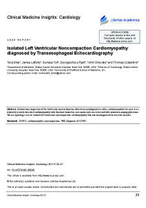
2. Magnetic resonance imaging Paper II The MRI image acquisition was performed in a Philips 1.5T Achieva scanner (Philips Healthcare, Best, the Netherlands), using a five-element cardiac synergy surface coil. Cine-MR, synchronised using retrospective ECG gating, was performed with a balanced steady-state free precession sequence (bSSFP). bSSFP uses a high number of excitation pulses to provide a high signal-tonoise ratio (SNR) and better definition of structure compared with gradient echo-sequences. The echo time (TE) was 1.5 ms, repetition time (TR) 3.0 ms, flip angle 60º and pixel size 1.25x1.25 mm2 for all image acquisitions. Three long axis planes of the left ventricle (2- and 4- chamber views as well as the apical long-axis view) were obtained for orientation. The short axis plane was planned on the LV, covering the entire heart with on average 19 (range 17-25) short axis, 8mm thick slices. Using sensitivity encoding (SENSE) reconstruction, each SA slice took 6 heart beats to acquire. A long axis was drawn in the RV cavity joining the middle of the tricuspid annulus to the RV apex. Six long axis slices were acquired, by incrementally rotating 30 º rotation around this original RV long axis. Every long axis slice was acquired during 12 hearts beats. Six slices were considered as a suitable compromise between potential spatial information and time spent for image acquisition. The order of the sequence of axial and short-axis acquisition was assigned randomly for each patient. In addition, two-dimensional cine through-plane PC-MRI velocity data were acquired in a plane perpendicular to the main flow direction in the ascending aorta (AoA), just downstream from the aortic valve (AoV). All scans were performed by a single operator. To test the accuracy of the volume estimation for different geometries, a phantom study was performed using both SA and RLA acquisitions. We used symmetrical objects such as a bottle and a pear, in addition to a banana simulating the irregular crescentric right ventricular cavity. For all phantoms, six RLA slices were acquired, and sufficient SA slices to cover the complete phantom. Imaging parameters were identical for both the RLA and SA acquisitions (Fig. 5). True phantom volumes were measured immediately after the MR investigation, by immersing the objects in a beaker of water. 42
42
Methods
Fig. 4. 3D visualization of the six slices rotated around the long axis. A. depicts a pear and B. the right ventricular chamber as seen from the apex with the septum at the left.
Image analysis All calculated volumes from the phantom studies were based on still images and were, as well as the cine images, evaluated using the freely available analysis software Segment (v1.702, http://segment.heiberg.se) (Fig. 3). Two observers measured all the data independently. For the short axis datasets, a consensus was reached before segmentation on which basal slice to include. The volume of added, as well as of deleted slices was analyzed independently and the stroke volume recalculated. For the left ventricular volumes, at least half of the periphery of each ventricular slice had to be present in each time frame to allow its inclusion in the volume. When this rule was applied, a consensus procedure was not necessary for the left ventricle. Aortic flow volume (stroke volume) was calculated by multiplying aortic cross sectional area with mean flow from a slice placed cranial to the coronary ostia.
43
43
Fig. 5 The right ventricular end diastolic ED (A-C) and end systolic ES(B-D) slices on short axis (upper) and rotated long axis (Lower) acquisitions.
A
B
C
D
44
44
Methods
3. Signal-averaged electrocardiogram Paper I, III, IV SAECG is recorded using bipolar XYZ ECG leads. The X lead is positioned at the fourth intercostal space in both midaxillary lines. The Y lead is positioned on the superior aspect of the manubrium and on either the upper left leg or left iliac crest and the Z lead between the fourth intercostal space (V2 position) and directly posterior on the left side of the vertebral column. The signal is filtered using a high and low pass-filter (25 or 40-250HZ). Between 250- 400 beats are averaged to improve the signal-to noise ratio and facilitate the detection of low-amplitude potentials. Analysis of the filtered QRS complex includes 1) the filtered QRS duration(fQRS) ; 2) the root-mean-square voltage in the last 40 ms of the filtered QRS (RMS 40ms); and 3) the duration of the high frequency low-amplitude signal of the filtered QRS not exceeding 40 μV (HFLA < 40 μV) Criteria for normal values in SAECG were published in 1991 and 1996 by a joint committee of the AHA and the ESC [76, 77] and are the following: 1) fQRS ≤ 114ms 2) RMS 40 >20 μV 3) HFLA 40 2cm/m2) 6/15 (40%) RVIT dilatation 10/15 (67%) Sacculation/bulging 2/14 (14%)
Follow-up n/total % 8/15 (53%) 3/15 (20%) 12/15 (80%) 10/15 (67%) 7/15 (47%) 11/15 (73%) 5/14 (36%)
Echocardiographic results of the right ventricular function during follow-up are shown in (Fig.6)
51
51
Fig. 6. Variation in tricuspid annular plane systolic excursion (TAPSE) and systolic velocity on tissue Doppler imaging at the first (1) examination and at the follow-up (2)
A
B
A. TAPSE: Tricuspid annular motion on M-mode at the first (1) and the last (2) examination B. TALs: Tricuspid lateral systolic annular velocity at the first (1) and the last (2) examination
The data indicate a progression of the recorded signs of disease. The rate of progression was generally slow, but with individual variation.
Paper II A method for the determination of RV volume using MRI was developed and tested in phantoms and healthy individuals. The phantom result shows no significant difference between volumes acquired with the two MRI methods and the reference method which based on the displacement of water (Table 3).
Table 3. Volume of 3 phantoms measured with MRI in short axis and rotated long axis orientations compared to the displaced volume of water.
Bottle Pear Banana
Short axis 515 ml 216ml 126ml
Rotated long axis 516 ml 214ml 132ml
52
52
Real volume 515ml 215ml 129ml
Results
A consensus between the two observers whether to include or exclude the basal slice from the RV volume was necessary in 9/23 (39%) controls leading to a difference in EDV of 6-20ml (8-21%) or ESV 12-21ml (20-27% of the total volume). After the consensus, EDV, ESV, SV and EF for the right ventricle did not differ between the SA and RLA views with p-values in excess of 0.05. Thus, it was confirmed that a problem with the short axis method is the difficulty to define the basal limits of the right ventricle and that in many cases, consensus was needed to obtain an acceptable reproducibility. This seems to be overcome with our axial method that is planned to be tested in different RV pathologies in the future.
Paper III Sensitive methods for the detection of early signs of ARVC in patients and their relatives are needed. The development of genetic methods cannot replace phenotypic characterization but provide additional building blocks for a deeper understanding of the disease. In this study, we compared ARVC patients with first degree relatives and healthy age- matched controls. Table 4. Clinical and electrocardiographic characteristics of the study population
Age (years) Arrhythmiarelated symptoms T-inversion beyond V1 Late potentials on SAECG
ARVC (n=17) Median (range or %)
Relatives (n=19) Median (range or %)
Controls (n=22) Median (range or %)
P-values
49 (32-70)
29 (19-73)
36 (24-66)
0,44
13 (76%)
2 (11%)
0
13(76%)
0
0
9/14* (64%)
3/19 (16%)
0
SAECG: Signal-averaged electrocardiogram. *SAECG was not recorded in 3 patients due to frequent arrhythmia or ongoing cardiac pacing.
53
53
Using discriminant analysis on left and right ventricular echocardiography parameters, an LV and an RV index was developed based on data from ARVC patients and controls (Fig. 7). Fig. 7. Distribution of first degree relatives in relation to patients and healthy volunteers. The lower the index (