A novel mathematical fluid dynamic of left ventricular dilated cardiomyopathy Poster No.:
450
Congress:
ESCR 2014
Type:
Scientific Poster
Authors:
M. Karvandi, S. A. Hassantash, S. Ranjbar; Tehran/IR
Keywords:
Cardiac, MR physics, Molecular imaging, Echocardiography, Echocardiography (transoesophageal), MR, Computer Applications-3D, Comparative studies, Computer ApplicationsDetection, diagnosis, Blood, Image verification, Pathology
Any information contained in this pdf file is automatically generated from digital material submitted to EPOS by third parties in the form of scientific presentations. References to any names, marks, products, or services of third parties or hypertext links to thirdparty sites or information are provided solely as a convenience to you and do not in any way constitute or imply ECR's endorsement, sponsorship or recommendation of the third party, information, product or service. ECR is not responsible for the content of these pages and does not make any representations regarding the content or accuracy of material in this file. As per copyright regulations, any unauthorised use of the material or parts thereof as well as commercial reproduction or multiple distribution by any traditional or electronically based reproduction/publication method ist strictly prohibited. You agree to defend, indemnify, and hold ECR harmless from and against any and all claims, damages, costs, and expenses, including attorneys' fees, arising from or related to your use of these pages. Please note: Links to movies, ppt slideshows and any other multimedia files are not available in the pdf version of presentations. www.escr.org
Page 1 of 28
Purpose Modifications in diastolic function occur in a broad range of cardiovascular diseases and there is an increasing evidence that abnormalities in left ventricular function may contribute significantly to the symptomatology. The flow inside the left ventricle duringthe diastole is here investigated by numerical solution of the Navier-Stokes equations under the axisymmetric assumption. The equation are written in a body-fitted, movingprolate spheroid, system of coordinates and solved usinga fractional step method. The system is forced by a given volume time-law derived from clinical data, and varying the twodegrees-of-freedom ventricle geometry on the basis of a simple model. The solution under healthy conditions is analysed in terms of vorticity dynamics, showing that the flow field is characterised by the presence of a vortex wake; it is attached to the mitral valve duringthe acceleratingphase of the E-wave, and it detaches and translate towards the ventricle apex afterwards. The flow evolution is discussed, results are also reported as an M-mode representation of colour-coded Doppler velocity maps. So demonstration of blood flow direction inside cardiac chambers can provide valuable information in normal subjects and pathologic cardiac processes. In this project we have attempted to define blood flow pattern in left ventricular dilated cardiomyopathy. Ranjbar S. et al. (2013) recently developed the first novel left ventricular myocardial model mathematically based on echocardiography, by MATLAB software and LSDYNA software in normal subjects, which dynamic orientation contraction (through the cardiac cycle) of every individual myocardial fiber could be created by adding together the sequential steps of the multiple fragmented sectors of that fiber. The left ventricular myocardial modeling of the heart shows that in normal cases myocardial fibers initiate from the posterior-basal region of the heart, continues through the left ventricular free wall, reaches the septum, loops around the apex, ascends, and ends at the superioranterior edge of left ventricle. In the presence of the left ventricular dilation, papillary muscles away from each other and the left ventrile is like a sphere (Figure 1,2,3 and 4) Images for this section:
Page 2 of 28
Fig. 1: Normal left ventricular chamber in MATLAB software
Page 3 of 28
Fig. 2: Lower figure shows that papillary muscles are in a normal position.Upper figure presents an abnormal variation between papillary muscles position.
Page 4 of 28
Fig. 3: Left ventricular dilation in LSDYNA software
Page 5 of 28
Fig. 4: Left ventricular dilation (at the left side). Normal left ventricular chamber (at the right side)
Page 6 of 28
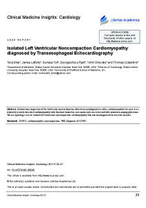
Methods and Materials The study was conducted on 20 patients (figure 5 to 11) whom their LV models were already determined, based on their echocardiograpic data. This information along with blood flow parameters including: local interacted force with the myocardial segments, velocity vectors (local radial, local longitudinal and local circumferential), local relative strength of blood with the 16 myocardial segments (radial, longitudinal and circumferential) and curvature index were defined and used as input into algorithm to solve the partial deferential Navier- Stocks equations. For all calculations average mean ± standard deviation was used. Images for this section:
Fig. 5: A left ventricular dilated case at early diastolic phase.
Page 7 of 28
Fig. 6: A left ventricular dilated case at early systolic phase.
Page 8 of 28
Fig. 7: Analyzing the same left ventricular dilated patient by using of the velocity vector imaging method to extract motion and deformation data.
Page 9 of 28
Fig. 8: Endocardial vector point velocity at the same case.
Page 10 of 28
Fig. 9: Endo long velocity at the same case.
Page 11 of 28
Fig. 10: Endo long strain at the same case.
Page 12 of 28
Fig. 11: Endo long strain rate at the same case.
Page 13 of 28
Results In patients with Dilated Cardiomyopathy (DCM), the vortex was located at the center of the LV throughout diastole and systole and did not redirect flow in a coherent, sequential fashion as in normal subjects. In normal subjects, the average vortex was compact, elliptically shaped, and was located apically. This vortex was persistent during diastole and directs vectors towards the LVOT. In patients with DCM, a spherical, centrally located vortex was observed with incoherent direction of LV flow (Figures 12 to 19) Images for this section:
Fig. 12: a left ventricular dilation at early diastolic phase
Page 14 of 28
Fig. 13: The same left ventricular dilated case at the end of diastolic phase.
Page 15 of 28
Fig. 14: The flow vortices inside the same left ventricular dilated at early diastolic phase.
Page 16 of 28
Fig. 15: The flow vortices inside the same left ventricular dilated case at diastasis phase. it shows a decreasing at vorticity in slow filling phase.
Page 17 of 28
Fig. 16: The irrotational flow vortices inside the same left ventricular dilated at atrial contraction time.
Page 18 of 28
Fig. 17: The rotational flow vortices inside the same left ventricular dilated at isovolumic contraction time
Page 19 of 28
Fig. 18: The irrotational flow direction into the LVOT at the same left ventricular dilated in ejection time.
Page 20 of 28
Conclusion In the presence of ventricle dilatation the mitral jet extends farther inside the ventricle, propagation velocity decreases, and the fluid stagnates longer at the apex. And blood flow pattern is followed in three distinct branches where two of them are helically back forwarded to the left atrium (mitral regurgitation) and third branch goes to aorta (Figures 20 to 25). (In compared with the normal fluid dynamic in LV) For the first time we have innovated a novel software that shows complete blood flow path inside the LV. Detailed information can be obtained about heart function by interfacing the related software within the echocardiograph machine. This innovation also opens doors to much more research in the heart chambers in normal and diseased hearts. Images for this section:
Page 21 of 28
Fig. 19: Two regurgitant branch flow inside the left ventricular dilated case.
Fig. 20: Velocity vectors of blood flow inside the left ventricular dilated case.
Page 22 of 28
Fig. 21: Velocity vectors of blood flow inside the left ventricular dilated case.
Page 23 of 28
Fig. 22: Velocity vectors of blood flow inside the left ventricular dilated case.
Page 24 of 28
Fig. 23: Velocity vectors of blood flow inside the left ventricular dilated case.
Page 25 of 28
Fig. 24: Velocity vectors of blood flow inside the left ventricular dilated case.
Page 26 of 28
Fig. 25: Velocity vectors of blood flow inside the left ventricular dilated case.
Page 27 of 28
References 1- Mersedeh Karvandi, Saeed Ranjbar, Seyed Ahmad Hassantash, Mahnoosh Foroughi. Brief communication: Mathematical concepts of mechanisms of left ventricular myocardium. International Journal of Medical Imaging, 2014; 2(2): 19-23. 2- Mersedeh Karvandi, Saeed Ranjbar, Seyed Ahmad Hassantash. Mechanical mitral valve modeling: Advancing the field through emerging science. International Journal of Medical Imaging, 2014; 2(2): 24-28. 3- Mersedeh Karvandi, Saeed Ranjbar, Seyed Ahmad Hassantash . An essential calculated power formula as a new index to study myocardial function of heart. International Journal of Clinical Medicine Research, 2014; 1(1):5-10.
4- Mersedeh Karvandi, Saeed Ranjbar, Seyed Ahmad Hassantash. Dynamic features creating (which cause) the blood direction inside the left ventricle. International Journal of Medical Imaging, 2014; 2(2): 14-18. 5- Mersedeh Karvandi, Saeed Ranjbar, Mehrdad Shahshahani, Arash Rastegar, Seyed Ahmad Hassantash, Mahnoosh Foroughi, Fariba Ranjbar. A research proposal in mathematical modeling applied to heart and mathematical concepts of mechanisms of heart. American Journal of Science and Technology, 2014; 1(1): 30-35.
US patents: 1- Saeed Ranjbar, Mersedeh Karvandi. System and method for modeling left ventricle of heart. US Patent Number: 8,414,490. 2- Saeed Ranjbar, Mersedeh Karvandi. Solution Navier-stocks equations of the blood as a non-Newtonian fluid in the left ventricle. NOTICE OF ALLOWANCE in January 27, 2014.US Publication number: US-2011-016646467-A1
Page 28 of 28