D E C E M B E R 2 0 06 Vo l u m e 10 , I s s u e 1 0
®
www.cardiologyrounds.org
Acute Stress Cardiomyopathy and Reversible Left Ventricular Dysfunction B y G E R A R D P. AU R I G E M M A , M D
Over the past three decades, in addition to permitting insight into common diseases (eg, valvular or ischemic heart disease), echocardiography has greatly improved the understanding of systolic and diastolic function. It has also allowed clinicians to recognize seemingly new diseases, such as diastolic dysfunction and heart failure,1 asymptomatic severe aortic stenosis, 2 mitral valve prolapse,3 and many of the cardiomyopathies, including isolated noncompaction of the left ventricle.4 Few, if any, of these disorders were commonly diagnosed before the widespread availability of echocardiography, nor did clinicians have the ability to follow the natural history or the response to treatment of these disorders. This issue of Cardiology Rounds reviews the insights from serial echocardiograms of a peculiar and increasingly recognized entity, stress cardiomyopathy.5 Diagnosis of reversible LV dysfunction: importance of echocardiography The noninvasive assessment of left ventricular (LV) systolic function that is possible with echocardiography provides a novel insight into conditions that cause reversible LV dysfunction.6 Table 1 lists conditions in which the ejection fraction can improve dramatically. Several of these disorders involve excessive afterload. Since the ejection fraction is inversely related to the level of afterload – which, by the Laplace relationship, is directly related to the product of systolic pressure and heart size – aortic stenosis, hypertension, aortic regurgitation, and even chronic-mitral regurgitation are all conditions where excess afterload can result in reduced fractional shortening.6,7 Accordingly, in these conditions, a reduction in afterload can be associated with a dramatic improvement in ejection fraction.6-8 Clinical features of acute, stress cardiomyopathy Stress cardiomyopathy is a more recently described form of reversible LV systolic dysfunction, and the most common form of stress cardiomyopathy is transient apical ballooning.9-13 Since 1990, there have been dozens of reports of this cardiomyopathy, with most, but not all, published in the Japanese literature.9,14 Synonyms for the syndrome of transient apical ballooning are found in Table 2. The Japanese have named this syndrome “takotsubo” because the shape of the ballooned apex in systole is similar to the shape of an octopus trap (Figure 1). However, in acute stress-related cardiomyopathy, LV dysfunction is not always confined to the apex. Variations of this syndrome include wall motion abnormalities involving the basal, midportion, and lateral walls of the left ventricle. This is the reason why the term “acute, stress cardiomyopathy” or, more simply, “stress cardiomyopathy” may be preferable to transient apical ballooning. As recently reviewed, the clinical background associated with the transient apical ballooning form of acute stress cardiomyopathy is remarkably similar in most cases.5,15 Salient features of this condition are indicated in Table 3. Typically, the patient is a woman in her 7 th decade, evaluated for the sudden onset of chest symptoms, such as dyspnea or chest pain (Figures 2 and 3). In most instances, these symptoms occur close in time to an identifiable “trigger.” The triggering event is commonly, but not exclusively, an emotionally traumatic one, but may also be an acute medical illness (eg, a severe asthmatic attack requiring intubation).5,15 In some cases, the trigger is unusually strenuous physical activity or a public performance. The majority of patients exhibit chest symptoms (not necessarily chest pain) and electrocardiogram (ECG) changes consistent with acute myocardial ischemia. Experience with patients with stress cardiomyopathy suggests some features may differentiate it from LV dysfunction related to an acute LAD territory MI.
Cardiovascular Division (Clinical) Dale Adler, MD Christine Albert, MD Michelle Albert, MD Elliott Antman, MD Kenneth Baughman, MD Joshua Beckman, MD Charles M. Blatt, MD Eugene Braunwald, MD Christopher Cannon, MD Ming Hui Chen, MD Alanna Coolong, MD Mark Creager, MD Akshay Desai, MD Elazer Edelman, MD, PhD Andrew Eisenhauer, MD Laurence Epstein, MD David Faxon, MD Mark Feinberg, MD Daniel Forman, MD Peter Ganz, MD J. Michael Gaziano, MD Thomas Gaziano, MD Marie Gerhard-Herman, MD Robert Giugliano, MD Michael Givertz, MD Samuel Z. Goldhaber, MD Thomas B. Graboys, MD Howard Hartley, MD Carolyn Ho, MD John Jarcho, MD Paula Johnson, MD Scott Kinlay, MD Jamil Kirdar, MD James Kirshenbaum, MD Bruce Koplan, MD Raymond Kwong, MD Michael J. Landzberg, MD Richard Lee, MD
Jane A. Leopold, MD Eldrin Lewis, MD James Liao, MD Peter Libby, MD (Division Chief) Leonard Lilly, MD Bernard Lown, MD Laura Mauri, MD Thomas Michel, MD, PhD David Morrow, MD Karen Moulton, MD Gilbert Mudge, MD Anju Nohria, MD Patrick O’Gara, MD Marc A. Pfeffer, MD, PhD (Editor) Jorge Plutzky, MD Jeffrey Popma, MD Shmuel Ravid, MD Frederic Resnic, MD Paul Ridker, MD Thomas Rocco, MD Maria Rupnick, MD, PhD Marc Sabatine, MD Arthur Sasahara, MD Benjamin Scirica, MD Christine Seidman, MD Andrew Selwyn, MD Laurence Sloss, MD Piotr Sobieszczyk, MD Regina Sohn, MD Scott Solomon, MD Lynne Stevenson, MD William Stevenson, MD Peter Stone, MD Michael Sweeney, MD Usha Tedrow, MD Stephen Wiviott, MD Justina Wu, MD
Brigham and Women’s Hospital Fax: (617) 732-5291 Website: www.heartdoc.org The editorial content of Cardiology Rounds is determined solely by the Cardiovascular Division of Brigham and Women’s Hospital. This publication is made possible by an educational grant.
Cardiology Rounds is approved by the Harvard Medical School Department of Continuing Education to offer continuing education credit
Table 1: Causes of reversible LV dysfunction encountered in clinical practice
• • • • • • •
Afterload excess conditions: aortic stenosis; aortic regurgitation; severe hypertension; mitral regurgitation with LV systolic dysfunction Toxic exposure: alcoholic cardiomyopathy Tachycardia-induced cardiomyopathy Myocarditis Ischemic heart disease: transient myocardial stunning Pheochromocytoma Stress cardiomyopathy
Stress cardiomyopathy patients: • usually do not develop Q-waves • have lower levels of biomarker release, especially total creatine kinase (CPK), usually not exceeding 400 IU • have wall motion abnormalities that appear to span a larger perfusion territory than one coronary artery (Figure 4). • do not demonstrate delayed hyperenhancement by magnetic resonance imaging (MRI) with gadolinium (Figure 5)11,13 • have wall motion abnormalities that improve on follow-up, some as early as 2 days following presentation (Figure 6) • have extensive wall motion abnormalities and even severe hemodynamic compromise, but relatively low mortality, (in the range of 0%-8%). Stress cardiomyopathy is a clinical diagnosis. To date, most of the reports on this syndrome have described the transient apical ballooning variety. The closest approximation to diagnostic criteria are those used by Tsuchihashi,9 who has published the findings of 88 patients with transient apical ballooning enrolled in a multicenter registry of acute MI, by far the largest reported series. Tsuchihashi designated the following criteria for the diagnosis of transient apical ballooning: acute chest symptoms; characteristic ballooning of the LV apex on ventriculography; deeply inverted T waves on the ECG; documented reversibility of the wall motion abnormality; no obstructive coronary disease by arteriography. Bybee et al have proposed the Mayo Clinic diagnostic criteria, shown in Table 4.15 These criteria, like those of Tsushihashi, require reversible dysfunction of the apex and midventricle. Although it was initially thought that this syndrome occurred only in Japan,9 it is now appreciated that there is a more global distribution.10,11,13,16-18 Until recently, however, no western group had assembled a series of more than 2-3 individuals, nor was there any understanding of prevalence. Pilliere et al were the first to estimate prevalence for the apical form of acute stress-related cardiomyopathy.18 These authors suggest that 0.7% of patients referred for coronary arteriography due to an acute coronary syndrome (ACS) have transient apical ballooning.18 The more recent of Sharkey’s 2 studies represents the largest non-Japanese series.11 In this report,22 Table 2: Nomenclature used for acute, reversible cardiomyopathy involving the apex of the LV
• • • • •
Takotsubo cardiomyopathy Apical ballooning syndrome Stress cardiomyopathy Ampulla cardiomyopathy “Broken heart” syndrome
Figure 1: Representative ventriculogram taken from the study of a patient with transient apical ballooning. The shape of the LV in systole is thought to resemble the shape of the octopus trap, here oriented to simulate a right anterior oblique projection. (Ventriculogram image taken from Tsuchihashi et al,9 used with permission.)
patients were prospectively identified in a community-based cardiology practice over approximately three years.19 Acute MI or an ACS was suspected in each case on the basis of a clinical presentation of typical chest pain and ECG changes. Each patient sought medical attention following the onset of symptoms after a stressful incident. Hemodynamic compromise requiring either vasopressor support or intra-aortic balloon counterpulsation was common and occurred in more than onethird of the subjects. However, LV systolic dysfunction and the associated apical regional wall motion abnormalities resolved rapidly, some in 2 to 3 days, and all by one month. In reports from the US,10-13 the salient features described by Japanese investigators were confirmed: over 90% were women; the presentation resembled that of an acute MI; there was a uniform lack of critical coronary artery stenosis; emotional stress usually preceded presentation; and resolution of the wall motion abnormalities was rapid. In both the Sharkey11 and the Wittstein studies,13 careful regional function analysis confirmed that, in the majority of patients, wall motion abnormalities extended beyond any single coronary vascular territory (Figure 4).13 Key differential features The salient features of acute MI and acute stress cardiomyopathy are compared and contrasted in Table 5. At this point there are no clear prospective identifying factors, either clinically, electrocardiographically, or by echocardiography. The apical portion of the left ventricle may show wall motion abnormality in both disorders; Bybee et al have demonstrated that approximately 30% of their patients with LV apical ballooning also have right ventricular apical involvement.20 In the past, circulating catecholamine levels in patients presenting with the syndrome have shown inconsistent results, but more recent data from Wittstein demonstrates significant elevation Figure 2: Representative electrocardiograms during (a) and 3 days after presentation (b) with transient apical ballooning. Note that there are nonspecific ST-T wave changes in electrocardiogram a which evolve into diffuse, symmetrically inverted T waves on the succeeding tracing. a
b
Table 3: Summary of recent findings in the transient apical ballooning form of stress cardiomyopathy Study Country Year Number of patients Age (years) Male/Female *Identifiable trigger (%) Chest pain (%) ST-segment elevation on presentation (%) T-wave inversion (%) Q-waves (%) QTc (msec) Creatine kinase (IU/L)
Tsuchihashi et al9 Kurisu et al21 Seth et al10 Japan Japan United States 2001 2002 2003 88 30 12 67±13 70±8 64±14 12/76 2/28 1/11 70 33 100 67 63 25
Troponin I LV ejection fraction (%) In-hospital mortality (%)
Desmet et al16 Sharkey et al11 Wittstein et al13 Belgium United States United States 2003 2005 2005 13 22 19 62 65±13 63 1/12 0/22 1/18 69 100 100# 62 90 95
90
100
33
38
59
11
44 19 NR 209-1625
39 NR NR 539 ± 631
NR
NR
41 ±14 1
49 ±12 0
100 0 578 ± 96 153 0.99 ± 0.66 ng/mL 32 0
92 23 450 (310-674) NR 18.7 µg/mL (2.0-97.6) NR 8
68 45 NR NR 0.42 ng/mL ( 40 patients) at our institution, despite the fact that most of our patients have received urgent Figure 4: Wall motion analysis in patients with transient apical ballooning. Note that in patients with apical ballooning, there is hypokinesis and akinesis in the midportion of segments; this does not follow a single coronary artery distribution. (Wittstein et al,13 used with permission.) 100
% of patients with segmental wall motion abnormality
Figure 3: Top panel: Representative 2-dimensional echocardiogram in a 59-year-old woman with transient apical ballooning. This patient developed chest discomfort and dyspnea following an emotionally stressful event. She underwent coronary arteriography and was found to have nonobstructive coronary disease. Panels A and B are from the study performed shortly after presentation and Panels C and D are from the study performed one month later. Panels A and B are diastolic and systolic frames, respectively. Note that in Panel B, there is lack of contraction of the LV apex (arrow). By contrast, the follow-up echocardiogram obtained one month after presentation is seen in the lower panels. Panel C demonstrates end diastolic and Panel D end-systolic frames. As in Panel B, the arrow in Panel D indicates the endocardial surface. Note the improvement in systolic function and reduction in systolic chamber size, when comparing Panel B with Panel D.
Dyskinesia Akinesia Hypokinesia
80 60 40 20 0 1
2
3 4 Basal
5
6
7
8
9 10 11 12 13 14 15 16 17 Mid Apical
LV myocardial segments
Figure 6: Graph showing improvement in the ejection fraction in a series of patients with transient apical ballooning. (Wittstein et al,13 used with permission).
0.60
Ejection fraction
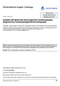
Figure 5: Gadolinium-contrast MRI image taken from a patient with acute stress cardiomyopathy (left panel) contrasted with an image taken from a patient with an apical infarction. Note that presence of a rim of bright signal, also known as hyperenhancement, in the apical region (arrow, right panel), indicative of irreversibly scarred myocardium. Such hyperenhancement is not present in the patient with stress cardiomyopathy. (Wittstein et al,13 used with permission).
0.40
0.20
0.00 Admission
Day 3-7 (inpatient)
2-4 Weeks (outpatient)
coronary arteriography. Ibanez proposed that transient apical ballooning is caused by spasm of a “wraparound” LAD artery,22 but we have not found this to be the case in our experience at the University of Massachusetts.
sole mechanism of myocardial damage in transient apical ballooning, it may contribute to the LV dysfunction (see scheme suggested by Ako in Figure 7).27
Endothelial dysfunction
Catecholamine-induced LV dysfunction
There are data to support a role for endothelial dysfunction in the apical form of acute stress cardiomyopathy. Thrombolysis in Myocardial Infarction (TIMI) frame counts, a measure of the time required by intracoronary contrast to reach distal landmarks, has been employed in the study of these patients by Kurisu et al and Bybee et al.23,24 Prolonged TIMI frame counts in all 3 major epicardial coronary arteries during the acute phase of the syndrome are suggestive of endothelial dysfunction.23,24 Kurisu and co-workers performed elegant clinical studies involving dual isotopes and provided evidence for endothelial dysfunction in these patients.25 They found severe abnormalities of fatty acid metabolism despite an improvement in myocardial perfusion and wall motion.25 Although microvascular ischemia is appealing mechanistically, it has not yet been demonstrated in the limited number of physiological studies in this population that have used Doppler flow-wire measurements or contrast echocardiographic techniques. However, it is possible that, while microvascular dysfunction may not be the
We believe that the available evidence best supports the catecholamine-injury hypothesis as the partial, if not the sole mechanism, for acute stress cardiomyopathy. Catecholamine-stress injury is likely to be present in a number of disorders whose common phenotypic expression is regional wall motion abnormality. These include subarachnoid hemorrhage,27 the endocrine crisis of pheochromocytoma,28 and acute respiratory crises, including asthma.29 A catecholamine-injury hypothesis, as opposed to a coronary spasm hypothesis, would also support the appearance of nonapical regional wall motion abnormalities that do not follow a single coronary distribution. It is also possible, as has been speculated by Ako,26 that catecholamine-excess may also promote endothelial dysfunction, which may contribute to the wall motion abnormality (Figure 7). The data of Wittstein, demonstrating significant elevations in all catecholamine levels tested, even compared with individuals suffering acute MI with hemodynamic compromise (Killip class 3 MI), strongly supports the catecholamine-injury hypothesis.13 In my opinion, the stress cardiomyopathy syndrome results from catecholamine-
Table 4: Proposed Mayo criteria for the clinical diagnosis of the transient left ventricular apical ballooning syndrome*
•
• • •
Transient akinesis or dyskinesis of the left ventricular apical and mid-ventricular segments with regional wall-motion abnormalities extending beyond a single epicardial vascular distribution Absence of obstructive coronary disease or angiographic evidence of acute plaque rupture New electrocardiographic abnormalities (either ST-segment elevation or T-wave inversion) Absence of : – Recent significant head trauma – Intracranial bleeding – Pheochromocytoma – Obstructive epicardial coronary artery disease – Myocarditis – Hypertrophic cardiomyopathy
Table 5: Suggested distinguishing features between transient apical ballooning and LAD territory MI
ST elevations CPK Troponin-1 Wall motion abnormality RV apical involvement Coronary arteries Akinesis
Transient apical ballooning Yes 150-400 Positive
LAD territory MI Yes > 500 Positive
> 1 territory
LAD territory
Variable
Variable
Normal or nonobstructive disease Resolves
Coronary occlusion usually present Does not resolve
LAD = left anterior descending coronary artery
Figure 7: A proposed mechanism for apical ballooning. (Ako et al,26 used with permission.)
Figure 8: Schematic showing various ventriculographic morphologies in patients with stress cardiomyopathy (Shimizu et al,37 used with permission.)
“Stress” Sympathetic nervous system activation ? Microvasculature endothelium
Myocardium norepinephrine release
Microvascular spasm Transient myocardial ischemia
Ca++ overload
Takotsubo type
MidReverse takotsubo ventricular type type
Localized type
Myocardial injury LV dysfunction (reversible) Contraction band necrosis (irreversible?)
associated stunning of the myocardium that is provoked by emotional or physiologic stress. The observations by Pollick et al 30 and Kono et al27 were major conceptual breakthroughs because they demonstrated that reversible apical dysfunction existed in patients recovering from subarachnoid hemorrhage. Prior to this study, it was thought that such “cerebral T waves” were not necessarily reflective of myocardial injury. The transient wall motion abnormalities in acute stress-related cardiomyopathy, generally, although not exclusively, involve the ventricular apex. However, the explanation for this finding has not been established.19 According to Owa et al31 and Akashi et al,32 there are definable abnormalities of cardiac sympathetic innervation and sympathetic hyperactivity in the LV apex.31,32 Mori et al speculated that there is increased adrenergic responsiveness or increased β-receptor density at the apex that compensates for relatively sparse sympathetic innervation in this region.33 There is some support for this mechanism from animal models where isoproterenol damage is predominantly endocardial and apical.34 Transient apical and midventricular wall-motion abnormalities in rats were produced by immobilization,35 and these abnormalities were not noted when the rats were pretreated with adrenergic blockade. Important unresolved issues Despite the flurry of recent reports from US and European investigators, and continued clinical research by Japanese investigators, a number of unanswered questions persist regarding transient apical ballooning. Classification There is variability in the site of LV dysfunction, both in the apical form of stress cardiomyopathy syndrome, described by Wittstein et al,13 as well as in patients suffering an acute cerebral hemorrhage. We, as well as others, have observed transient dysfunction involving the basal portion of the left ventricle (“reverse takotsubo”) or the midportion of the left ventricle (Figure 8).37 These findings have been summarized recently by Shimizu and co-workers.37 For that reason, as stated earlier in this review, we believe that the term “stress cardiomyopathy” may prove to be preferable to “transient apical ballooning” or “Takotsubo syndrome,” since it encompasses the different phenotypic expressions.
Prospective identification At this point, there are insufficient data available to enable the clinician to prospectively distinguish transient apical ballooning or other forms of stress cardiomyopathy from acute MI caused by occlusion of the left anterior descending artery. Until research permits prospective distinction, the diagnosis of stress cardiomyopathy should remain an exclusion. Certainly, a clinical history of an emotional or medical “trigger” in a middle-aged or older woman should alert the clinician to the possible diagnosis of stress cardiomyopathy. It is possible that further research with biomarkers, echocardiography, and prospective validation may eventually enable the clinician to make the diagnosis of stress cardiomyopathy without necessarily performing coronary arteriography. Why women? Why now? Why is this important? Why middle-aged/elderly women appear particularly susceptible to this disorder is not clear. Further, is this a new disorder or one “discovered” by serial echocardiography? The lack of any publisheddescription despite decades of left ventriculographic studies before the early 1990s, in my opinion, is explained by the fact that serial 2-dimensional echocardiography was not available until relatively recently (M-mode echocardiography does not visualize apical ballooning). Finally, what is the clinical relevance of this uncommon syndrome in day-to-day practice? Acute stress cardiomyopathy syndrome must now be considered by internists/cardiologists, intensivists, emergency department physicians, and anesthesiologists in the differential diagnosis of a patient who presents with a suspected acute coronary syndrome or ST-segment elevation MI, or who develops arrhythmia or T wave inversions during the preoperative or intraoperative period. As noted above, this cardiomyopathy may account for a small percentage (eg, 1% -3%) of ACSs.18,21 However, a recent report, summarizing a prospective evaluation of consecutive patients admitted to a medical intensive care unit (ICU), demonstrates a remarkably high incidence of apical dysfunction; 26 of 92 patients had apical ballooning and this was often associated with sepsis.36 Stress cardiomyopathy shouldbe considered, especially, when the extent of ischemic ECG abnormalities exceeds biomarker evidence for myocardial necrosis and coronary angiography confirms noncritical atherosclerotic disease. The most effective long-term management of this syndrome remains to be defined, although we favor chronic beta-blocker therapy (without clinical trial validation) in individuals who have had stress cardiomyopathy.
It is also hoped that further investigations using echocardiography in concert with biomarkers may help clinicians diagnose the syndrome prospectively with greater certainty. However, until stress cardiomyopathy can be conclusively distinguished from acute MI, using prospectively validated clinical criteria, we emphasize that it must first be assumed that acute coronary occlusion is responsible for the patient’s symptoms, until coronary angiography excludes this more common cause of acute regional LV dysfunction. References 1. Aurigemma GP, Gaasch WH. Clinical practice. Diastolic heart failure. N Engl J Med 2005;351:1097-105. 2. Pellikka PA, Nishimura RA, Bailey KR, Tajik AJ. The natural history of adults with asymptomatic, hemodynamically significant aortic stenosis. J Am Coll Cardiol 1990;15: 1018-20. 3. Freed LA, Levy D, Levine RA, et al. Prevalence and clinical outcome of mitral-valve prolapse. N Engl J Med 1999;341:1-7. 4. Agmon Y, Connolly HM, Olson LJ, Khandheria BK, Seward JB. Noncompaction of the ventricular myocardium. J Am Soc Echocardiogr 1999;10:859-63. 5. Aurigemma GP, Tighe DA. Echocardiography and reversible left ventricular dysfunction. Am J Med 2006;119:18-21. 6. Aurigemma GP, Gaasch WH, Villegas B, Meyer TE. Noninvasive assessment of left ventricular mass, chamber volume, and contractile function. Curr Probl Cardiol 1995; 20:361-440. 7. Ross J. Afterload mismatch in aortic and mitral valve disease: implications for surgical therapy. J Am Coll Cardiol 1985;5:811-826. 8. Goldfine H, Aurigemma GP, Zile MR, Gaasch WH. Left ventricular length-force-shortening relations before and after surgical correction of chronic mitral regurgitation. J Am Coll Cardiol 1998;31:180-185. 9. Tsuchihashi K, Ueshima K, Uchida T, et al. Transient apical ballooning without coronary artery stenosis: a novel heart syndrome mimicking acute myocardial infarction. Angina Pectoris-Myocardial Infarction Investigators in Japan. J Am Coll Cardiol 2001; 38:11-18. 10. Seth PS, Aurigemma GP, Krasnow JM, Tighe DA, Untereker WJ, Meyer TE. A syndrome of transient left ventricular apical wall motion abnormality in the absence of coronary disease: a perspective from the United States. Cardiology 2003;100:61-66. 11. Sharkey SW, Lesser JR, Andrey G, et al Acute and reversible cardiomyopathy provoked by stress in women from the United States. Circulation 2005;111:472-479. 12. Dec GW. Recognition of the apical ballooning syndrome in the United States. Circulation 2005;111:388-90. 13. Wittstein IS, Thiemann DR, Lima JA, et al. Neurohumoral features of myocardial stunning due to sudden emotional stress. N Engl J Med 2005;352:539-48. 14. Dote K, Sato H, Uchinda AT, Ishihara M. Myocardial stunning due to simultaneous multivessel coronary spasms: a review of 5 cases. J Cardiol 1991;21:203-214. 15. Bybee KA, Kara T, Prasad A, et al. Systemic Review: Transient left ventricular apical ballooning: A syndrome that mimics ST-segment elevation myocardial infarction. Ann Intern Med 2004;141:858-865. 16. Desmet WJR, Adriaenssens BFM, Dens JAY. Apical ballooning of the left ventricle: first series of white patients. Heart 2003;89:1027-1031. 17. Witzke C, Lowe HC, Waldman H, Palacios IF. Transient left ventricular apical ballooning. Images in Cardiovascular Medicine. Circulation 2003;108:2014. 18. Pilliere R, Mansencal N, Digne F, Lacombe P, Joseph T, Dubourg O. Prevalence of takotsubo syndrome in a large urban agglomeration. Am J Cardiol 2006;98(5):662-5. 19. Sharkey SW, Shear W, Hodges M, Herzog CA. Reversible myocardial contraction abnormalities in patients with an acute noncardiac illness. Chest 1998;11:98-105. 20. Bybee KA, Prasad A, Barsness GW, et al. Clinical characteristics and thrombolysis in myocardial infarction frame counts in women with transient left ventricular apical ballooning syndrome. Am J Cardiol 2004;94:343-6. 21. Kurisu S, Sato KT, Ishihara M, et al. Tako-tsubo–like left ventricular dysfunction with ST segment elevation: a novel cardiac syndrome mimicking acute myocardial infarction. Am Heart J 2002;143:448-455. 22. Ibanez B, Navarro F, Cordoba M, M-Alberca P, Farre J. Tako-tsubo transient left ventricular apical ballooning: is intravascular ultrasound the key to resolve the enigma? Heart 2005;91:102-4. 23. Kurisu S, Inoue I, Kawagoe T, et al. Myocardial perfusion and fatty acid metabolism in patients with tako-tsubo-like left ventricular dysfunction. J Am Coll Cardiol 2003;41(5): 743-8. 24. Bybee KA, Prasad A, Barsness GW, et al. Clinical characteristics and thrombolysis in myocardial infarction frame counts in women with transient left ventricular apical ballooning syndrome. Am J Cardiol 2004;94:343-6. 25. Kurisu S, Inoue I, Kawagoe T, et al. Myocardial perfusion and fatty acid metabolism in patients with tako-tsubo-like left ventricular dysfunction. J Am Coll Cardiol 2003;41: 743-8. 26. Ako J, Sudhir K, Farouque HM, Honda Y, Fitzgerald PJ. Transient left ventricular dysfunction under severe stress: brain-heart relationship revisited. Am J Med 2006; 199(1):10-17. 27. Kono T, Morita H, Kuroiwa T, Onaka H, Takatsuka H, Fujiwara A. Left ventricular wall motion abnormalities in patients with subarachnoid hemorrhage: neurogenic stunned myocardium. J Am Coll Cardiol 1994;24:636-40.
28. Shaw TRD, Bafferty P, Tait GW. Transient shock and myocardial impairment caused by pheochromocytoma crisis. Br Heart J 1987;57:194-198. 29. Levine GN, Powel C, Bernard SA, Sherman D, Faling LG, Davidoff R. Acute reversible left ventricular dysfunction in status asthmaticus. Chest 1995;107: 1469-73. 30. Pollick C, Cujec B, Parker S, Tator C. Left ventricular wall motion abnormalities in subarachnoid hemorrhage: an echocardiographic study. J Am Coll Cardiol 1988;12:600-5. 31. Owa M, Aizawa K, Urasawa N, et al. Emotional stress-induced “ampulla cardiomyopathy”: discrepancy between the metabolic and sympathetic innervation imaging performed during the recovery course. Jpn Circ J 2001;65:349-52. 32. Akashi YJ, Nakazawa K, Sakakibara M, Miyake F, Musha H, Sasaka K. 1231-MIBG myocardial scintigraphy in patients with “takotsubo” cardiomyopathy. J Nucl Med 2004; 45:1121-7. 33. Mori H, Ishikawa S, Kojima S, et al. Increased responsiveness of left ventricular apical myocardium to adrenergic stimuli. Cardiovasc Res 1993;27:192-8. 34. Teerlink JR, Pfeffer JM, Pfeffer MA. Progressive ventricular remodeling in response to diffuse isoproterenol-induced myocardial necrosis in rats. Circ Res 1994;75:105-13. 35. Ueyama T, Kasamatsu K, Hano T, Yamamoto K, Tsuruo Y, Nishio I. Emotional stress induces transient left ventricular hypocontraction in the rat via activation of cardiac adrenoceptors: a possible animal model of ‘tako-tsubo’ cardiomyopathy. Circ J 2002; 66:712-3. 36. Park JH, Kang SJ, Song JK, et al. Left ventricular apical ballooning due to severe physical stress in patients admitted to the medical ICU. Chest 2005;128: 296-302. 37. Shimizu M, Kato Y, Masai H, Shima T, Miwa Y. Recurrent episodes of Takotsubo-like transient apical ballooning occurring in different regions: a case report. J Cardiol 2006;48:101-107.
Dr. Gerard P. Aurigemma was born in Brooklyn, NY. He graduated from Harvard College (1975) and Harvard Medical School (1979). He completed his medical residency and served as Chief Medical Resident at the UCSF, completed a cardiology fellowship at the University of Pennsylvania, and joined the faculty of the University of Massachusetts Medical School in 1987. He has a longstanding interest in LV systolic and diastolic function in hypertension, valvular heart disease, and diastolic heart failure and the application of noninvasive imaging techniques to these disorders. His recent interest has been the study of reversible LV dysfunction, including the intriguing syndrome of stress cardiomyopathy, an entity he first encountered in 1985. Dr. Aurigemma is the author of over 100 articles and reviews on LV function and other cardiology topics and served as an associate editor of the textbook, Cardiology. He serves on the editorial board of several cardiology journals and has served on the board of directors of the American Society of Echocardiography (ASE). He has served as course director for the ASE Board Review Course. Dr. Aurigemma is currently a Professor of Medicine and Radiology at the University of Massachusetts Medical School and has directed its cardiology fellowship program since 1990. He is director of Noninvasive Cardiology at UMass Memorial Health Care, Worcester, Massachusetts. Please contact Dr. Aurigemma by email if you are interested in possible collaboration on a registry of cases of stress cardiomyopathy:
[email protected]
Disclosure statement: Dr. Aurigemma has received research grant support from Omron Healthcare, Toshiba Medical, and Novartis.
Brigham and Women’s Hospital, Cardiovascular Division website: www.heartdoc.org
This publication is made possible by an educational grant from
Novartis Pharmaceuticals Corporation © 2006 Brigham and Women’s Hospital, Boston, Massachusetts, which is solely responsible for the contents. The opinions expressed in this publication do not necessarily reflect those of the publisher or sponsor, but rather are those of the author based on the available scientific literature. Publisher: SNELL Medical Communication Inc. in cooperation with Brigham and Women’s Hospital, Boston, Massachusetts. ®Cardiology Rounds is a registered trade mark of SNELL Medical Communication Inc. All rights reserved. The administration of any therapies discussed or referred to in Cardiology Rounds should always be consistent with the recognized prescribing information as required by the FDA. Snell Medical Communication Inc. is committed to the development of superior Continuing Medical Education.
SNELL
302-061