Application Brief: Nephrology Introduction 1 in 9 adults in North America suffer from chronic kidney diseases.1 Furthermore, acute renal failure is not an uncommon adverse event associated with many drugs. It is no wonder that nephrology research is currently one of the key focal points in biomedical research. One of the difficulties associated with kidney disease, especially chronic forms, is that in vivo studies must be used to track disease progression longitudinally. Furthermore, techniques that can visualize renal blood flow will greatly complement research in this area. However, nephrology research in animal models has long been hindered by the lack of a fast, portable, high-resolution, research and animal focused imaging system that can visualize 2D and 3D kidney images, blood flow and tissue perfusion in vivo, in real-time, and most importantly, non-invasively. In order to ameliorate this problem, VisualSonics has introduced a revolutionary micro-ultrasound and photoacoustic imaging system that allows researchers to collect a plethora of important data over the lifespan of animals, thereby significantly reducing the number of animals needed. Numerous satisfied nephrology researchers using Vevo® micro-ultrasound systems, from institutions such as Johns Hopkins University, Princess Margaret Hospital and NIH are publishing articles in leading journals such as The American Journal of Pathology, Journal of the American Society of Nephrology and American Journal of Physiology – Renal Physiology. This is a testament of the power and versatility of high-resolution micro-ultrasound.
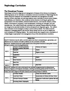
Figure 1 - Mouse kidney 3D Vasculature
A pplication Brief: Nephrology ve r1.2
Page 1
Micro-Ultrasound in Nephrology Research The use of micro-ultrasound in nephrology research has been regarded by leading researchers as an attractive technique for non-invasive, in vivo imaging of kidney microstructures,2 monitoring drug safety in kidneys3 and kidney blood flow and perfusion.4,5,6 Most research in this area is done using mice, because of their wide availability, variety of strains and ease of handling. Micro-ultrasound is especially suitable to study mice as they are the perfect size to take advantage of the maximum resolution Vevo systems offer. For example, Sullivan et al. recently reported their findings in the American Journal of Physiology – Renal Physiology of using the Vevo high-resolution system with enhanced contrast agents to measure renal blood flow.4 The authors were limited by the fact that conventional methods such as laser Doppler flowmetry are highly invasive and location-restricted and thus explored the use of micro-ultrasound as an exciting alternative. 4 The authors found that renal cortical and medullary rates of perfusion in response to endothelin-1 infusion were readily measured by micro-ultrasound and the results could be verified by laser Doppler flowmetry. They concluded that the Vevo system offers a rapid, non-invasive way of obtaining hemodynamic measurements with little risk to experimental subjects.4 Dieterle et al. recently published in Current Opinion in Drug Discovery & Development their review of different methods to monitor drug-induced nephrotoxicity in preclinical research.3 The researchers found that the Vevo 770 system is capable of high-resolution, non-invasive imaging of not only renal artery blood flow, but even microvascular blood flow that is paramount in studies of changes such as renal artery stenosis and renal remodeling.3 They concluded that micro-ultrasound is non-invasive, does not require radiation, does not suffer from motion artifacts and thus allows the performance of longitudinal studies of disease progression and regression. 3 Another very interesting paper was published in Ultrasound in Medicine & Biology by Wang et al.2 The authors examined the use of micro-ultrasound to image conscious rats.2 Conscious imaging not only speeds up the screening process, but also decreases confounding variables such as anesthesia interactions in new drug discovery research. In this example, the authors used the Vevo system to identify unilateral congenital hydronephrosis of the right kidney in rats, which was later confirmed via histology.2 They further visualized kidney capsule, cortex, corticomedullary junction, medulla, papilla, pelvis and hilum vasculature via micro-ultrasound in 24 kidneys.2 It was concluded that the Vevo system allowed imaging of organ and tissue in the living state where normal tissue dynamics and physiological processes are intact and that conscious imaging of animals was possible without causing stress.2
A pplication Brief: Nephrology ve r1.2
Page 2
Many other researchers in recent years have also been using Vevo systems as an easily accessible, applicable, fast and superior way to image kidney structure and blood flow. For a complete list of publications, please refer to Top Nephrology Research Papers using the Vevo Systems.
Figure 2 – Rat Kidney Blood Flow
A pplication Brief: Nephrology ve r1.2
Page 3
Photoacoustics in Nephrology Research Photoacoustic imaging has recently been recognized in the field of nephrology research as a hybrid technology capable of retaining the sensitivity and specificity of optical imaging while overcoming poor spatial resolution through the depth and scalability of ultrasonic imaging8. Song and Wang (2008) demonstrated the feasibility of photoacoustic imaging for imaging deep organ tissues, specifically the kidney in both a rat and rabbit. Because of the depth penetration offered by ultrasound, combined with the optical imaging abilities of photoacoustic imaging, both anatomical and functional imaging can be achieved within a tissue on the same plane. Specifically, oxygen saturation10, blood volume11 and hemoglobin content can be detected & quantified (Figure 3). Finally, photoacoustics further expands the molecular imaging capabilities for researchers through the use of nanoparticles and contrast agents, which can be targeted to specific intracellular receptors and biomarkers. This capability is useful for monitoring disease progression and therapeutic intervention. 100 1mm
% sO 2
Cortical region 50
Figure 3 –Oxygen saturation map (62% in outlined region) in mouse kidney inhaling 100% O 2 under anesthesia. Red corresponds to regions to high oxygenation levels, blue low. Outlined area is an overlay of the cortex.
A pplication Brief: Nephrology ve r1.2
Page 4
What Can The Vevo® LAZR Photoacoustic Imaging System Do For Me? High-frequency ultrasound and photoacoustics are unique imaging modalities for small animals non-invasive imaging in real-time. With Vevo systems, researchers now have access to a tool to conduct longitudinal studies in vivo. It has been demonstrated by Loveless et al. that microultrasound produces images eclipsing MRI resolution and that the two imaging systems can be combined to validate each other’s results.7 Numerous unique tools for quantification of perfusion, as well as an open data platform allows the nephrology researcher to have full control over his/her findings. Below is a summary of the unique value proposition VisualSonics delivers to researchers with the Vevo micro ultrasound systems: 1. Non-invasive, in vivo, real-time imaging for processes that happen over a period of time, such as chronic kidney disease, cyst development and tumor progression. 2. High-spatial resolution up to 30 μm allows for excellent delineation of renal and renalrelated structures such as medulla, cortex, renal vein and artery, ureter, urethra and bladder in 2D and 3D. 3. Quantification of renal function and flow, such as pulsatility and resistivity indices, including flow in small vessels of diameter >50 μm. 4. Unique microbubble microcirculation.
contrast
agents
allow
quantification
of
kinetics
in
the
5. Targeted contrast agents for quantification of biomarkers involved in inflammation and angiogenesis. 6. Image-guided injection of cancer cells, stem cells, therapeutic compounds, etc. into the kidney and/or surrounding tissue without surgery. 7. Gene transfection and enhanced drug delivery into renal cells using the Vevo SoniGene™ system through sonoporation. 8. Dedicated animal platform to monitor ECG, heart rate, body temperature and respiration rates. 9. Data export capabilities in excel. Image/movie export capabilities in TIFF, BMP, AVI, etc. 10. Measurement of oxygen saturation for studies in ischemia. 11. Nanoparticle detection and quantification in vivo for targeted cellular and molecular studies.
A pplication Brief: Nephrology ve r1.2
Page 5
The Vevo LAZR Photoacoustic Imaging System
A pplication Brief: Nephrology ve r1.2
Page 6
References 1. Craig B. Langman. The Epidemic of Chronic Kidney Disease. MedGenMed 2006; 8(3): 55. 2. Wang YX, Betton G, Floettmann E, Fantham E, Ridgwell G. Imaging kidney in conscious rats with high-frequency ultrasound and detection of two cases of unilateral congenital hydronephrosis. Ultrasound Med Biol 2007 Mar;33(3):483-6. 3. Dieterle F, Marrer E, Suzuki E, Grenet O, Cordier A, Vonderscher J. Monitoring kidney safety in drug development: emerging technologies and their implications. Curr Opin Drug Discov Devel 2008 Jan;11(1):60-71. 4. Sullivan JC, Wang B, Boesen EI, D'Angelo G, Pollock JS, Pollock DM. Novel use of ultrasound to examine regional blood flow in the mouse kidney. Am J Physiol Renal Physiol 2009 Jul;297(1):F228-35. 5. Andonian S, Coulthard T, Smith AD, Singhal PS, Lee BR. Real-time quantitation of renal ischemia using targeted microbubbles: in-vivo measurement of P-selectin expression. J Endourol 2009 Mar;23(3):373-8. 6. Hakroush S, Moeller MJ, Theilig F, Kaissling B, Sijmonsma TP, Jugold M, Akeson AL, Traykova-Brauch M, Hosser H, Hähnel B, Gröne HJ, Koesters R, Kriz W. Effects of increased renal tubular vascular endothelial growth factor (VEGF) on fibrosis, cyst formation, and glomerular disease. Am J Pathol 2009 Nov;175(5):1883-95. 7. Loveless ME, Whisenant JG, Wilson K, Lyshchik A, Sinha TK, Gore JC, Yankeelov TE. Coregistration of ultrasonography and magnetic resonance imaging with a preliminary investigation of the spatial colocalization of vascular endothelial growth factor receptor 2 expression and tumor perfusion in a murine tumor model. Mol Imaging 2009;8(4):187-98. 8. Ntziachristos V. et al. Looking and listening to light: The evolution of whole-body photonic imaging. Nat. Biotechnol. 2005 (23):313–320 9. Song KH and Wang, LV. Noninvasive photoacoustic imaging of the thoracic cavity and the kidney in small and large animals. Med Phys. 2008 Oct;35(10):4524-9. 10. Wang X et al. Noninvasive laser-induced photoacoustic tomography for structural and functional in vivo imaging of the brain. Nat. Biotechnol. 2003 (21):803–6. 11. Stein EW, Maslov K, and Wang LV. Noninvasive mapping of the electrically stimulated mouse brain using photoacoustic microscopy. Proc. SPIE (2008):685-6.
A pplication Brief: Nephrology ve r1.2
Page 7