Home SVCC
Area: English
- Español - Português
Abnormal Morphologic Features of Hypertrophic Cardiomyopathy Jamshid Shirani, MD Departments of Medicine (Division of Cardiology) and Pathology, Albert Einstein College of Medicine, Bronx, New York, USA INTRODUCTION The first detailed description of HCM was published by Teare in 1958 who noted "asymmetric hypertrophy of the heart" in several family members who died suddenly (1). Teare also observed abnormal arrangement and variable size of cardiac muscle fibers in the ventricular septum (1). In the following years, obstruction to LV outflow tract was considered the principal clinical manifestation of the disease leading to its designation as "idiopathic hypertrophic subaortic stenosis" (2). The last two decades have witnessed major contributions to the understanding of HCM, now considered a primary and genetically transmitted cardiac disease with extremely heterogeneous clinical and morphologic profile (3). In recent years, an increasing number of mutations in genes encoding cardiac sarcomeric proteins ( Table 1 ) have been identified in individuals and families expressing the cardiac disease phenotypes (4, 5). These mutations are thought to be responsible for some of the prominent and characteristic structural alterations in HCM, such as cardiac myocyte hypertrophy and disorganization (6). Other frequently found cardiac morphologic abnormalities in patients with HCM, such as myocardial fibrosis (7) and abnormalities of intramural coronary arteries (8) involve the connective tissue rather than cardiac myocytes. In addition, other cardiac morphologic abnormalities, such as direct insertion of papillary muscle into mitral leaflet and solitary papillary muscle, have been reported in association with HCM (9). Table 2 lists the most prominent gross and microscopic cardiac morphologic abnormalities reported in individuals with HCM. In the following sections, each particular cardiac morphologic abnormality is discussed and the interrelation of these morphologic findings and the clinical presentation of HCM explored.
DEFINITION HCM is classically defined as "hypertrophied and non -dilated left ventricle in the absence of any other cardiac or systemic disease capable of producing the degree of left ventricular hypertrophy present in that patient" (3). However, marked heterogeneity in cardiac morphologic features and clinical presentation of HCM has made it difficult, if not impossible, to provide an all-inclusive definition of the disease. This is further complicated by the observations that: i) age, sex, and other environmental, hormonal as well as genetic modifying factors significantly alter the disease phenotype; ii) none of the gross and histomorphologic features of the disease are in fact specific for HCM. Rather, they are present in greater proportions than found in other cardiac diseases; and 3) as previous studies unavoidably have focused on patients with the most dramatic presentation of the disease, the full spectrum of cardiac morphologic abnormalities of HCM has not been unraveled (10). In addition, the diagnosis of HCM poses a special problem in infants (11), children (12), elderly (13), competitive athletes (14) and in patients with systemic hypertension (15). Finally, the definition of the disease requires broadening in order to address those with the disease genotype in whom clinically recognizable cardiac morphologic abnormalities are absent or only mild. CARDIOMEGALY AND LEFT VENTRICULAR HYPERTROPHY Heart weight, as measured at necropsy, is increased in most patients (>95%) with HCM and at times may exceed 1,000 grams. Normal heart weight in HCM is more often found in i) asymptomatic young patients with the disease genotype who have not yet expressed the phenotypic characteristics of the disease (16), or ii) in those who die suddenly in the absence of known cardiac symptoms during life (17). The primary gross morphologic feature of HCM is hypertrophy of LV walls and often the right ventricle (18). However, echocardiographic and necropsy studies have demonstrated marked variations in the extent and patterns of LVH in HCM (19,20). In most patients LVH is diffuse although the magnitude of segmental hypertrophy often differs significantly among various regions. The ventricular septum usually shows the greatest magnitude of hypertrophy followed by large portions of the anterolateral LV free wall.
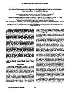
The posterior LV free wall is the least affected region in HCM. Asymmetric septal hypertrophy (defined as a septal to posterior wall thickness ration exceeding 1.3) is observed in up to 90% of patients with HCM (Figure 1 ). In these patients, septal hypertrophy may be limited to the basal most portion of the septum (in 25% of the patients), may extend down to the level of papillary muscles (in another 25%), or may involve the entire ventricular septum (in the remaining 50%). When the entire ventricular septum is thickened, the hypertrophy often extends to regions of the anterolateral LV free wall. Nevertheless, segmental LV hypertrophy in HCM may be confined to the anterior or posterior septum, anterolateral free wall, posterior free wall or even the most apical portion of the LV (19, 21-23). The latter, called apical HCM, is often associated with deep, symmetrically inverted T waves in precordial leads of surface 12-lead electrocardiogram in its early stage (24). However, gradual LV dilation, apical wall thinning and even aneurysm formation have been reported in some patients with apical HCM in association with loss of inverted T waves, reduction in R wave amplitudes, and development of Q waves presumably due to apical scarring (25). A distinct subtype of patients with HCM shows hypertrophy limited to the LV papillary muscles (26).
Figure 1. Gross photograph of the heart if a patient with hypertrophic cardiomyopathy demonstrating marked asymmetric left ventricular (LV) hypertrophy with preferential wall thickening in the ventricular septum (VS) and anterior free wall.
The magnitude and distribution of LVH in HCM often determine the clinical features of HCM. Asymmetric, marked hypertrophy of the basal septum is a prerequisite for development of dynamic LV outflow tract (subaortic) obstruction whereas HCM patients with mid-LV cavity obstruction show marked symmetric (concentric) LVH especially at the papillary muscle level. Systolic mid-LV cavity obstruction results from complete apposition of the hypertrophied walls and papillary muscles, leads to separation of the apical and basal LV regions and may be associated with apical ischemia, scarring and even aneurysm formation (27, Figure 2 ). Patients with milder concentric LVH often show LV cavity obliteration due to increased ejection fraction without focal intracavitary or outflow tract obstruction. Marked LVH has been identified as a risk factor for sudden death in HCM (28). However, LV diastolic dysfunction and presence of symptoms of CHF do not closely parallel the severity of LVH in this disease (29,30). In some patients with HCM, asymmetric hypertrophy of the infundibular region may result in right ventricular outflow tract obstruction.
Figure 2. Gross photograph of the heart in a patient with midventricular obstruction and left ventricular apical aneurysm (A). LA=left atrium;PW=posterior wall;VS=ventricular septum.
CARDIAC MYOCYTE DISORGANIZATION Histologic examination of myocardial biopsy samples and necropsy material has revealed marked disorganization of myocardial fibers (involving at least 5% of the tissue surface) in about 95% of patients with HCM (6,20). This cellular disarray involves >25% of myocardium in 50% and >50% of the tissue in 25% of the patients and is characterized by oblique and perpendicular arrangement of adjacent muscle fibers (6, Figure 3 ). Myofiber disorganization in HCM is not necessarily confined to the most hypertrophied LV segments. Indeed, there is little correlation between wall thickness and extent of cellular disorganization (31). Thus, cellular disorganization involves an average of 40% of the ventricular septum and 33% of the LV free wall despite differences in the magnitude of hypertrophy in these regions (31,32). The extensive cellular disorganization in HCM is in contrast to other cardiac diseases such as aortic and mitral valve disease, hypertension, and congenital lesions where myocyte disarray involves