HYPERTROPHIC CARDIOMYOPATHY (HCM)
: A Review Last Updated: Jan 24, 2008
Dr. Satish Kumar, MD Consultant Dept of Medicine Bokaro General Hospital
[Synonyms and related keywords: Idiopathic Hypertrophic Subaortic Stenosis (IHSS), Asymmetric Septal Hypertrophy (ASH), Muscular Subaortic Stenosis, Hypertrophic Obstructive Cardiomyopathy (HOCM, HCM)] Hypertrophic Cardiomyopathy (HCM): A Review | Dr. Satish Kumar; Jan’08
Page |2
INTRODUCTION Background: Hypertrophic cardiomyopathy (HCM) consists of genetically abnormal, usually hypercontractile and asymmetric myocardium that may obstruct output and cause sudden death if the hypertrophy is localized in the upper septum. The disease includes asymmetric septal hypertrophy, and idiopathic subaortic stenosis (IHSS), but the term HCM is preferred because the majority (75%) of patients do not present with outflow obstruction (Braunwald, 1997), and 30% do not exhibit asymmetric hypertrophy. Pathophysiology: The heredity pattern is variable, with half of patients showing transmission to first-degree relatives; usually, the disease is inherited as an autosomal dominant trait. Various genetic abnormalities may be found; these usually involve abnormal sarcomere proteins, including the following: beta-Myosin heavy chain (chromosome 14), troponin T (chromosome 1), troponin I (chromosome 19), myosin light chains (chromosomes 2 and 12), alpha tropomyosin (chromosome 15), and myosin-binding protein C (chromosome 11). Also, an abnormality of chromosome 7 has been found in combined HCM and Wolf-Parkinson-White (WPW) syndrome. The myofibrils are abnormally short, broad, and hypertrophied, and they may run in different direction with complex intercellular bridging resulting in the formation of whorls (Maron, 1979; Maron, 1980; Becker, 1994). The left ventricle (LV) is usually more involved in hypertrophy than is the right ventricle. The atria may be dilated, and they are often hypertrophied. The characteristic feature is disproportionate thickening of the interventricular septum (IVS) and the anterolateral wall of the LV compared with the posterior free wall (Maron, 1993). Other patterns include concentric hypertrophy; this is sometimes difficult to differentiate from physiologic hypertrophy, which occurs in some highly trained athletes (Maron and Isner et al, 1994; Maron and Pelliccia et al, 1993; Maron, 1995). Some patients have significant hypertrophy in unusual locations, such as the posterior portion of septum, the posterobasal free wall of the LV, or at the midventricular level (Maron, 1993). One unusual type involves marked posterior wall hypertrophy and virtually no septal hypertrophy. These patients are young are very symptomatic (Lewis, 1991). Apical HCM with predominant involvement of apex is most common in Japan (Maron, 1990). Hypertensive HCM in elderly patients is characterized by severe concentric LV hypertrophy (LVH), a small LV cavity, and hypertension (Shapiro, 1990; Karam, 1989). It may look similar to symmetric HCM, but it responds better to beta-blockers at doses sufficient to control the hypertension, and then, it has a better prognosis. Hypertrophic Cardiomyopathy (HCM): A Review | Dr. Satish Kumar; Jan’08
Page |3
HCM results in impaired diastolic relaxation. This relaxation can produce symptoms of heart failure despite a normal and usually supernormal ejection fraction due to high filling pressures, which result in pulmonary congestion. During systole, approximately 25% of patients have LV outflow obstruction with a dynamic pressure gradient secondary to systolic anterior motion of mitral valve, which further narrows an already small outflow tract because of septal hypertrophy. Myocardial ischemia is also common in HCM despite normal epicardial coronary arteries. The causes are multifactorial and include increased muscle mass, inadequate capillary density, elevated diastolic filling pressure, abnormal intramural coronary arteries, impaired vasodilatory reserve, systolic compression of ventricles, and increased myocardial oxygen demand secondary to increased stress (Braunwald, 1997). Frequency: In the US: The prevalence of HCM is 0.1-0.2%. However, the incidence may be higher in select populations. Internationally: In Japan, Kibira et al reported a prevalence of 170-574 cases per 100,000 population with mass screening. Apical HCM with predominant involvement of apex is most common in Japan (Maron, 1990). The proportion of HCM patients with APH can vary depending on the population; it is highest in Japanese populations (23%, as reported by Hada et al). It is less common in other populations worldwide. Mortality/Morbidity: The annual mortality rate is approximately 1% when all patients are included, although it is about 3% in large referral centers, which tend to have more severe cases (Spirito, 1989; Maron and Spirito, 1993; Kofflard, 1993). Clinical progression: The clinical course of HCM is variable. The progression of HCM to LV dilatation and dysfunction without a gradient (ie, dilated cardiomyopathy) occurs in 10-15% of patients (Maron, 1993; Spirito, 1994). This progression is related, at least in part, to thinning and scarring of the myocardium from small-vessel ischemic changes (Maron, 1993; Factor, 1991). Atrial fibrillation develops in 10% of patients and has been reported to be a sign of advanced disease (Marian, 1995). Sudden death: In children, the risk of sudden death can be as high as 6% per year (Clark, 1993). Aside from sudden death, the rate of clinical deterioration is slow, and symptoms are poorly related to the severity of hypertrophy or the gradient (Maron, 1993). In many patients, symptoms are mild or absent, and the percentage of patients with severe symptoms increases in older patients (McKenna, 1988). Most sudden deaths are thought to be due to an electrical instability, most commonly ventricular fibrillation (Slade, 1996). In children, the mechanism of Hypertrophic Cardiomyopathy (HCM): A Review | Dr. Satish Kumar; Jan’08
Page |4
sudden death may be different than in older patients because ventricular arrhythmias and inducibility at electrophysiologic testing is less common (Fananapazir, 1991). Ischemia may play role in these patients (Dilsizian, 1993; Botvinick, 1993; Nienaber, 1993; Takata, 1993; Pedrinelli, 1993). Race: Apical HCM with predominant involvement of apex is most common in Japan (see also Frequency above). Age: LVH usually develops in patients aged 5-15 years (Fananapazir, 1997). Sudden death occurs more commonly in those aged 12-35 years or in those older than 65 years than in others. LVH rarely occurs in children aged 10 years or younger. Clinical Details: Most patients with HCM are either asymptomatic or only mildly symptomatic (Spirito, 1989), and they are often identified during screening of relatives of known patients with HCN. Patients may first present with exertional dyspnea, angina, syncope, or atrial fibrillation and systemic embolism. The most common symptom is dyspnea, which occurs in 90% of symptomatic patients (Maron, 1993). Angina pectoris occurs in about 75% of symptomatic patients. Fatigue, syncope, and presyncope (graying-out spell) are other common symptoms. Sudden death can be the first clinical manifestation; it is common in children and young adults and often occurs during or after physical exertion (Brunwald, 1994). Most patients with gradients have a double or triple apical impulse, a rapidly rising carotid arterial pulse, and a fourth heart sound (Brunwald, 1994). A tall A-wave on venous pulsations reflects impaired diastolic relaxation, as does S3 and/or S4. The apical precordial impulse may be shifted laterally, and it is usually forceful and enlarged. The auscultatory hallmark of HCM is a harsh midsystolic murmur, which is best heard between the apex and left sternal border that commences well after the first heart sound. The murmur becomes louder with a Valsalva maneuver and standing, unlike most other murmurs (except that of mitral valve prolapse). Likewise, vasodilators, dehydration, and inotropes increase the murmur. The potentiated beat after an extra systole also increases the outflow gradient. The murmur often decreases with a hand-grip exercise. Mitral regurgitation often accompanies HCM, resulting in a holosystolic apical murmur. The murmur of aortic regurgitation occurs in 10% of the patients, although mild aortic regurgitation can be present in as many as one third of patients at Doppler echocardiography (Kar, 1993). Hypertrophic Cardiomyopathy (HCM): A Review | Dr. Satish Kumar; Jan’08
Page |5
Preferred Examination: Electrocardiography (ECG): Findings usually are abnormal in HCM (Maron, 1993); findings are normal in only 15% of patients. Common findings are LVH and widespread, deep, Q waves, which suggest an old myocardial infarction. Many patients have arrhythmias, both atrial and ventricular. Electrophysiologic studies (EPSs): The role of EPSs in identifying HCM patients at risk of sudden death is controversial (Maron, 1993). The predictive value of inducible sustained ventricular arrhythmias during EPS is low, unlike its value in ischemic heart disease (Maron and Isner et al, 1994; Maron and Cecchi et al, 1994). Echocardiography: This is the usual method of diagnosis. It can be used to confirm the size of the heart, the pattern of ventricular hypertrophy, the contractile function of the heart, and the severity of the outflow gradient. It has the advantages of high resolution and no known risk. Chest radiography: The cardiac silhouette can vary from normal to markedly enlarged in rare cases. Thallium-201 myocardial imaging: This test, particularly with single photon emission CT (SPECT) for cross-sectional imaging, can be used to assess myocardial perfusion and the relative thickness of IVS and free ventricular walls. Gated radionuclide ventriculography permits evaluation of ventricular size, ejection fraction, and septal and wall motion. Positron emission tomography (PET): This test can also be used as an early diagnostic tool. ECG-gated MRI: The high contrast resolution provides excellent information about cardiac anatomy. Spin-echo MRI or cine magnetic resonance angiography (MRA) can be used demonstrate ventricular anatomy and wall thickness. Cine MRA is used to evaluate ventricular function, ventricular enddiastolic and end-systolic volumes, valvular dysfunction, and outflow tract obstruction. In some cases, the signal intensity through the thickened myocardium varies. A major recent development in MRI is myocardial tagging, which involves localized radiofrequency (RF) saturation of myocardial tissue before image acquisition to permit monitoring of the progressive distortion of myocardial wall during the cardiac cycle (van der Wall, 1995). It can provide unique information about regional myocardial strain and function, and it is particularly useful in diseases with regional heterogeneity such as HCM. Magnetic resonance spectroscopy: This is a tool for the evaluation of cardiac metabolism with direct measurement of ischemia-induced changes of highenergy phosphates and intracellular pH (van der Wall, 1995). The technique Hypertrophic Cardiomyopathy (HCM): A Review | Dr. Satish Kumar; Jan’08
Page |6
is still in the research phase. ECG-gated CT: This test can be used to evaluate the patterns of LVH and wall motion HCM. Cardiac catheterization and angiography: These can be performed to evaluate hemodynamic and morphologic abnormalities associated with HCM, along with associated coronary arteries anomalies. However, these are invasive procedures and should be used only if other tests cannot provide adequate information or if alcohol injection of septal branches is performed to reduce an outflow gradient by means of tissue ablation. DIFFERENTIAL DIAGNOSIS Aortic Stenosis Other Problems to be Considered: Hypertensive heart disease Subaortic membrane Amyloidosis DIAGNOSTIC MODALITIES RADIOGRAPHY Findings: Chest radiographic findings of HCM are variable and nonspecific. The cardiac silhouette can be normal or enlarged. In most cases, cardiomegaly is due to LVH and/or left atrial enlargement (Maron, 1993). Significant mitral regurgitation leads to left atrial enlargement. Degree of Confidence: Cardiomegaly is a nonspecific finding on the chest radiograph and does not usually suggest the cause. It should be interpreted in context of the clinical information, in which case it can be a useful guide. CT SCAN Findings: Electron-beam CT (EBCT) is an excellent method for observing irregular wall hypertrophy, apical morphology, and wall motion dynamics (Yoshida, 1997). However, this modality is seldom used because of the radiation and contrast media and also because MRI provides the same, and additional, information. The criteria for LV wall hypertrophy are a LV wall thicker than 13 mm. Right ventricular hypertrophy is considered when right ventricle wall is thicker than 6 mm. Yoshida et al used EBCT to evaluate 51 patients with HCM. Asymmetrical hypertrophy was present in 26 (51%) of 51 cases. Of these, 21 had asymmetrical septal hypertrophy; 3, lateral hypertrophy; and 2, posteroseptal and lateral hypertrophy. Diffuse LVH was present in 9 (18%); apical hypertrophy, in 14 (27%); and papillary Hypertrophic Cardiomyopathy (HCM): A Review | Dr. Satish Kumar; Jan’08
Page |7
muscle hypertrophy without LVH, in 2 (4%). The proportion of HCM patients with apical hypertrophy can vary depending on the population; the rate is highest in Japanese populations (23%, as reported by Hada et al). It is less common in other populations worldwide. In the study by Yoshida et al, right ventricular hypertrophy was present in 6 patients, 5 of whom had significant LVH of more than 20 mm. Midventricular obstruction was present in 9 patients, 8 of whom had papillary muscle hypertrophy. Wall thickening during systole can be calculated with EBCT. Most patients (71%) have decreased wall thickening at the hypertrophic site and normal or increased thickening at the nonhypertrophic site (Yoshida, 1997). In 1990, Naito et al studied the enhancement characteristics of the myocardium at EBCT. Approximately 47% patients with HCM had late enhancement; this finding suggests the presence of abnormal tissue with a capillary architecture different from that of normal myocardium. The degree of regional wall thickening also is significantly less in areas of late enhancement; this result reflects the abnormal myocardial architecture (Saito, 1991). MRI Findings: Spin-echo MRI and cine MRA Cardiac morphology can be evaluated by using either ECG-gated spin-echo MRI (see Image 1) or cine MRA (see Image 2). The 2 most common views are 4-chamber (see Images 1-2) and short-axis (see Images 3-4) views (Park, 1992).
Image - 1 Axial electrocardiographically (ECG) gated spin-echo MRI in a patient shows marked septal (S) and less-prominent posterior wall thickening.
Hypertrophic Cardiomyopathy (HCM): A Review | Dr. Satish Kumar; Jan’08
Page |8
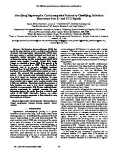
Image - 2 Oblique axial cine magnetic resonance angiogram in the same patient as in Image 1 shows a spade-shaped left ventricle with relative sparing of the apical myocardium (arrow).
Spin-echo MRI can be used to accurately characterize the distribution and degree of myocardial hypertrophy. Sardinelli et al found good correlation between MRI and 2dimensional (2D) echocardiography in demonstrating asymmetric septal hypertrophy. MRIs depicted apical and posterolateral myocardial hypertrophy not depicted on 2D echograms in 7 of 22 patients. Posma et al examined 52 unselected patients with HCM; MRIs provided accurate comprehensive quantitative information that could be used to calculate hypertrophic scores in all the patients. In comparison, echocardiography could not display all desired segments of the LV wall; image quality was adequate in 36% of the patients. In patients with asymmetric septal hypertrophy, the basal IVS at end diastole is disproportionately thickened (see Images 3-4), and the ratio of IVS thickness to posterolateral wall thickness is significantly increased. Spin-echo magnetic resonance examination of patients with asymmetric septal hypertrophy produces consistent findings (Sardinelli, 1993; Arrive, 1994; Higgins, 1985). Patients with asymmetric septal hypertrophy also have decreased systolic myocardial thickening, probably due to disarray and disorganization of myocardial fibers (Maron, 1987)
Hypertrophic Cardiomyopathy (HCM): A Review | Dr. Satish Kumar; Jan’08
Page |9
Image - 3 Short-axis cine end-diastolic magnetic resonance angiogram shows asymmetric hypertrophy with septal thickening (S).
Image - 4 Short-axis cine end-systolic, magnetic resonance angiogram obtained in the same patient as in Image 3 shows marked myocardial thickening that affects the entire myocardium.
MRI is superior to echocardiography in depicting hypertrophy of the apical portion of IVS and apical hypertrophy (Posma, 1996; Casolo, 1989; Suzuki, 1993). Long-axis MRIs accurately show the typical spade-shaped deformity of the LV cavity and the apical distribution of myocardial hypertrophy in patients with asymmetric septal hypertrophy (see Image 2). Hypertrophic Cardiomyopathy (HCM): A Review | Dr. Satish Kumar; Jan’08
P a g e | 10
With MRI, assessment of the thickness of the free wall of right ventricle and measurement of right ventricular mass are possible (Suzuki, 1988). For estimation of LV mass, spin-echo MRI, ECG-gated MRI, or multilevel cine MRA may be used. Studies with human cadaveric hearts (Allison, 1993; Katz, 1988) and canine hearts (Caputo, 1987) revealed no significant difference between LV mass estimations determined with MRI and the actual weights of the specimens. LVH in HCM often decreases the LV volume and increases the ejection fraction, without a significant change in stroke volume. Cine MRA can be used to calculate these parameters. If volume calculation is performed throughout the cardiac cycle, a timevolume curve can be obtained for more detailed functional analysis. An obstruction of the LV outflow tract (LVOT) obstruction resulting in a subaortic pressure gradient can be detected as an area of signal void in the normal area of flowing blood with high-signal-intensity on the cine MRAs (see Image 5). Although areas of physiologic signal void are depicted on images of healthy individuals, such signal voids are larger and persist longer in the cardiac cycle in pathologic conditions that cause obstruction (Mirowitz, 1990). Difficultly in differentiation between the 2 are rare.
Image - 5 Oblique cine magnetic resonance angiogram (outflow 2-chamber view equivalent to a long-axis echocardiogram) shows an area of signal intensity loss (white arrow) in the left ventricular outflow tract, where an obstruction between the hypertrophied septum and anterior mitral leaflet is present. The obstruction is well below the aortic valve ring (between black arrows).
Systolic anterior motion (SAM) of anterior mitral leaflet toward the IVS can be recognized on cine MRAs as a cause of LVOT obstruction. Mitral regurgitation is a common finding in patients with HCM and appears on cine MRAs as a signal void in the left atrium during ventricular systole. It may be associated with mitral valve prolapse Hypertrophic Cardiomyopathy (HCM): A Review | Dr. Satish Kumar; Jan’08
P a g e | 11
(see Images 6-7). Mirowitz et al described a small round area of physiologic signal void within the left atrium immediately behind and between the 2 mitral leaflets in 11 of 20 healthy individuals. In pathologic conditions, the signal void is larger and persists longer through ventricular systole. The area and extent of the signal void is well correlated with the grade of mitral regurgitation, as estimated with angiography and echocardiography (Aurigemma, 1990; Utz, 1988; Wagner, 1989).
Image - 6 Oblique cine magnetic resonance angiogram (outflow 2-chamber view) shows prolapse (arrow) of the posterior mitral leaflet in early systole.
Figure - 7 Oblique cine magnetic resonance angiogram (outflow 2-chamber view obtained during the same study as Image 6) shows mitral prolapse of the posterior mitral leaflet with a small signal intensity loss due to mitral regurgitation (arrow).
Hypertrophic Cardiomyopathy (HCM): A Review | Dr. Satish Kumar; Jan’08
P a g e | 12
Myocardial structural abnormality resulting from fiber disarray and disorganization can result in abnormal signal intensity. Fattori et al reported areas of reduced signal intensity, probably due to myocardial fibrosis in 16 (43%) of 37 unselected patients with HCM. This group also had higher maximum septal thickness (25 mm ± 7 vs 21 mm ± 6) and maximum posterior left wall thickness (15 mm ± 9 vs 7 mm ± 8). In patients with HCM, cine MRA also can be used to demonstrate nonuniform regional LV function (ie, LV asynchrony resulting in abnormal diastolic relaxation). As mentioned earlier, amyloidosis can resemble HCM. MRI has the capability to depict amyloidosis; the tissue may be characterized by its diffuse high signal intensity on T2weighted spin-echo and short–inversion time inversion recovery (STIR) MRIs. The signal intensity of cardiac amyloidosis was significantly lower with echo times of 20 ms and 60 ms, compared with that of HCM and that of healthy volunteers (Fattori, 1998). Magnetic resonance spectroscopy Alterations in myocardial metabolism are considered to be causes for contractile dysfunction in HCM. Proton-decoupled phosphorus-31 nuclear magnetic resonance spectroscopy depicts alterations of myocardial metabolism in asymptomatic patients with HCM. These alterations may contribute to the understanding of the pathophysiology and natural history of the disease (Jung, 1998). Patients with HCM have a significantly reduced ratio of phosphocreatine (PCr) to adenosine triphosphate (ATP) compared with that of healthy control subjects (Jung, 1998; de Roos, 1992). In addition, patients with severe hypertrophy of the IVS have a significantly increased inorganic phosphate (Pi)–to-PCr ratio compared with that of control subjects (Jung, 1998; Sieverding, 1997; de Roos, 1992). Both abnormalities are similar to those in ischemic myocardia. Also, significantly increased phosphomonoester (PME)-to-PCr ratios are present in patients with HCM; this finding indicates altered glucose metabolism (Jung, 1998). Myocardial pH is lower in patients with HCM relative to that of control subjects (de Roos, 1992). MRI myocardial tagging MRI myocardial tagging can be used to quantify the severity and extent of subtle regional heart wall motion abnormalities. The 3 stages of myocardial tagging are the following: (1) placement of a saturation band pattern (either a grid or parallel tag lines) over the myocardium with spatially selective RF pulses; (2) MRI acquisition, during which tag motion is observed; and (3) detection of myocardial tag motion. The motion of the saturation pattern is then used to compute the regional myocardial function. The fundamental improvements in regional function assessment with MR tagging techniques rather than echocardiography and nontagged MRI are 2-fold: (1) The same volume of myocardium can be tracked throughout the heart cycle to map function in a specific region, and (2) precise quantitative estimates of myocardial shortening and wall thickening can be computed from the images. The position of a myocardial tag can be estimated within approximately 150 m. Hypertrophic Cardiomyopathy (HCM): A Review | Dr. Satish Kumar; Jan’08
P a g e | 13
Maier et al found, with myocardial tagging, that the wall motion of the hypertrophied septum was significantly reduced in HCM, and Kramer et al showed depressed circumferential myocardial segment shortening in the septum and in the anterior and inferior regions. Three-dimensional analysis of tagged images showed that although circumferential and longitudinal ventricular strains were reduced in patients with HCM, the magnitude of the maximal contraction strain was reduced only in the basal septum and anterior walls (Young, 1994). This finding suggests that a major portion of the mechanical work in HCM contributes to wall shearing and not cavity reduction. Dong et al reported that the myocardium in patients with HCM is heterogeneously thickened and that the fractional thickening and circumferential shortening of the abnormally thickened myocardium are reduced.
ECHOCARDIOGRAPHY Findings: The classic echocardiographic definition of HCM includes LV wall thickness of more than 15 mm without LV dilatation in the absence of other cardiac and systemic causes of increased mass (Maron, 1979). However, no definitive criterion or single echocardiographic feature pathognomonic for HCM exists today (Fananapazir, 1994; Solomon, 1993). The typical echocardiographic feature in HCM is hypertrophy of the septum and LV anterolateral free wall (see Images 8-9); however, the degree and pattern of hypertrophy vary. Maximum hypertrophy of the septum often occurs midway between the base and the apex. On echocardiograms, asymmetrical septal hypertrophy is defined as a ratio of septal thickness to posterior wall thickness of at least 1.3-1.5. Although the average LV wall is thicker than 20 mm (ie, almost twice the normal thickness), it can vary from 13-15 mm in mild hypertrophy to 50 mm in massive hypertrophy (Maron, 1995). Image - 8 M-mode echocardiogram recorded at the level of the tips of the mitral valve (horizontal arrows) to assess left ventricular dimensions shows moderate thickening of both the septum (S) and posterior wall of the left ventricle (PW).
Hypertrophic Cardiomyopathy (HCM): A Review | Dr. Satish Kumar; Jan’08
P a g e | 14
Image - 9 Axial 2-dimensional echocardiogram obtained in the same patient as in Image 8 shows asymmetric septal thickening (23 mm) and a small left ventricular cavity (LV).
Often, the echocardiographic feature of a ground-glass appearance is noted either visually or by using quantitative texture analysis in both hypertrophied and nonhypertrophied regions of the ventricle (Lattanzi, 1991). It can be used to distinguish HCM from other causes of secondary hypertrophy (Naito, 1994). Another common echocardiographic feature in HCM is narrowing or obstruction of the LVOT caused by IVS and the anterior leaflet of the mitral valve. The abnormal geometry of LVOT results in a dynamic pressure gradient. Abnormal SAM of the anterior leaflet (see Images 10-11), and occasionally, the posterior leaflet of the mitral valve, is present (Klues, 1993). About 66% of mitral valves excised from patients with HCM have structural malformations, including an increased leaflet area, elongation of the leaflet, and an anomalous insertion of papillary muscle directly into the anterior mitral leaflet (about 15% of HCM cases) (Klues, 1992; Klues, 1991). Recognition of these anatomic abnormalities during the preoperative assessment is important.
Hypertrophic Cardiomyopathy (HCM): A Review | Dr. Satish Kumar; Jan’08
P a g e | 15
Image - 10 M-mode echocardiogram recorded at the level of the mitral valve shows a small ventricular cavity (arrow) and systolic anterior motion of the anterior mitral valve leaflet (*).
Image - 11 M-mode echocardiogram recorded at the level of the mitral valve in a patient with extreme septal hypertrophy (>40 mm) shows a small ventricular cavity (3 cm) and systolic anterior motion of the anterior mitral valve leaflet (*).
Hypertrophic Cardiomyopathy (HCM): A Review | Dr. Satish Kumar; Jan’08
P a g e | 16
Other echocardiographic findings may include a small ventricular cavity, reduced septal motion and systolic thickening, normal or increased motion of the posterior wall, abnormal rate of closure of mitral valve in middiastole (secondary to decreased LV compliance or abnormal transmitral diastolic flow), mitral valve prolapse, and partial systolic closure or coarse systolic fluttering of the aortic valve (see Image 12).
Image - 12 M-mode echocardiogram recorded at the level of the aortic valve in the same patient as in Image 8 shows high-frequency flutter on the aortic leaflets (arrows).
Doppler ultrasonographic findings have confirmed mitral regurgitation in virtually all patients with HCM and an outflow gradient (Maron, 1993). Doppler ultrasonography can be used to accurately measure the gradient (Louie, 1994) and to demonstrate the characteristic velocity profile due to dynamic outflow obstruction (see Image 13).
Image - 13 Continuous-wave Doppler image shows a typical concave profile (arrows) compared with the systolic waveform recorded from the left ventricular outflow; this finding represents subvalvular dynamic outflow obstruction.
Hypertrophic Cardiomyopathy (HCM): A Review | Dr. Satish Kumar; Jan’08
P a g e | 17
Athlete's heart Athletes with a LV wall that is thicker than 16 mm are likely to have pathologic hypertrophy such as HCM (Pelliccia, 1991), but smaller degrees of hypertrophy cannot always be differentiated from HCM. Maron et al (1995) published criteria that can help in the distinction of HCM from athlete's heart. Echocardiographic features that suggest HCM are an unusual pattern of LVH, asymmetry, end-diastolic LV dimension less than 45 mm, left atrial enlargement, and abnormal Doppler diastolic indices of LV filling. Other associated features suggestive of HCM are bizarre ECG findings, female sex, and family history of HCM. Also, athletic heart hypertrophy usually regresses within 3 months on cessation of exercise. Dilation of the LV chamber LV chamber dilatation and systolic dysfunction occur in about 1.5% of patients of HCM per year (McIntosh, 1992). This dilatation can evolve into a phase resembling dilated cardiomyopathy. NUCLEAR MEDICINE Findings: 201
TI myocardial tests TI myocardial tests, particularly those with SPECT, permit direct determination of the relative thickness of septum and free wall, and they may be useful in cases in which echocardiography is technically limited. Typically, Tl-enhanced images demonstrate a small LV cavity with marked Tl uptake in the hypertrophied myocardium. 201
Reversible perfusion defects (see Image 14), which presumably reflect myocardial ischemia, are common in HCM without coronary artery disease (Cannon, 1991). These defects are common in adult patients with HCM and in young patients with a history of syncope and sudden death (Dilisizian, 1993). Fixed defects occur in patients with impaired systolic function and likely represent myocardial scarring. Also, technetium-99m–labeled perfusion agents can be used, with similar results.
Image - 14 Stress (top row) and rest (bottom row) technetium-99m Sesta-2-methoxy-isobutylisonitrile (MIBI) perfusion images of hypertrophic cardiomyopathy shows a reversible septal perfusion defect that is not related to coronary obstruction. The septum is markedly thickened (4 cm on the echocardiogram). Hypertrophic Cardiomyopathy (HCM): A Review | Dr. Satish Kumar; Jan’08
P a g e | 18
Gated radionuclide ventriculography Gated radionuclide ventriculography with bloodpool labeling permits evaluation of the size and diastolic filling of the ventricular cavity and of the motion of the septum and ventricular wall. Myocardial scintigraphy Myocardial scintigraphy with iodine-123–m-iodobenzylguanidine (123I-MIBG) demonstrates decreased uptake and increased clearance in the hypertrophied myocardium. Nakajima et al performed myocardial scintigraphy with MIBG and 201TI in patients with HCM. Nonhypertensive patients were classified into 3 groups according to septal thickness, as determined with ultrasonography: group 1, wall thickness less than 16 mm; group 2, wall thickness 16-20 mm; and group 3, wall thickness more than 20 mm. The regional myocardial uptakes of both 201Tl and 123I-MIBG (percentage of injected dose per myocardial volume in cubic centimeters) were higher in the more hypertrophic septa. However, when regional MIBG uptake at 3 hours was divided by the 201TI uptake to calculate the MIBG uptake per unit of blood flow, the hypertrophic septa had lower mean values in groups 3 and 2. The regional MIBG clearance rate in the septum was significantly higher in group 3 compared with that of group 1. These findings suggested that flow-independent changes in sympathetic innervation or activity may exist in patients with HCM (Nakajima, 1990). Positron emission tomography Alteration in fatty acid metabolism is frequently seen in HCM at PET with carbon-11 palmitate (Seggewiss, 1998). In Japanese patients, studies performed with 123I-labeled 15-(p-iodophenyl)-3-R,S-methylpentadecanoic acid (BMIPP) have revealed heterogeneous distribution of this compound independent of thallium perfusion (Kurala, 1992; Nishimura, 1993; Ohtsuki, 1994; Takeiski, 1992; Taki, 1993). The finding seems to be an early sign of HCM, although more clinical experience is necessary for confirmation. ANGIOGRPHY Findings: Cardiac catheterization demonstrates decreased LV compliance and, in some patients, a subaortic systolic pressure gradient (see Image 15). The pressure gradient may be labile, varying 0-175 mm Hg in the same patient under different conditions. Increased myocardial contractility can worsen the gradient, particularly in patients with midventricular gradient, because of a direct muscular sphincteric action. Conversely, reduction in contractility or increases in preload or afterload (which increase the LV cavity size) reduce or eliminate the outflow gradient. This dynamic characteristic of HCM distinguishes it from other forms of ventricular outflow obstruction. The arterial pressure tracing may demonstrate a spike-and-dome configuration (Louie, 1994) (see Image 15). Approximately 25% patients have pulmonary hypertension, at least partly due to decreased LV compliance and elevated left atrial pressure. A right ventricular outflow tract pressure gradient occurs in 15% of patients who have LVOT obstruction (Frank, 1968), and this likely results from a markedly hypertrophied right ventricle (Maron and McIntosh et al, 1993). Hypertrophic Cardiomyopathy (HCM): A Review | Dr. Satish Kumar; Jan’08
P a g e | 19
Image - 15 Pressure tracing obtained as the catheter is pulled back from the center of the left ventricle to the aortic root shows a reduction in systolic pressure (arrow 1) in the left ventricle; this finding indicates a subaortic gradient. The waveform changes at the level of the aortic valve, but the systolic pressure does not change (arrow 2). Note the spike-and-dome configuration of the left ventricular pressure tracing.
Left ventriculography reveals a hypertrophied ventricle with vigorous ejection (see Images 16-20). The papillary muscles often are prominent, filling the LV cavity at the end of systole. In patients with apical involvement, extensive hypertrophy may result in a spadelike configuration of the LV cavity (Smolders, 1993). Associated mitral regurgitation may be present (see Image 18, Image 20). Simultaneous right ventriculography in cranially angulated left anterior oblique projections can be performed for optimal evaluation of the IVS. The left septal surface is flat or it bulges into the LV cavity at its middle or lower portion, in contrast to the normal curve toward the right ventricle. Image - 16 End-diastolic right anterior oblique digital subtraction left ventriculogram shows the normal size and shape of the left ventricle in a patient with hypertrophic cardiomyopathy.
Image - 17 End-diastolic right anterior oblique digital subtraction left ventriculogram obtained in the same study as Image 16 shows a small cavity, with prominent papillary muscles (arrows) projecting into the remains of the ventricular cavity.
Image - 18 Images 16-17 are used to calculate function and left ventricular dimensions. The outline of the end-diastolic image (Image 16) has been superimposed on the systolic image (Image 17). Ejection fractions were calculated by using the area-length method (ejection fraction, 86%) and the Simpson rule (ejection fraction, 84%). The videodensitometric technique shown is inaccurate because of incorrect background registration. Hypertrophic Cardiomyopathy (HCM): A Review | Dr. Satish Kumar; Jan’08
P a g e | 20
Coronary angiographic findings usually are normal, but images may show myocardial bridging. The distance between the coronary arteries on the epicardial surface and the ventricular cavity is increased, indicating myocardial hypertrophy (see Images 19-21).
Image - 19 Conventional end-diastolic right anterior oblique left ventriculogram acquired during cardiac catheterization shows the normal size and shape of the left ventricle in a patient with hypertrophic cardiomyopathy. Note the distance between the ventricular cavity and the coronary arteries (arrows), which define the epicardial surface of the heart. This distance indicates considerable thickening of the myocardium.
Image - 20 Conventional end-systolic right anterior oblique left ventriculogram acquired during the same cardiac catheterization study as in Image 19 shows a small left-ventricular cavity with mild mitral regurgitation (M). Note the increased distance between the ventricular cavity and the coronary arteries (arrows), which define the epicardial surface of the heart as the myocardium becomes thickened in systole.
Image - 21 Conventional right anterior oblique aortogram acquired during cardiac catheterization in the same patient as in Image 19-20 shows unobstructed coronary arteries.
Hypertrophic Cardiomyopathy (HCM): A Review | Dr. Satish Kumar; Jan’08
P a g e | 21
INTERVENTION The management of HCM involves identifying and reducing the risk of sudden death, performing therapeutic evaluations, and providing medical and/or invasive treatments for the purpose of alleviating symptoms and preventing complications. Family members of patients should undergo echocardiographic screening to facilitate early diagnosis and management. Risk factors for sudden death Risk factors for sudden death include young age (