Internet Journal of Food Safety, Vol.9, 2007, p. 56-62 Copyright© 2007, Food Safety Information Publishing
A Multiplex PCR for Detection of Virulence Associated Genes in Listeria monocytogenes Rawool, D. B.1*, Malik, S. V. S.1, Barbuddhe, S. B.2, Shakuntala, I.1 and Aurora, R.3 1
Division of Veterinary Public Health, Indian Veterinary Research Institute, Izatnagar 243 122 India 2 ICAR Research Complex for Goa, Ela, Old Goa 403 402, India 3 Dayalbagh Educational Institute (Deemed University), Dayalbagh, Agra 282005, India. Introduction
Abstract: Listeriosis is an important food-borne infection caused by Listeria monocytogenes. A multiplex PCR assay was developed for detection of four virulence-associated genes of L. monocytogenes namely, plcA, hlyA, actA and iap. The method was used to detect L. monocytogenes in milk samples with and without enrichment and simultaneously comparing it with conventional cultural method. The assay could detect as low as 15 cells after enrichment. Later, the multiplex PCR was successfully employed to detect virulence-associated genes in L monocytogenes isolated from various sources.
Listeriosis is an important food-borne infection caused by Listeria monocytogenes. L. monocytogenes has been isolated from various foodstuffs including milk (Barbuddhe et al., 2002). Identification of listeriae from samples contaminated with multiple species relies on selective enrichments and subsequent biochemical analyses. It is laborious and requires at least five days for confirmation. In vitro amplification of specific DNA sequences by PCR allows direct detection and identification of the pathogen (Herman et al. 1995). Multiple key virulence factors such as hemolysin (hlyA), phosphatidylinositol phospholipase C (plcA), actin polymerization protein (actA) and invasive associated protein (iap) are important in L. monocytogenes pathogenesis (Furrer et al., 1991, Portnoy et al., 1992). Therefore, detection of just one virulence-associated gene by PCR is not always sufficient to identify L. monocytogenes (Nishibori et al., 1995). In addition, it is plausible that some L. monocytogenes strain may lack one or more virulence determinants because of spontaneous mutations (Cooray et al., 1994). Therefore, simultaneous detection of virulent genes in a single step will be desirable as it reduces time, labour and it will be useful in a large scale survey for detecting virulent strain of Listeria. Direct detection of pathogens from foods by PCR may be problematic due to the presence of a PCR-inhibitory factor in the food (Fluit et al., 1993). Various sample preparation methods have been developed to remove or to reduce the effects of PCR inhibitors in food stuffs (Lantz et al. 1994). A pre-enrichment method (Niederhauser et al., 1992) has been reported to overcome the problem of inhibitors. In the present study, we tried to find a procedure enabling the detection of individual virulence-associated genes of L. monocytogenes by PCR in milk samples with and without enrichment and simultaneously comparing it with conventional cultural method. Later, we developed a multiplex PCR for simultaneous detection of four virulenceassociated genes of L. monocytogenes namely, plcA, hlyA, actA and iap and employed for PCR analysis of virulence-
Key words: Milk, Listeria monocytogenes; multiplex PCR; virulence –associated genes
*
Corresponding author. mailing address: Division of Veterinary Public Health, Indian Veterinary Research Institute, Izatnagar 243 122 India, E-mail:
[email protected]
56
associated genes in L. monocytogenes isolated from various sources.
mM KCl, 15 mM MgCl2 and 0.01% gelatin), 0.2 mM dNTP mix, 2 mM MgCl2 and 10 µM of a primer set containing forward and reverse primers (a final concentration of 0.1 µM of each primer), 1 unit of Taq DNA polymerase, 5 µl of cell lysate and sterilized milliQ water to make up the reaction volume. The cycling conditions for PCR included an initial denaturation of DNA at 95°C for 2 min followed by 35 cycles each of 15 s denaturation at 95°C, 30 s annealing at 60°C and 1 min 30 s extension at 72°C, followed by a final extension of 10 min at 72°C and hold at 4°C. All the four set of primers for virulence-associated genes were amplified under the similar PCR conditions and amplification cycles. The resultant PCR products were further analyzed by agarose gel electrophoresis (1.5%; low melting temperature agarose L); stained with ethidium bromide and visualized by a UV trans-illuminator (UVP Gel Seq Software, England).
Materials and Methods Bacteria. The strains of L monocytogenes 4b (MTCC 1143), Staphylococcus aureus (MTCC 1144), Rhodococcus equi (MTCC 1135), Streptococcus faecalis (MTCC 439), Bacillus cereus (MTCC 1272), Escherichia coli (MTCC 443), Aeromonas hydrophila (MTCC 646) used in the study were obtained from Institute of Microbial Technology, Chandigarh, India. The reference strains of Listeria namely, L. monocytogenes 4b (NCTC 11994), L. monocytogenes 1/2a (NCTC 7973), L. monocytogenes 1/2b (NCTC 10887), L.ivanovii (NCTC 11846), L. innocua (NCTC 11288), L. seeligeri (NCTC 11856), L. grayi (NCTC 10812), L. welshimeri (NCTC 11857) were kindly provided by Prof. K.L. Morgan, University of Liverpool, U.K. The strains of Salmonella (1117) and Vibrio cholerae (0139) were procured from Division of Veterinary Public Health, Indian Veterinary Research Institute, India. The strains were tested for their purity besides morphological and biochemical characteristics.
Sensitivity of PCR. The standardized PCR was assessed for its sensitivity. Briefly, L. monocytogenes 4b (MTCC 1143) culture grown overnight in BHI broth was centrifuged at 6000 rpm for 10 min. and the pellet obtained was washed once with phosphate buffered saline (PBS), pH 7.2. The concentration of cells was adjusted to 3 x 10⁸/ml using McFarland’s nephelometric tube No.1 and 10-fold serial dilutions were made upto 3 cell/ml of PBS. The template DNA was prepared by subjecting 1ml aliquot of each dilution to snap-chill method as described above.
Polymerase chain reaction. For standardization of PCR a virulent strain of L. monocytogenes MTCC 1143 was grown overnight in Brain Heart Infusion broth (BHI) at 37°C. The DNA was extracted as per the methodology of Makino et al., (1995). The obtained culture (approximately 1 ml) was centrifuged in a microcentrifuge (Sigma, USA) at 6000 rpm for 10 min. The recovered pellet was resuspended in 100 µl of sterilized DNAse and RNAse-free milliQ water (Millipore, USA), heated in a boiling water bath for 10 min. and then snap chilled in crushed ice. The obtained lysate (5µl) was used as a DNA template in PCR reaction mixture. Bacterial DNA was also extracted employing DNA extraction kit (Mini Prep, Sigma). PCR was standardized for the detection of individual virulence associated genes namely, plcA, hlyA, actA and iap using primer pairs of 5'-CTG CTT GAG CGT TCA TGT CTC ATC CCC C-3' and 5'-ATG GGT TTC ACT CTC CTT CTA C-3' specific for plcA (Notermans et al. 1991), 5'-GCA GTT GCA AGC GCT TGG AGT GAA-3’ and 5’GCA ACG TAT CCT CCA GAG TGA TCG-3' for hlyA (Paziak-Domanska et al. 1999), 5'-CGC CGC GGA AAT TAA AAA AAG A-3' and 5'- ACG AAG GAA CCG GGC TGC TAG - 3' for actA (Suarez and Vazquez-Boland 2001) and 5'-ACA AGC TGC ACC TGT TGC AG-3' and 5'-TGA CAG CGT GTG TAG TAG CA-3' for iap (Furrer et al. 1991) of L. monocytogenes by optimizing the different conditions that affect the sensitivity and specificity of the reaction. Based on optimization trials, the standardized PCR protocol for 50 µl reaction mixture included 10 X PCR buffer (100 mM Tris-HCl buffer, pH 8.3 containing 500
Specificity of PCR. The specificity of the standardized PCR was tested by screening the standard strains of L. monocytogenes, Listeria species as well as some other commonly prevalent and cross-reacting bacterial species (Table 1) with the primers used in this study. The DNA template preparation from the test organisms and other PCR conditions were similar to those described earlier. Multiplex PCR. The multiplex PCR was standardized for detecting four virulence associated genes of L. monocytogenes namely, plcA, hlyA, actA and iap in a single reaction tube containing all the four primer sets for these genes. The DNA template preparation from pathogenic strain of L. monocytogenes, PCR reaction and agarose gel electrophoresis of the PCR products were done as per the methods employed for the detection of virulence genes of L. monocytogenes described earlier. The standardized PCR protocol for 50 µl reaction mixture included 10 X PCR buffer (100mM Tris-HCl buffer, pH 8.3 containing 500 mM KCl, 15 mM MgCl2 and 0.01% gelatin), 1 mM dNTP mix, 6 mM MgCl2 and 10µM of four primer sets (containing forward and reverse primers a final concentration of 0.1 µM of each primer), 4 units of Taq DNA polymerase, 5 µl of cell lysate and sterilized milliQ water to make up the reaction volume. The cycling conditions for multiplex PCR
57
Table 1. Standard bacterial strains used to check the specificity of PCR Virulence genes detected Description of the organism hlyA
plcA
actA
+ + + + + -
+ + + + + -
+ + + + + -
L. monocytogenes 4b (MTCC 1143) L. monocytogenes 4b (NCTC 11994) L. monocytogenes 1/2a (NCTC 7973) L. monocytogenes 1/2b (NCTC 10887) L. ivanovii (NCTC 11846) L. welshimeri (NCTC 11857) L. innocua (NCTC 11288) L. grayi (NCTC 10812) L. seeligeri (NCTC 11856) Escherichia coli (0157H7) Vibrio cholerae (0139) Salmonella (1117) Bacillus cereus (MTCC 1272) Streptococcus faecalis (MTCC 439) Escherichia coli (MTCC 443) Staphylococcus aureus (MTCC 1145) Rhodococcus equi (MTCC 1135) Aeromonas hydrophila (MTCC 646)
iap + + + + -
spiked milk samples in set 1 were centrifuged at 12000 rpm for 10 min. The pellet obtained was washed once with PBS (1ml) and recovered by centrifugation at 12000 rpm for 10 min. The washed pellet was dissolved in sterilized milliQ water (100µl), subjected to vigorous heating in boiling water bath for 10 minutes and then snap-chilled in crushed ice for 2 min. The cell lysate was centrifuged at 3000 rpm for 10 min and the supernatant was used as DNA template for detection of the test strain by PCR. Simultaneously, 1 ml of each dilution of the spiked samples in set 2 was spreaded on Dominguez-Rodriguez isolation agar (Dominguez-Rodriguez et al. 1984) and incubated at 37°C for 24 h. Similar procedure was adopted for detection of L. monocytogenes suspended in milk after enrichment in UVM-1 by PCR as well as cultural method.
were similar as described earlier for the detection of individual virulence gene of L. monocytogenes. Artificially contaminated milk samples. Using the abovementioned PCR setting we tried to detect L. monocytogenes suspended at serial concentrations ranging from 3 x 10 to 3 × 10⁸ cells ml-1 in commercially available pasteurized cow milk. The pasteurized milk samples those turned out to be culturally negative for Listeria were pooled and stored at – 20˚C till used for experimental work. Aliquots (3 ml) drawn from the pooled milk sample were spiked with standard strain of the pathogen and processed for (with and without enrichment) detection of the pathogen by cultural method and PCR. Briefly, milk aliquots (3 ml) drawn from the pooled milk sample were inoculated with the different concentration of test strain ranging from 30 to 3 x 10⁸ cells/ml. Each dilution of the sample was distributed in three test tubes, 1ml/test tube designated as set 1, set 2 and set 3. Accordingly 9 test tubes including negative control were included in each set. Of these, two sets not subjected to enrichment (set1 and 2) were analyzed immediately for the detection of the test strain, i.e. one (set1) by the PCR targeted at virulence-associated genes of L. monocytogenes and another (set 2) by cultural method. The remaining set (set 3) was enriched in University of Vermont medium 1 (UVM-1) in the ratio of 1: 10 (i.e. 1 part of milk and 9 parts of UVM-1 broth) and incubated overnight at 37°C before attempting detection of the test strain by PCR as well as cultural method. Aliquots (1 ml) from each dilution of the
Listeria strains. Twenty strains of L. monocytogenes isolated from milk (10), buffaloes with reproductive disorders (5) and fresh water fishes (5) were analysed for the presence of virulence associated genes employing the multiplex PCR.
Results and Discussion Rapid isolation and confirmation methods for L. monocytogenes in foods are still being sought (Beumer and Hazeleger, 2003). But there are certain strains of L. monocytogenes which behave phenotypically quite typical and inconspicuous but are non-pathogenic (Hof and
58
Rocourt, 1992). Thus, to address the pathogenic potential of Listeria isolates, in vivo methods namely, chick embryo and mouse inoculation tests remain the most reliable and mandatory approach in order to link these isolates with the cases of listeriosis. However, the in vivo methods remain objectionable from ethical point of view and need skilled personnel to perform. In view of this situation, it has been suggested that diagnosis of pathogenic Listeria spp. and listeric infection should ideally be based on virulence markers (Notermans et al., 1991). Moreover, the importance of PCR has been investigated for detection of L. monocytogenes from foods (Gouws and Liedemann, 2005). Thus, in the present study we first attempted to standardize a PCR protocol, which could detect individual virulent associated genes (plcA, hlyA, actA and iap) of L. monocytogenes in milk samples with and without enrichment and simultaneously comparing it with conventional cultural method. Later, we developed a multiplex PCR for simultaneous detection of all the virulence-associated genes of L. monocytogenes under study for rapid detection of virulent strains of L. monocytogenes.
the DNA marker (Fig. 1). These findings commensurate with the published work for detection of hlyA gene (PaziakDomanska et al., 1999), plcA and prfA genes (Notermans et al., 1991b), iap gene (Furrer et al., 1991) and actA gene (Suarez and Vazquez-Boland, 2001) with respective sets of primers giving no cross-reactions with other bacteria.
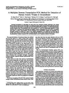
Figure 2. Multiplex PCR of four virulence associated genes for standard Listeria monocytogenes (MTCC 1143). M: DNA Marker (150 to 2000 bp), Lane 1: Negative control, Lane 2: Amplified products of four genes (plcA, actA, hlyA and iap).
Each of the primer was found to be specific to the target gene as it specifically amplified the PCR product of that gene. All the four genes were detected in standard strains of L. monocytogenes, two genes namely, plcA, and actA in the standard strain of L. ivanovii and the hlyA gene in the standard strain of L. seeligeri whereas, none of the genes was detected in the cultures of the remaining Listeria spp. and other bacterial species cultures (Table 1). The amplification of plcA gene in L. ivanovii can be explained based on the reports that plcA and plcB genes are present in L. monocytogenes and L. ivanovii (Vazquez-Boland et al., 2001). The actA gene from L. ivanovii, i-actA, encodes a protein larger than actA from L. monocytogenes. The similarity between the two proteins is also limited (34% identity), however, actA and i-actA have a similar overall structure (Gerstel et al., 1996). The amplification of haemolysin gene (hly A) in L. seeligeri genome could be explained on the basis of homology at both the protein (8691%) and in the nucleotide (76-78%) of gene sequence of LLO, ILO and seeligerilysin O (LSO) (Mengaud et al., 1988; Haas et al., 1992). However, our amplification
Figure 1. Virulence associated genes of standard Listeria monocytogenes (MTCC 1143). M: DNA Marker (150 to 2000 bp), Lane 1: plcA gene, Lane 2: actA gene, Lane 3: hlyA gene, Lane 4: iap gene, Lane 5: Negative control.
In the present study the primer sets for all the four genes allowed amplification of 1484 bp (plcA), 839 bp (actA), 456 bp (hlyA) and 131 bp (iap) PCR products, respectively, each represented by a single band in the corresponding region of
59
system does not yield DNA fragments of hlyA gene in standard strain of L. ivanovii, which can be explained on the basis of report that the genetic information for haemolysin production in L. ivanovii could be divergent from L. monocytogenes hemolysin gene despite high homology in partial amino acid sequence analysis (Kreft et al., 1989). Enrichment of food samples prior to PCR analysis overcomes most of the problems and has been recommended by several workers (Wang et al., 1992, Boer and Beumer 1999, Olsen 2000). In the present study, the sensitivity of PCR was compared for the detection of the pathogen in spiked milk samples before and after their enrichment in UVM-1 at 37°C for 24 h. It was possible to detect as low as 3x10⁶ cells/ml suspended in PBS by using each of the primer set. Compared with the detection limit for bacteria suspended in PBS, the enrichment of milk samples in UVM-1 at 37°C for 24 h has increased the sensitivity of PCR from 3 x 10⁶ cells/ml (before enrichment) to 3 x 10³ cells/ml. With the PCR, setting fewer than 15 cells could be detected after enrichment. Similar type of increase in the sensitivity of cultural method was also observed following 24 h enrichment from 3 x 10³ cells/ml (before enrichment) to 30 cells/ml of the milk samples in UVM-1. The poor sensitivity of PCR as compared to culture method might be because only 5.0µl of the DNA was used in PCR reaction whereas 1ml of sample (200 times more) was used for plating in cultural method. Our results are in agreement with Cartyyvels et al., (1996) who observed similar results of poor sensitivity of PCR for detection of Mycobacterium tuberculosis. They explained that greater sensitivity of the culture method could be due to 40-times greater sample volume used in the culture method. In the present study, the better detection of the pathogen (as less as 3 x 10³ cells/ml) by PCR in the enriched spiked samples compared to the detection level (3 x 10⁶ cells/ml) in samples not subjected to enrichment in UVM-1 might be attributed to the significant increase in the number of the pathogen following enrichment of the samples compared to the relatively lesser number of L. monocytogenes present in the milk samples before enrichment. This approach is in agreement with reported detection of L. monocytogenes by the PCR in artificially contaminated milk samples (Cooray et al., 1994). Since only 5 µl of lysate was taken as DNA template into the PCR mixture, it indicated that fewer than 15 cells could be detected after enrichment. The present level of sensitivity in the detection appeared to be applicable to the practical survey of milk and milk products for L. monocytogenes. This assay may prove useful for rapid detection of L. monocytogenes in milk and milk products and can be adopted for meat and clinical samples. In the food industry the contamination of surfaces, equipment, and food with non-pathogenic Listeria spp. is common. Therefore, the ability to rapidly detect pathogenic Listeria strains without
cross-reactions with non-pathogenic and related strains can potentially improve upon current approaches. In the present study L. monocytogenes strains isolated from various sources were analyzed for the presence of virulence-associated genes employing the multiplex PCR. Different combinations of genes were detected in different isolates (data not shown). The choice of the target gene is of utmost importance for detection of virulent strains of L. monocytogenes by PCR. The use of primers specific for plcA, hlyA, actA and iap seems to be reasonable and unique to pathogenic Listeria (Furrer et al., 1991, Notermans et al. 1991, Paziak-Domanska et al., 1999, Suarez and VazquezBoland 2001). However, it is plausible that some L. monocytogenes strain may lack one or more virulence determinants because of some mutation (Cooray et al., 1994). Moreover, our previous studies have also revealed that using PCR assay for detection of single virulence associated gene is neither sufficient to identify the L. monocytogenes isolates nor to reveal its true pathogenic potential as majority of L. monocytogenes isolates showed different gene profiles (Rawool et al., 2007; Shakuntala et al., 2006). Therefore, simultaneous detection of virulent genes namely plcA, hlyA, besides actA and iap genes in a single step will be desirable as it reduces time, labour and also it would be useful in a large scale survey aiming at detection of virulent strain of Listeria.
Summary The multiplex PCR was successfully employed for the detection of different genes in L. monocytogenes. The PCR method reported here could be completed in 6 h without enrichment however since the detection level was poor without enrichment, the authors would like to recommend the use of this multiplex PCR after enrichment for a large scale survey to detect of virulent strains of Listeria from milk samples.
Acknowledgements We thank Director, Indian Veterinary Research Institute, Izatnagar, India for providing facilities for the research work. Thanks are due to Dr. A.R. Datta, US FDA for critically checking the manuscript
References Barbuddhe, S.B., Chaudhari, S.P. and Malik, S.V.S. (2002) The occurrence of pathogenic Listeria monocytogenes and antibodies against listeriolysin-O in buffaloes. J. Vet. Med. B 49: 181-184.
60
Lantz, P., Tjerneld, F., Borch, E., Hahn-Hagerdal, B. and Radstrom, P. (1994) Enhanced sensitivity in PCR detection of Listeria monocytogenes in soft cheese through use of an aqueous two-phase system as a sample preparation method. Appl. Environ. Microbiol. 60, 3416-3418. Makino, S., Okada, Y., & Maruyama, T. (1995). A New Method for Direct Detection of Listeria monocytogenes from Foods by PCR. Applied and Environmental Microbiology, 61, 3745-3747. Mengaud, J., Vincente, M.F., Chenevert, J., Periera, J.M., Geoffroy, C., Gicquel-Sanzey, B., Baquero, F., PerezDiaz, J.C. and Cossart, P. (1988). Expression in Escherichia coli and sequence analysis of listeriolysin O determinant of Listeria monocytogenes. Infect. Immun., 56: 766-772. Niederhauser,C., Candrian, C., Hofelein, M., Buhler, H.P. and Luthy, J. (1992) Use of polymerase chain reaction for detection of Listeria monocytogenes in food. Appl. Environ. Microbiol. 58, 1564-1568. Nishibori, T., Cooray, K., Xiong, H., Kawamuru, I., Fujita, M. and Mitsuyama, M. (1995) Correlation between the presence of virulence associated genes as determined by PCR and actual virulence to mice in various strains of Listeria spp. Microbiol. Immunol. 39, 343-349. Notermans, S.H.W., J. Dufrenne, M. Leimeister-Wachter, E. Domann, and Chakraborty, T. (1991) Phosphatidylinositol-specific phospholipase C activity as a marker to distinguish between pathogenic and nonpathogenic Listeria species. Appl. Environ. Microbiol. 57, 2666-2670. Olsen, J.E. (2000) DNA-based methods for detection of foodborne bacterial pathogens. Food Res. Int. 33, 257266. Paziak-Domanska, B., Bogulawska, E., Wiekowska-Szakiel, M., Kotlowski, R., Rozalska, B., Chmiela, M., Kur, J., Dabrowski, W. and Rudnicka, W. (1999) Evaluation of the API test, phosphatidylinositol-specific phospholipase C activity and PCR method in identification of Listeria monocytogenes in meat foods. FEMS Microbiol. Lett. 171, 209-214. Portnoy, D.A., Chakraborty, T., Goebel, W. and Cassart, P. (1992) Molecular determinants of Listeria monocytogenes pathogenesis. Infect. Immun. 60, 12631267. Rawool, D.B., Malik, S.V.S., Shakuntala, I., Sahare, A.M. and Barbuddhe, S.B. (2007). Detection of multiple virulence-associated genes in Listeria monocytogenes isolated from bovine mastitis cases. Int. J. of Food Microbiol. 113:201-207. Shakuntala, I., Malik, S.V.S., Barbuddhe, S.B. and Rawool, D.B., (2006). Isolation of Listeria monocytogenes from buffaloes with reproductive disorders and its confirmation by polymerase chain reaction. Vet. Microbiol. 117:229-234.
Beumer R.R. and Hazeleger, W.C. (2003). Listeria monocytogenes: diagnostic problems, FEMS Immunol. Med. Microbiol. 35: 191–197. Boer, B.D. and Beumer, R.R. (1999) Methodology for detection and typing of food borne microorganisms. Int. J. Food Microbiol. 50: 119-130. Cartuyvels R, Ridder C, Jonckheere S, Verbist L, van Eldere J. (1996). Prospective clinical evaluation of Amplicor Mycobacterium tuberculosis PCR test as a screening method in a low-prevalence population. J Clin Microbiol 34: 2001-2003. Cooray, K. J., Nishibori, T., Xiong, H., Matsuyama, T., Fujita, M. and Mitsuyama, M. (1994). Detection of multiple virulence-associated genes of Listeria monocytogenes by PCR in artificially contaminated milk samples. Appl. Environ. Microbiol.. 60: 3023-3026. Dominguez-Rodriguez, L., Suarez-Fernandez, G., Fernandez-Garayzobal, J. and Rodriguez-Ferri, E. (1984). New methodology for the isolation of Listeria monocytogenes from heavily contaminated environments. Appl. Environ. Microbiol. 47: 1188-1190. Fluit, A. C., Torensma, R., Visser M. J. C., Aarsman, C.J.M., Poppeiler, M. J. J. G., Keller, B. H. I., Klapwijk, P. and Verhoef, J. (1993). Detection of Listeria monocytogenes in cheese with the magnetic immunopolymerase chain reaction assay. Appl. Environ. Microbiol. 59: 1289-1293. Furrer, B., Candrian, U., Hoefelein, Ch. and Luethy, J. (1991). Detection and identification of Listeria monocytogenes in cooked sausage products and in milk by in vitro amplification of haemolysin gene fragments. J. Appl. Bacteriol. 70: 372-379. Gerstel, B., Gröbe, L., Pistor, S., Chakraborty, T. and Wehland, J. (1996). The ActA polypeptides of Listeria ivanovii and Listeria monocytogenes harbor related binding sites for host microfilament proteins. Infect. Immun. 64:1929-1936. Gouws, P. A. and Liedemann, I. (2005). Detection of L. monocytogenes in Food Products. Food Technol. Biotechnol. 43 (2): 201–205 Haas, A., Dumbsky, M. and Kreft, J. (1992). Listeriolysin genes: complete sequence of ilo from Listeria ivanovii and of iso from Listeria seeligeri. Biochim. Biophys. Acta, 1130: 81-84. Herman, L.M.F., de Block, J.H.G.E. and Moermans, R.J.B. (1995) Direct detection of Listeria monocytogenes in 25 milliliters of raw milk by a two-step PCR with nested primers. Appl. Environ. Microbiol. 61: 817-819. Hof, H. and Rocourt, J. (1992). Is any strain of Listeria monocytogenes detected in food a health risk? Int.l J. Food Microbiol., 16: 173 182. Kreft, J., Funke, D., Haas, A., Lottspeich, F. and Goepel, W. (1989). Production, purification and characterization of haemolysins from L. ivanovii and L. monocytogenes. Sv.4b.FEMS Microbiol. Let. 57: 197-202.
61
Suarez, M. and Vazquez-Boland, J. A. (2001) The bacterial actin nucleater protein actA is involved in epithelial cell invasion by Listeria monocytogenes. www.ncbi.nlm.nih.gov/pubmed [Accession No. AF103807]. Vázquez-Boland, J.A., Kuhn, M., Berche, P., Chakraborty, T., Domínguez-Bernal, G., Goebel, W., González-Zorn, B., Wehland, J. and Kreft, J. (2001). Listeria pathogenesis and molecular virulence determinants. Clin Microbiol Rev 14: 584–640. Wang, R.F., Cao, W.W. and Johnson, M.G. (1992) Development of cell surface protein associated gene probe specific for Listeria monocytogenes and detection of the bacteria in food by PCR. Mol. Cell. Prob. 6, 119129 ADEWOYE , S.O. and OMOTOSHO ,J.S. (1997): Nutrient Composition of some freshwater fishes in Nigeria Biosci. Res. Commun. 11 (4) 333-336.
Proceedings of the 2nd Annual Conference of fisheries society of Nigeria (FISON), Calabar, 25th -27th, January. LAGLER, K.F., BARDACH, J.E. and. MILLER, R.R (1977): “Lethology, the study of fishes. Wiley, New York 156-163pg. LENNTECH, (2006): “Lenntech Water Treatment and air purification holding B.V.” Retrieved June 2006, from http://www.lenntech.com/feedback2.htm MILLS, C.F. (1980): The mineral nutrition of livestock (Underwood, E.J. 1981 Ed.) Common Wealth Agricultural Bureaux Pg 9. EIL, W., TREMBLAY and ANDREW, P. G. (1995): “Human Health and Great lakes, and Environmental Pollution: A 1994 perspectives”. Retrieved June 26, 2006, from http://www.ehp.com. SADIKU, S.O.E. and OLADIMEJI, A..A. (1991): “Relationship of proximate composition of Lates niloticus (L), synodontis schall REs. Commun. 3 (1), 2940.
ADEWOYE, S. O., FAWOLE, O. O. and OMOTOSHO, J. S. (2003). Concentrations of selected elements in some fresh water fishes in Nigeria. Science Focus. Vol. 4, pp 106-108. BENTLY, P. J. (1971). Endocrine and osmoregulation, Springer-Verdag Heidelberg. Pp. 220-230.
SHUL’MAN, G.E. (1974): Life cycle of fish: Physiology and Biochemistry, Halsted Press a division of John Wiley and Son Inc. N.Y. (1st Ed.) Pg 101-104.
AKO, P.A. and SALIHU, S.O. (2004): “Studies on Some Major and Trace Metals in Smoked and Over-Dried Fish”, Journal of Applied Sciences and Environmental Management, Vol. 8, No.2 Dec. pp 5-9.
TAYLOR, D.J.; GREEN, N.P.O. and. STOUT, G.W. (2002): Biological Science, 3rd edition, Cambridge CB21RP, Cambridge University Press. WATERMAN, J..J. (2000): Composition and Quality of Fish, Edinburgh, Torry Research Station.
A.O.A.C., (1975): Official Methods of Analysis 12th Edition. (W. Hortuntzed). Association of Official Analytical Chemists, Washington, D.C.
WINDOW H.; STEIN, D.; SCHELDON, R.; and SMITH, J. R. (1987): “Comparison of trace metal concentrations in muscle of a benthopelagic fish (Cory phaenoides armatus) from the Atlantic and Pacific oceans”. Deep seaResearch 34 (2): 213-220.
BOYD, C.E. and. DAVIS J.A (1978): Concentration of selected element and ash in Bluegill (Lepomis macrochirus) and certain other freshwater fish. Trans. Am. Fish Soc. 6: 862-867. BURGRESS, G.H.O (1975): “Increasing the direct consumption of fish. In: WW Pirie (Edu). Food Protein Sources. International Biological Programme 4. Cambridge University Press, Cambridge, pp 187-200. FORAN, J.A., CARPENTER, D. O., Hamilton, M.C., Knuth, B.A., and SCHWAGER, S.J., (2005): “Riskbased consumption advice for farmed Atlantic and wild pacific salmon contaminated with dioxins and dioxinlike compounds”. Environmental health perspective 33:552-556. KHAN, A.H., ALI, M., BIASWAS, S. K., and HADI, D.A. (1987): “Trace elements in marine fish from the Bay of Bengal”. The science of the total environment 61: 12130. LADIPO, O. O., SONAIKE, O.O. and OLUDIMU, O.L. (1982): A statistical investigation of fish in Nigeria.
62