Urethral Stents: Review of Technology and Clinical Applications Mordechai Duvdevani, Ben H. Chew, and John D. Denstedt
Summary. Urethral stents are a minimally invasive therapy used in the treatment of benign prostatic hyperplasia, urethral stricture, or detrusor sphincter dyssynergia. This chapter reviews the different types of urethral stents, indications for their use, and clinical results. Urethral stents may be positioned in the urethra or prostatic urethra and are classified as temporary or permanent. Temporary stents are further subdivided into biodegradable and nonbiodegradable. This form of therapy is particularly useful in patients who are at high anesthetic risk and are unable to undergo surgical procedures considered to be the gold standard, such as transurethral prostatectomy or open urethroplasty for prostatic enlargement and urethral stricture disease. Urethral stents can provide an effective alternative to transurethral and open procedures in many urological disorders that affect the prostate and urethra. Keywords. Urethra, Stent, Benign prostatic hyperplasia, Detrusor sphincter dyssynergia, Stricture
Introduction The term “stent” is defined as “a thread, rod, or catheter, lying within the lumen of tubular structures, used to provide support during or after an anastomosis, or to assure patency of an intact but contracted lumen” [1]. Urethral stents are typically made of a metal alloy or polymeric or biodegradable material in a variety of designs that are rigid enough to maintain urethral patency. Urethral stents are designed to relieve bladder outlet obstruction caused by various etiologies. Indications in appropriate patients for urethral stent placement include urethral stricture disease, benign prostatic hyperplasia (BPH), detrusor-sphincter dyssynergia (DSD), and bladder outlet obstruction second-
Division of Urology, University of Western Ontario, London, Ontario, Canada 191
192
M. Duvdevani et al.
Table 1. Characteristics of the ideal urethral stent 1. Easy to insert 2. Easy to remove 3. Biocompatible (i.e., induces no reaction to surrounding tissue and is not altered by the in vivo environment) 4. Radiopaque (to facilitate stent insertion using fluoroscopy and confirm position during followup radiography) 5. Rigid enough to relieve urethral obstruction 6. Resistant to encrustation and infection even after prolonged indwelling times 7. Resistant to migration 8. Comfortable 9. Internal lumen large enough to alleviate the obstruction and to facilitate cystoscopy if needed
ary to locally advanced prostate cancer [2,3]. Patients with BPH who have failed medical management or patients with locally advanced prostate cancer causing bladder outlet obstruction who are not medically suitable for anesthesia are potential candidates for urethral stent insertion as opposed to an indwelling Foley catheter or intermittent catheterization [3]. Patients with neurogenic bladder and DSD may also benefit from a urethral stent [4,5]. Urethral stents are placed endoscopically under either radiologic or cystoscopic control and should be easily inserted or removed and be of large enough diameter to relieve urethral obstruction as well as to facilitate cystoscopy if necessary. Characteristics of the “ideal” urethral or prostatic stent are listed in Table 1. To date, however, no stent encompasses all these factors.
Indications and Contraindications for Urethral Stent Placement There are several accepted indications for placement of a temporary or permanent urethral stent, which include patients with enlargement of the prostate gland and significant obstruction of urinary flow (BPH or patients with locally advanced prostate cancer) who are unsuitable for surgical procedures requiring anesthesia. Other indications for placement of urethral stents include patients with mechanical obstruction of the urethra due to urethral stricture disease or with functional obstruction of the bladder outlet due to DSD. Likewise, several contraindications for urethral stent insertion exist including acute prostatitis, an active infection of the urethra or bladder, cystolithiasis, penile urethral stricture, stricture involving the external urethral sphincter, or recurrent bladder tumors (these patients require repeated cystoscopy for followup, which may be problematic after stent insertion). Before placement of a urethral stent, patients should be evaluated with investigations appropriate for the underlying disease process. Irregardless of the etiology of the stricture, a thorough anatomical and functional evaluation of the
Urethral Stents Review
193
urethra should be performed to delineate the anatomical location and length of the diseased area via retrograde or antegrade (if a suprapubic catheter is present) urethrography, magnetic resonance imaging (MRI), uroflowmetry, videourodynamic studies and cystoscopy. Urinary tract infection should be ruled out with a urinalysis and culture.
Stents for the Treatment of Obstructing Prostate Tissue: Benign Prostatic Hyperplasia (BPH) and Prostate Cancer Benign prostatic hyperplasia (BPH) is a common and well-known cause of lower urinary tract symptoms (LUTS) in men. The treatment options for symptomatic BPH include oral alpha-blockade alone or in conjunction with 5-alpha reductase inhibitors, surgical resection of the obstructing adenoma (either open prostatectomy or transurethral prostatectomy), or minimally invasive procedures such as transurethral needle ablation, transurethral microwave treatment, laser prostatectomy which is available in a variety of forms, and prostatic stent placement [6,7]. Minimally invasive procedures are generally used in patients who are unfit for surgery because serious comorbidities place them at greater anesthetic risk [3,8,9]. Another potential indication for prostatic stent insertion is in the patient with serious comorbidities and a greater anesthetic risk with locally advanced prostate cancer resulting in bladder outlet obstruction.
Urethral Stricture Urethral strictures can be classified according to their location (proximal or distal) or their etiology, such as iatrogenic or secondary to other pathology. Iatrogenic causes are related to previous surgical urethral manipulation including cystoscopy, ureteroscopy, transurethral prostatectomy or resection of bladder tumor, catheter manipulation, pelvic irradiation, and any surgery involving the urethra. Secondary urethral strictures may be due to previous infection such as sexually transmitted diseases (especially gonoccocal urethritis); malignancies such as prostate, bladder, or urethral cancer, or pelvic trauma with pelvic bone fracture. Urethral strictures can be also idiopathic. The treatment for urethral strictures is generally surgical and involves urethral dilatation, direct visual internal urethrotomy or open urethroplasty. The recurrence rate of strictures is 50%–75% within 2 years after endoscopic treatment [10]. Transurethral treatment of urethral strictures is not suitable for every type of stricture, particularly long strictures, strictures in conjunction with spongiofibrosis, or patients that have had recurrent urethral strictures and prior failed treatments. For these patients, open urethroplasty offers the highest success rate of over 90% and is considered the gold standard of therapy [11]. Patients with serious comorbidities who are at great anesthetic risk with recurrent or long urethral strictures are good candidates for urethral stent placement.
194
M. Duvdevani et al.
Detrusor Sphincter Dyssynergia (DSD) Traumatic suprasacral injury to the spinal cord can cause neurogenic bladder associated with DSD, leading to elevated bladder pressure, voiding dysfunction, vesicoureteral reflux, nephropathy, and even loss of renal function. The primary goal in the treatment of DSD is to lower bladder pressure to preserve renal function using either medical or surgical therapies. The standard surgical solution for patients with DSD has been transurethral sphincterotomy, which is irreversible. Permanent sphincter stenting can be considered as an appropriate alternative, which would improve symptoms and preserve bladder and renal function.
Types of Urethral Stents A variety of urethral stents is available and can be broadly classified as temporary or permanently implantable.
Temporary Stents Temporary urethral stents maintain urethral patency and are not incorporated into the wall of the urethra. The aim of such stents is to provide an alternative to an indwelling urethral or suprapubic catheter for the short-term relief of bladder outlet obstruction [7]. Temporary urethral stents enable normal micturition with a success rate ranging from 50% to 90% [12]; however, cystoscopy or urethral catheterization cannot usually be carried out with these stents in place due to the small luminal size. Temporary stents are made of stainless steel, biodegradable polymers [13], or a nickel titanium alloy (Table 2). Temporary urethral stents are replaced every 6–36 months depending on the manufacturer’s recommendations. Other temporary stents consist of poly-D/L-lactic acid, which is biodegradable and dissolves spontaneously over time [14]. Such stents are used postoperatively in conjunction with minimally invasive surgery involving the urethra or prostate such as transurethral microwave therapy (TUMT) [15] and visual laser ablation of the prostate (VLAP) [16] to provide temporary drainage and slowly dissolve thereafter, thus precluding the need for an indwelling urethral catheter or a subsequent procedure to remove the prostatic stent.
Urospiral and Prostakath The Urospiral (Porges, Paris, France) is a 21 Fr stainless steel coil that is constructed in three segments including a proximal portion in the prostatic urethra extending up to 10 mm into the bladder, a midsection at the sphincteric level, and a distal end positioned in the bulbar urethra distal to the external sphincter. The Urospiral was one of the first temporary stents that was designed for the relief of urinary obstruction in patients with BPH. The Prostakath (Engineers &
40–70 40–80
21
24
Urethrospiral (Porges, Paris, France) UroCoil (Instent Israel, Haifa, Israel)
35–95
22
40–80
35–95
21
24–30
40–80
21
Length (mm)
ProstaCoil (Instent, Eden Prairie, MN)
Spiral stents Urospiral (Porges, Paris, France) Prostakath (Engineers & Doctors, Copenhagen, Den-mark) Memokath (Doctors & Engineers, Kvistgaard, Denmark)
Stent name
External caliber (Fr) Composition
Nitinol
Stainless steel
Nitinol
Nitinol
Gold-plated stainless steel
Stainless steel
Table 2. Temporary prostatic and urethral stents
36
12
36
36
12
12
Maximum indwelling time (months)
Urethra
Urethra
Prostate
Prostate
Prostate
Prostate
Location
(1) Heat expandable (2) Mounted on a delivery catheter under ultrasound or using flexible endoscope under direct vision (3) Permits the passage of flexible cystoscopes (1) Self-expanding (2) Mounted on a delivery catheter under fluoroscopy (3) Permits the passage of flexible cystoscopes
(1) Inserted with 21 Fr Endoscope under direct vision or over a catheter with ultrasound guidance (2) High complication rates
Notes
Urethral Stents Review 195
Biodegradable stents Biofix (Bionx Implants, Tampere, Finland)
Barnes stent (Angiomed, Bard, UK) Trestle stent (Boston Scientific Microvasive, Natick, MA)
Polyurethane stents Intraurethral Catheter
Stent name
Table 2. Continued
45–85
75
22
21
50
25–80
Length (mm)
16
16–18
External caliber (Fr)
Polyglycolic acid, polylactic acid
Polyurethane
Polyurethane
Puroflex
Composition
6
6
3
6
Maximum indwelling time (months)
Prostate
Prostate
Prostate
Prostate
Location
(1) Degrades with time—does not require removal (2) Used short term after minimally invasive procedures in the prostate or urethra
(1) Consists of two tubes and an interconnecting string (2) Suitable for prostates of less than 80 ml (3) The connecting string lies across the sphincter (maintains continence) (4) Inserted under topical anesthesia (5) Positoned under transrectal ultrasound control
(1) Inserted under topical anesthesia using 22 Fr cystoscope (1) Inserted using a curved introducer and a cystoscope
Notes
196 M. Duvdevani et al.
Urethral Stents Review
197
Doctors, Copenhagen, Denmark) is similar to the Urospiral but is coated with gold in an attempt to prevent encrustation. These stents are inserted using a 21 Fr endoscope either under direct vision or using ultrasound guidance over a catheter.
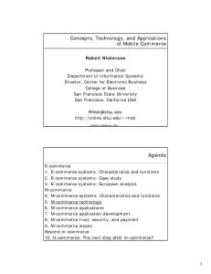
Memokath The Memokath (Engineers & Doctors, Hornbaek, Denmark) is a nickel-titanium alloy stent mounted on a polyurethane insertion catheter with an inflatable balloon that is used to expand and deploy the stent within the urethra. The shaft is 24 Fr, and the lower cone expands to 44 Fr when heated to 55°C and has “shape memory” due to its nickel-titanium alloy construction (Fig. 1). Deployment can be performed under ultrasound guidance or flexible cystoscopy using a 22 Fr insertion stent. Removal takes an average of 11 min, even in patients who have had the stent for a mean indwelling time of 12.9 months [17]. Removal involves flushing the stent with cool water (10°C or less), which alters the spiral to become soft and pliable to facilitate transurethral removal.
ProstaCoil The ProstaCoil (Instent, Minneapolis, MN, USA) is designed to be inserted under fluoroscopic guidance. Retrograde urethrography is used to measure the prostatic urethra and mark the bladder base and urethral sphincter. The stent is then inserted with the patient conscious and able to cooperate. Before the end of the procedure, the patient is asked to voluntarily stop the urinary stream during micturition to ensure that the stent is not interfering with sphincteric function. Antibiotic coverage is started 2–3 days before the procedure and continued for 2 weeks after stent placement. To remove the ProstaCoil stent, a 21 Fr
Fig. 1. Memokath
198
M. Duvdevani et al.
cystoscope and endoscopic grasper are inserted transurethrally. The distal end of the stent is grasped and pulled out of the urethra atraumatically. A second established method for ProstaCoil stent removal is via insertion of a 12–14 Fr Foley catheter through the stent lumen to its proximal end. The balloon is inflated with 2–3 ml saline and pulled out of the urethra under fluoroscopic guidance. Removal of the ProstaCoil stent is typically atraumatic to the anterior urethra.
Polyurethane Stents Three main types of temporary stents are made from polyurethane, including the IntraUrethral Catheter (IUC), the Barnes stent, and the Trestle Catheter. The intraurethral catheter (IUC) is a 16–18 Fr device that has a similar shape to a double-Malecot catheter and is available in lengths of 25–80 mm. The device is inserted under local anesthesia and direct vision using a 22 Fr, cystoscope. The removal of the IUC is easily achieved by pulling a nonabsorbable nylon string attached to its distal end. The Barnes stent (Angiomed, Bard, UK) is a 16 Fr urethral device with a length of 75 mm. The proximal end of the stent is similar to a regular urethral catheter and the distal end resembles a Malecot catheter that is positioned proximal to the verumontanum. Insertion is accomplished by using a special introducer that advances the stent into the bladder, and then a cystoscope is used to retract the stent into the urethra to its correct position using nylon threads attached to the distal end of the device. Removal of the Barnes stent is easy and achieved under local anesthesia by pulling the strings. The Trestle Catheter (Boston Scientific Microvasive, Natick, MA, USA) has two 22 Fr tubes that are connected by a compressible thread which is positioned across the sphincter, thus maintaining continence. The catheter is inserted under local anesthesia and positioned under transrectal ultrasound control.
Permanent Stents Permanent urethral stents are manipulated into the urethral lumen and become incorporated into the wall of the urethra as urothelium covers the device. Permanent stents are used to alleviate bladder outlet obstruction in cases of urethral stricture, DSD, or anastomotic stricture after radical prostatectomy [18,19]. The initial enthusiasm for the use of permanent stents has waned in recent years [20]. The common permanent stents are detailed in Table 3.
UroLume One of the most widely used permanent stents is the UroLume, which is constructed as a nickel superalloy wire mesh configured as a flexible expandable tube. Originally reported in the use of bulbar urethral strictures [21], it rapidly found use in patients with BPH [22] and DSD [23].
Urethral Stents Review
199
Table 3. Permanent prostatic and urethral stents External caliber (Fr)
Length (mm)
UroLume (American Medical Systems, Minnetonka, MN, USA)
42
Memotherm (Bard, Covington, GA, USA)
Ultraflex (Boston Scientific, Natick, MA, USA)
Stent name
Composition
Location
Notes
20–30
Biocompatible superalloy woven tubular mesh
Prostate or urethra
42
15–80
Nitinol woven single wire
Prostate or urethra
42
20–60
Nitinol
Prostate or urethra
(1) Inserted with 21 Fr endoscope under direct vision (2) Gradual epithelization over the wires of the mesh (1) Heat expandable (2) Inserted with endoscope under direct vision (1) Heat expandable
The UroLume is inserted using an introducer that resembles a cystoscope. The procedure is performed under general, regional, or local anesthesia. The correct length of stent is chosen by measuring the urethra from the bladder neck to the distal urethral sphincter under direct vision. The stent is placed under direct vision distal to the bladder neck and proximal to the distal urethral sphincter so that the patient maintains urinary continence. Although it is meant to be permanent, removal of the UroLume stent is possible when necessary. To remove the UroLume stent, a standard resectoscope and loop cautery or Colling’s knife is used to resect the overlying urothelium and push the stent into the bladder, after which it is extracted transurethrally through a larger sheath.
Memotherm The Memotherm is a 42 Fr coil-shaped stent made of nickel-titanium alloy (NiTinol). The Memotherm is positioned within the urethra using an endoscope and insertion catheter under direct vision. The stent is heat expandable with the ability of changing from one configuration to another at different temperatures. After the stent is positioned in the desired location, it is flushed with 45°C water, causing the NiTinol stent to expand. Removal of the Memotherm stent is achieved by irrigating with 15°C water, which softens the metal, causing it to uncoil.
200
M. Duvdevani et al.
Clinical Results with Urethral Stents Stents for the Treatment of Benign Prostatic Hyperplasia (BPH) The prostatic stent was first described by Fabian in 1980, who named it the “partial catheter” [24]. The use of prostatic stents for the treatment of bladder outlet obstruction is known to be safe and effective [8], offers immediate relief, and has 7-year follow-up data [6]. Several stent types have been investigated in patients with bladder outlet obstruction from BPH. Poulsen et al. investigated the use of the Memokath stent and report an 83% success rate in 30 patients with BPH without problems of stent migration, but stent encrustation occurred [25]. Most patients, however, were satisfied with this minimally invasive outpatient procedure for BPH. The first use of the UroLume stent in urology was to treat bulbar urethral strictures in patients who had failed internal urethrotomy [21]. Since then, the indications for UroLume stent insertion have widened to include patients with symptomatic BPH [26–28] and in particular those patients who are unfit for anesthesia and surgical treatment [22].
Urethral Stricture Several trials have reported the utilization of stents for urethral stricture disease. Shah et al. reported the long-term results of the UroLume endourethral prosthesis in the treatment of recurrent bulbar urethral strictures in a multicenter North American trial [29]. The study included 24 patients with recurrent bulbar urethral strictures treated with a UroLume stent and 11 years of follow-up. Preoperative evaluation included uroflowmetry (peak and average urinary flow rates), a urinary symptom questionnaire, and cystoscopy to determine the length and location of the stricture. They found a dramatic improvement in the mean flow rates after stenting (9.5 to 20.8 ml/s) and in the mean urinary symptom scores. Complete epithelialization of more than 90% of the surface area of the stent was seen in the majority of patients (90%) at 1 year follow-up and was persistent through 11 years. The authors recommend this stent for patients with bulbar urethral strictures of less than 3 cm and after at least two recurrences following endoscopic dilation or incision. The distance from the external sphincter should be at least 10 mm to conserve urinary continence. Badlani et al. reviewed the long-term results of the North American Multicenter UroLume Trial for the treatment of recurrent bulbar urethral strictures [30]. This multicenter prospective controlled trial included 175 patients with a bulbar urethral stricture who failed prior treatment attempts including urethral dilatation, visual internal urethrotomy, or urethroplasty (25%). Etiology of the strictures was attributed to prior instrumentation (21.6%), urethral catheterization (14.9%), trauma (18.9%), inflammation/congenital problems
Urethral Stents Review
201
(8.1%), or idiopathic (36.5%). The mean stricture length was 2.34 cm. Follow-up was undertaken at 6 weeks, 6 months, 1 year, and annually after urethral stent insertion. The study demonstrated a continuous improvement in symptom score values and peak and mean urine flow rates. Fifteen percent of the patients required further treatment for recurrent stricture within (44%) or adjacent (56%) to the stent. Only seven patients (4%) required stent removal, and 14.3% required adjuvant treatment after 1 year compared to 75.2% of controls. There was no significant difference in the rate of urinary tract infection before and after stent insertion, and the only predictive factor of postinsertion infection was a positive preoperative urine culture. Stent migration occurred in 4% of patients and occurred predominantly in the first 6 weeks after insertion. Pain and discomfort in stented patients decreased progressively with time from 62% after 6 weeks to 11% at 2 years of follow-up. Severe urinary incontinence was found in 4.3% and 2.5% of the patients after 6 weeks and 2 years of follow-up, respectively. Mild hematuria was noted postoperatively in 26% of the patients but improved to 4% at 2 years. Five patients (3%) had urinary retention after stent insertion. One of these patients developed hyperplastic tissue between two previously placed stents and was treated by insertion of a third stent to bridge the gap between the two existing stents. The other episodes of urinary retention occurred as a result of a new or preexisting urethral stricture adjacent to the stent. Patients with longer urethral strictures or those that have failed a previous stent insertion may require the placement of more than one stent. Tillem et al. reported on the use of multiple UroLume stents in complex bulbar urethral strictures in 41 patients from the 175 (23%) patients enrolled in the UroLume endourethral prosthesis study for recurrent bulbar urethral strictures [31]. Patients who required multiple stents generally had longer strictures with a mean length of 3.6 cm (range, 1.5–6.0). Of the 41 subjects, 32, 6, and 3 patients required two, three, and four stents, respectively. Multiple stents were inserted either simultaneously during the primary procedure (61%) or in a subsequent procedure (39%). Peak urine flow rates and symptom scores were significantly improved in these patients, who showed a similar benefit to other patients in this study who underwent single stent insertion. Patients with urethral strictures longer than 2.5 cm are more likely to require the insertion of multiple stents. Furthermore, patients with multiple stents are more likely to require retreatment, but fortunately the success rate after retreatments is equal to those patients with a single stent. Indications for multiple stent insertion includes strictures longer than 2.5 cm, strictures that were underestimated in length, malpositioned stents, stent migration, recurrent stricture separate from the previously stented region, or simple urethral narrowing adjacent to the stent. Wilson et al. reported on a small series where all DSD patients (n = 4) stented with a UroLume suffered urosepsis within 10 months and required hospitalization [20]. All six patients with urethral strictures had recurrences, and required subsequent surgery, and stent removal was not always straightforward.
202
M. Duvdevani et al.
Detrusor Sphincter Dyssynergia (DSD) The use of urethral stents for DSD was reported in 1990 [23], and a subsequent study demonstrated equivalent long-term results compared to surgical sphincterotomy [32]. Shah et al. assessed 14 male patients with suprasacral spinal cord injury and documented DSD with elevated detrusor pressures and postvoiding residual (PVR) volumes [33]. Patients underwent Memokath stent insertion and were reviewed at 1 month and every 3 months thereafter to assess for urinary tract infection (UTI), autonomic dysreflexia, erectile function, PVR volume, bladder stones, and signs of upper tract obstruction. They found a significant reduction in the PVR volume and improvement in hydronephrosis and autonomic dysreflexia after stent insertion. Six of eight (75%) patients who had a history of recurrent UTIs experienced a decrease in UTI occurrence following stent insertion, presumably from improved PVR volumes. Denys et al., in a study of 47 consecutive male patients with DSD secondary to a spinal cord lesion, reviewed the efficacy of the Ultraflex urethral stent (Boston Scientific, Boston, MA, USA) [34]. The stent was inserted endoscopically under local, neuroleptic, or general anesthesia. Twenty-one patients (44.6%) with a history of recurrent symptomatic infections had significantly fewer UTIs postoperatively (P = 0.001). Improvement in autonomic dysreflexia occurred in 5 of 8 stented patients, as well as an improvement in preoperative hydronephrosis in 7 of 8 patients (P = 0.005). Voiding function is also improved in DSD and spinal cord injury patients [35]. Patients who used an indwelling urinary catheter or intermittent catheterization for urinary drainage were able to void spontaneously into a condom catheter after stent placement. These results were also accompanied by a significant decrease in the occurrence of autonomic dysreflexia, symptomatic UTIs, and hydronephrosis. Hamid et al. evaluated the Memokath urethral stent, in 25 patients with DSD [36] with a mean age was 45.5 years (range, 32–65 years). Preoperatively, the majority of patients (80%) were draining their bladder using a reflex voiding with a condom drainage system, and the remaining patients used an indwelling suprapubic catheter or clean intermittent urethral selfcatheterization. After stent insertion, the patients demonstrated a significant reduction in maximum detrusor pressure, duration of detrusor contraction, and residual urine volume. In fact, bladder function and urinary drainage improved to the point that preexisting hydronephrosis in 4 patients resolved. Not all studies have found beneficial effects using urethral stents. Mehta et al. reviewed 29 patients with 33 Memokath stents who suffered from spinal cord injury patients and DSD [37]. These authors found the working life of the stent to be 21 months, with a high complication rate. Their overall experience with Memokath stents was disappointing, which has led them to abandon the use of this stent. Moreover, removal is necessary when there is extensive mucosal proliferation leading to lumenal obstruction [38]. Chronic infection and migration
Urethral Stents Review
203
have been other issues limiting the indwelling time of this stent in DSD patients [39].
Complications The use of urethral stents in urology is not free of complications. Patients who require cystoscopy to treat and follow certain urological conditions such as urinary stone disease, transitional cell carcinoma, or any other problems that require recurrent endoscopic manipulations should be precluded from stent insertion with certain stent types. Urethral stents are a foreign body and may cause irritative urinary symptoms such as frequency, urgency, dysuria, or urge incontinence. Other potential complications include encrustation, stent fracture, migration, UTI, hematuria, and clot retention [2,35]. Stent positioning is important, and complications may occur when a stent is placed distal to the bulbous urethra, resulting in incontinence or pain while sitting or during intercourse [40]. Shah et al. reviewed the data related to the explantation of UroLume urethral stents in the North American Study Group, which included 465 patients [41]. A total of 73 stents (15.6%) were removed from 69 patients (14.8%). Characteristics of the patients that underwent stent removal were examined: the explantation rate was 23%, 5%, and 22% from patients with BPH, bulbar urethral stricture, and DSD, respectively. Thirty-two stents (44.4%) were removed during the first year after insertion, with stent migration being the primary reason in 38.4% of explantations. Other reasons for stent removal included worsening symptoms, stent encrustation, and incomplete luminal epithelialization.
Conclusions There are several possible indications for urethral stent placement, including urethral stricture disease, BPH, and DSD. Much progress has been made during the last decade in the field of urethral stenting. Currently, one can choose an appropriate stent from a wide variety of temporary and permanent urethral stents. Although it is often not considered a definitive treatment, urethral stenting offers an alternative minimally invasive procedure to relieve the symptoms of bladder outlet obstruction in high surgical risk patients and as an alternative to open urethroplasty in select populations.
References 1. The American Heritage Dictionary of the English Language, edn (2006) Houghton Mifflin, Boston 2. Badlani GH (1997) Role of permanent stents. J Endourol 11:473–475
204
M. Duvdevani et al.
3. Slutzker D, Richter S, Lang R, Nissenkorn I (1994) The use of a prostatic stent in high risk patients over 80 years old with benign prostatic hyperplasia and chronic retention. J Am Geriatr Soc 42:1004–1005 4. Juma S, Niku SD, Brodak PP, Joseph AC (1994) Urolume urethral wallstent in the treatment of detrusor sphincter dyssynergia. Paraplegia 32:616–621 5. Soni BM, Vaidyanatham S, Krishnan KR (1994) Use of Memokath, a second generation urethral stent for relief of urinary retention in male spinal cord injured patients. Paraplegia 32:480–488 6. Kapoor R, Liatsikos EN, Badlani G (2000) Endoprostatic stents for management of benign prostatic hyperplasia. Curr Opin Urol 10:19–22 7. Ogiste JS, Cooper K, Kaplan SA (2003) Are stents still a useful therapy for benign prostatic hyperplasia? Curr Opin Urol 13:51–57 8. Lam JS, Volpe MA, Kaplan SA (2001) Use of prostatic stents for the treatment of benign prostatic hyperplasia in high-risk patients. Curr Urol Rep 2:277–284 9. Nagae H, Mugiya S (2002) Other less invasive surgical therapy for BPH (urethral stent). Nippon Rinsho 60 (suppl 11):408–412 10. Steenkamp JW, Heyns CF, de Kock ML (1997) Internal urethrotomy versus dilation as treatment for male urethral strictures: a prospective, randomized comparison. J Urol 157:98–101 11. Santucci RA, McAninch JW, Mario LA, Rajpurkar A, Chopra AK, Miller KS, Armenakas NA, Tieng EB, Morey AF (2004) Urethroplasty in patients older than 65 years: indications, results, outcomes and suggested treatment modifications. J Urol 172:201–203 12. Fitzpatrick JM, Mebust WK (2002) Minimally invasive and endoscopic management of benign prostatic hyperplasia. In: Walsh PC, Retik AB, Vaughan ED, Wein AJ (eds) Campbell’s Urology, 8th edn. Saunders, Philadelphia, p 1381 13. Isotalo T, Nuutinen JP, Vaajanen A, Martikainen PM, Laurila M, Tormala P, Talja M, Tammela TL (2005) Biocompatibility and implantation properties of 2 differently braided, biodegradable, self-reinforced polylactic acid urethral stents: an experimental study in the rabbit. J Urol 174:2401–2404 14. Talja M, Valimaa T, Tammela T, Petas A, Tormala P (1997) Bioabsorbable and biodegradable stents in urology. J Endourol 11:391–397 15. Djavan B, Ghawidel K, Basharkhah A, Hruby S, Bursa B, Marberger M (1999) Temporary intraurethral prostatic bridge-catheter compared with neoadjuvant and adjuvant alpha-blockade to improve early results of high-energy transurethral microwave thermotherapy. Urology 54:73–80 16. Petas A, Talja M, Tammela TL, Taari K, Valimaa T, Tormala P (1997) The biodegradable self-reinforced poly-dl-lactic acid spiral stent compared with a suprapubic catheter in the treatment of post-operative urinary retention after visual laser ablation of the prostate. Br J Urol 80:439–443 17. Barber NJ, Roodhouse AJ, Rathenborg P, Nordling J, Ellis BW (2005) Ease of removal of thermo-expandable prostate stents. BJU Int 96:578–580 18. Zivan I, Stein A (2001) New modality for treatment of resistant anastomotic strictures after radical prostatectomy: UroLume urethral stent. J Endourol 15:869–871 19. Elliott DS, Boone TB (2001) Combined stent and artificial urinary sphincter for management of severe recurrent bladder neck contracture and stress incontinence after prostatectomy: a long-term evaluation. J Urol 165:413–415 20. Wilson TS, Lemack GE, Dmochowski RR (2002) UroLume stents: lessons learned. J Urol 167:2477–2480
Urethral Stents Review
205
21. Milroy EJ, Chapple CR, Cooper JE, Eldin A, Wallsten H, Seddon AM, Rowles PM (1988) A new treatment for urethral strictures. Lancet 1:1424–1427 22. Williams G, Jager R, McLoughlin J, el Din A, Machan L, Gill K, Asopa R, Adam A (1989) Use of stents for treating obstruction of urinary outflow in patients unfit for surgery. BMJ 298:1429 23. Shaw PJ, Milroy EJ, Timoney AG, el Din A, Mitchell N (1990) Permanent external striated sphincter stents in patients with spinal injuries. Br J Urol 66:297–302 24. Fabian KM (1980) The intra-prostatic “partial catheter” (urological spiral) (author’s transl). Urologe A 19:236–238 25. Poulsen AL, Schou J, Ovesen H, Nordling J (1993) Memokath: a second generation of intraprostatic spirals. Br J Urol 72:331–334 26. Anjum MI, Chari R, Shetty A, Keen M, Palmer JH (1997) Long-term clinical results and quality of life after insertion of a self-expanding flexible endourethral prosthesis. Br J Urol 80:885–888 27. Masood S, Djaladat H, Kouriefs C, Keen M, Palmer JH (2004) The 12-year outcome analysis of an endourethral wallstent for treating benign prostatic hyperplasia. BJU Int 94:1271–1274 28. Milroy E, Chapple CR (1993) The UroLume stent in the management of benign prostatic hyperplasia. J Urol 150:1630–1635 29. Shah DK, Paul EM, Badlani GH (2003) 11-year outcome analysis of endourethral prosthesis for the treatment of recurrent bulbar urethral stricture. J Urol 170: 1255–1258 30. Badlani GH, Press SM, Defalco A, Oesterling JE, Smith AD (1995) Urolume endourethral prosthesis for the treatment of urethral stricture disease: long-term results of the North American Multicenter UroLume Trial. Urology 45:846–856 31. Tillem SM, Press SM, Badlani GH (1997) Use of multiple urolume endourethral prostheses in complex bulbar urethral strictures. J Urol 157:1665–1668 32. Chancellor MB, Bennett C, Simoneau AR, Finocchiaro MV, Kline C, Bennett JK, Foote JE, Green BG, Martin SH, Killoran RW, Crewalk JA, Rivas DA (1999) Sphincteric stent versus external sphincterotomy in spinal cord injured men: prospective randomized multicenter trial. J Urol 161:1893–1898 33. Shah NC, Foley SJ, Edhem I, Shah PJ (1997) Use of Memokath temporary urethral stent in treatment of detrusor-sphincter dyssynergia. J Endourol 11:485–488 34. Denys P, Thiry-Escudie I, Ayoub N, Even-Schneider A, Benyahya S, Chartier-Kastler E (2004) Urethral stent for the treatment of detrusor-sphincter dyssynergia: evaluation of the clinical, urodynamic, endoscopic and radiological efficacy after more than 1 year. J Urol 172:605–607 35. Juan Garcia FJ, Salvador S, Montoto A, Lion S, Balvis B, Rodriguez A, Fernandez M, Sanchez J (1999) Intraurethral stent prosthesis in spinal cord injured patients with sphincter dyssynergia. Spinal Cord 37:54–57 36. Hamid R, Arya M, Wood S, Patel HR, Shah PJ (2003) The use of the Memokath stent in the treatment of detrusor sphincter dyssynergia in spinal cord injury patients: a single-centre seven-year experience. Eur Urol 43:539–543 37. Mehta SS, Tophill PR (2006) Memokath stents for the treatment of detrusor sphincter dyssynergia (DSD) in men with spinal cord injury: The Princess Royal Spinal Injuries Unit 10-year experience. Spinal Cord 44(1):1–6 38. Vaidyanathan S, Soni BM, Oo T, Sett P, Hughes PL, Singh G (2002) Long-term result of Memokath urethral sphincter stent in spinal cord injury patients. BMC Urol 2:12
206
M. Duvdevani et al.
39. Low AI, McRae PJ (1998) Use of the Memokath for detrusor-sphincter dyssynergia after spinal cord injury—a cautionary tale. Spinal Cord 36:39–44 40. Chapman SM, Wemyss-Holden GD, Clarke NW (1994) Urinary incontinence following urethral Wallstent insertion. Br J Urol 73:586 41. Shah DK, Kapoor R, Badlani GH (2003) Experience with urethral stent explantation. J Urol 169:1398–1400