2.1 INTRODUCTION Blood is collected from the vein for various hematological investigations. In order to obtain accurate and precise results in the laboratory which will help the clinician to make a correct diagnosis of the patient’s disease, it is of paramount importance to collect the blood sample in a correct manner. Each sample is sent to the laboratory accompanied by a laboratory requisition form filled in by the clinician. Brief clinical details and any other relevant information must be mentioned on the form. Prior to blood sample collection it is essential to check the patient’s identity and make sure that it corresponds to the name and other details mentioned on the requisition form. Blood can be withdrawn from the vein, usually the antecubital vein on the forearm (Venous blood) or from the finger or heel (Capillary blood). Venous blood is preferred. It can be collected using a syringe and needle or a vacuum tube. Both these methods will be described seperately.
OBJECTIVES After reading this lesson, you will be able to: z
describe the technique of collection of venous blood using a syringe and needle and a vacuum tube
z
describe the method of collection of capillary blood
z
explain the various anticoagulants used in hematology laboratory and their action.
HEMATOLOGY AND BLOOD BANK TECHNIQUE
9
MODULE Hematology and Blood Bank Technique
Technique of Blood Collection
2.2 COLLECTION OF VENOUS BLOOD Blood is usually withdrawn from the antecubital vein or any other vein which is well identified on the forearm. The vein selected should be large, readily accessible, and sufficiently close to the surface to be seen and palpated Preparation of venipuncture site
Notes
Clean the skin of the area around the identified vein with 70% isopropyl alcohol in a circular fashion beginning at the site and moving outward. Allow to dry spontaneously. Do not touch the venipuncture site after it has been cleaned. Apply a tourniquet 3-4 inches above the venipuncture site. Ask the patient to make a fist a few times. Veins suitable for puncture will then become more apparent. Veins can become distended and easier to enter by allowing the arm to hang down for 2 or 3 minutes or by gently slapping the site of puncture.
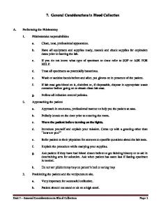
Fig. 2.1: Clean the area of the venipuncture site using circular anticlockwise motion
Collection of venous blood using a syringe 1. Clean hands thoroughly with soap and water. 2. Write the name and hospital number of the patient on the tube in which blood is to be collected. A printed label with these particulars can also be used for patient identification. 3. Place the needle into the syringe. Keep the cap over the needle capped till it is used. Check that the syringe works smoothly. 4. With the needle bevel up and parallel to the surface of the skin insert it into the vein. Appearance of blood in the hub of the needle indicates that the needle has successfully entered the vein. Release the tourniquet as soon as blood enters the syringe. 10
HEMATOLOGY AND BLOOD BANK TECHNIQUE
Technique of Blood Collection
5. Withdraw the piston slowly to avoid frothing.
MODULE Hematology and Blood Bank Technique
6. After obtaining the requisite amount of blood, place a sterile gauze pad over the point where the needle entered the skin and deftly withdraw the needle simultaneously while applying pressure over the site. 7. Deliver the blood gently into the specified receiver. Cap it firmly to prevent leakage. 8. Maintain light pressure on the gauze pad over venipuncture site till the bleeding stops and then cover the puncture site with a small adhesive dressing.
Notes
9. Destroy the needle in a special device (needle destroyer) immediately after use. DONOT break, bend or recap needles after use. 10. Place the used swab, syringe and any other contaminated material in a puncture resistant container for adequate disposal. Collection of venous blood using a vacuum tube The Vacutainer system consists of a double-pointed needle, a plastic holder or adapter, and several vacuum tubes with rubber stoppers of various colors depending on the sample to be collected. The color of the vacuum tube indicates the anticoagulant which it contains. The blood is directly collected into the tube from the vein. Procedure 1. Place the identification details of the patient on each vacuum tube and ensure that they tally with the form. 2. Identify the vein and clean the area as described before. Apply a tourniquet 3-4 inches above the identified vein. Do not touch the venipuncture site after cleaning. 3. Place the vacuum tube in a reusable plastic holder and attach a disposable needle to it. Insert the tube into the holder till the top of the stopper is level with the marked guideline. 4. Place the patient’s arm in a downward position to reduce the risk of backflow of any anticoagulant into the patient’s circulation. 5.
Insert the needle into the vein. Push the tube into the needle, puncturing the stopper/vacuum seal.
6.
Remove the tourniquet as soon as the blood appears in the tube. Do not allow contents of the tube to come into contact with the stopper
7. Do not allow the contents of the tube to come into contact with the stopper during the procedure. HEMATOLOGY AND BLOOD BANK TECHNIQUE
11
MODULE Hematology and Blood Bank Technique
Notes
Technique of Blood Collection
8. If more than one sample is required, successive tubes may be fitted into the holder after removing the previous tube and blood collected. The needle remains in the vein. While each successive tube is filling, the previous tube may be inverted gently to mix the sample well. Do not shake vigorously as this may lead to hemolysis of the blood sample. 9. After completion of blood collection, remove the holder and cover the site with a sterile swab and apply pressure till bleeding stops completely. 10. Destroy the needle in the destroyer without recapping it.
Fig. 2.2: Collection of venous blood using a vacuum tube
2.3 TYPES OF VACUUM TUBES Vacuum tubes with different colored caps are available. Each contains a different anticoagulant and is used for various hematological tests as described below. Color of cap
Anticoagulant
Test
Purple
EDTA
complete blood counts
Red
-
for tests which need serum
Blue
Sodium citrate
coagulation tests
Grey
fluoride
blood sugar
2.4 COLLECTION OF CAPILLARY BLOOD This can be obtained by skin puncture with a needle or lancet and is specially used in small children or very obese adults in whom venepuncture fails. Samples can be used for making peripheral blood films, performing hematocrit/Hb and point of care testing. In adults capillary blood sample can be obtained from the lateral side of tip of the 3rd or 4th finger while in infants the sample can be obtained by a deep puncture of the plantar surface of the heel. 12
HEMATOLOGY AND BLOOD BANK TECHNIQUE
MODULE
Technique of Blood Collection
Hematology and Blood Bank Technique
Procedure 1. Clean the area with 70% alcohol and allow it to dry spontaneously. Puncture the skin to a depth of 2-3 mm with a sterile disposable lancet/needle. 2. Wipe the first drop of blood and squeeze gently to allow free flow of blood and collect the sample. In an adequate puncture, large drops of blood should exude spontaneously. Do not squeeze firmly as this gives unreliable results.
Notes
INTEXT QUESTIONS 2.1 Match the following Test
Colour of cap
1. Complete blood count
(a) Grey
2. Coagulation test
(b) Purple
3. Blood sugar
(c) Red
4. Tests which need serum
(d) Blue
2.5 ANTICOAGULANTS USED IN HEMATOLOGY The anticoagulants used commonly in the hematology laboratory are EDTA: Ethylene Diamine Tetra Acetic acid Sodium citrate Heparin EDTA: This is used for complete blood counts. The sodium and potassium salts can both be used but the dipotassium salt is preferred as it is more soluble. The dilithium salt can also be used but like the disodium salt is less soluble. The dipotassium salt is used in solid form. The International Council for Standardization in Hematology recommends use of the dipotassium salt at a concentration of 1.50±0.25mg/ml of blood. Coating the inside of a blood collection vial with a thin layer of EDTA improves the speed of uptake by blood. It exerts it’s action by removing calcium which is essential for coagulation. If EDTA is used in excess, it causes shrinkage of red cells and leucocytes. A significant decrease in hematocrit and increase in MCHC also occur. Platelets swell and disintegrate causing a false high count. It is thus important to add the correct amount of blood to the vial.
HEMATOLOGY AND BLOOD BANK TECHNIQUE
13
MODULE Hematology and Blood Bank Technique
Notes
Technique of Blood Collection
Trisodium citrate: This is used in coagulation studies in a ratio of 9 volumes of blood to one volume of anticoagulant (32g/L) (0.5ml citrate+4.5ml blood). It binds calcium thus preventing coagulation. It can also be used for estimation of ESR by Westergren method in a ratio of 4 volumes of blood to 1 volume of sodium citrate. Heparin: The sodium or lithium salt of heparin is used at a concentration of 1020IU/ml of blood for osmotic fragility and for red cell enzyme studies like glucose-6-phosphate dehydrogenase. It does not change red cell size and is the best anticoagulant for osmotic fragility. It can also be used for immunophenotyping. Heparin is not suitable for complete blood counts as it induces platelet and leucocyte clumping. It should also not be used for making peripheral smears as it gives a faint blue background color after staining smears by Romanowsky dyes. Types of Samples Collected for Hematological Investigations Whole blood: This is used for performing complete blood counts including reticulocyte count and for making peripheral blood films. Serum: If blood is allowed to clot at room temperature, a straw colored fluid appears which is called serum. This may be admixed with red cells which can be removed by centrifugation at 1200g for 10 minutes. It is used for various biochemical tests and also serum protein electrophoresis to diagnose plasma cell disorders such as multiple myeloma. Serum lacks coagulation factors. Plasma: This is obtained by centrifugation of anticoagulated blood and is used for coagulation studies.
INTEXT QUESTIONS 2.2 1. The anticoagulant used for complete blood counts is (a) EDTA (b) Heparin (c) Sodium citrate (d) Any of the above 2. For coagulation studies the ratio of blood to anticoagulant is (a) 9 : 1 (b) 7 : 1 (c) 8 : 1 (d) Any of the above 14
HEMATOLOGY AND BLOOD BANK TECHNIQUE
Technique of Blood Collection
3. Complete the sentence
MODULE Hematology and Blood Bank Technique
EDTA exerts it’s action as an anticoagulant by ...................
WHAT HAVE YOU LEARNT z
Blood is collected from the vein for various hematological investigations
z
Blood can be withdrawn from the vein, usually the antecubital vein on the forearm or for capillary blood form the finger or heel
z
Blood can be collected using a syringe and needle or a vacuum tube
z
The vein should be large, readily accessible, and sufficiently close to the surface of the skin to be seen and palpated
z
Write the patient name and hospital number on the tube
z
The skin area around the identified vein should be cleaned with 70% isopropyl alcohol
z
Apply a tourniquet 3-4 inches above the venipuncture site
z
Insert the needle into the vein and withdrawn the required amount of blood slowly
z
Deliver the blood in the specific container
z
Maintain even pressure on the venipuncture site till bleeding stops
z
Destroy the needle used immediately
z
Vacuum tubes with different coloured caps are available and each contains different anticoagulant used for various hematological tests
z
Capillary blood can be obtained from the lateral side of tip of 3rd or 4th finger and for infants from plantar surface of heel
z
The anticoagulants used commonly in the hematology laboratory are Ethylene Diamine Tetra Acetic acid (EDTA), Sodium citrate, Heparin
Notes
TERMINAL QUESTIONS 1. Explain the different types of vacuum tubes 2. What the commonly used anticoagulants in hematology