Biochimica et Biophysica Acta 1739 (2005) 179 – 197 http://www.elsevier.com/locate/bba
Review
Tau protein as a differential biomarker of tauopathies Nicolas Sergeant, Andre´ Delacourte, Luc Bue´e* INSERM U422, 1, Place de Verdun, 59045 Lille cedex, France Received 3 June 2004; accepted 16 June 2004 Available online 20 July 2004
Abstract Microtubule-associated Tau proteins are the basic component of intraneuronal and glial inclusions observed in many neurological disorders, the so-called tauopathies. Many etiological factors, phosphorylation, splicing, and mutations, relate Tau proteins to neurodegeneration. Molecular analysis has revealed that hyperphosphorylation and abnormal phosphorylation might be one of the important events in the process leading to tau intracellular aggregation. Specific set of pathological tau proteins exhibiting a typical biochemical pattern, and a different regional and laminar distribution, could characterize five main classes of tauopathies. A direct correlation has been established between the regional brain distribution of tau pathology and clinical symptoms; for instance progressive involvement of neocortical areas is well correlated to the severity of dementia in Alzheimer’s disease, overall suggesting that pathological tau proteins are reliable marker of the neurodegenerative process. Recent discovery of tau gene mutations in frontotemporal dementia with parkinsonism linked to chromosome 17 has reinforced the predominant role attributed to tau proteins in the pathogenesis of neurodegenerative disorders, and underlined the fact that distinct sets of tau isoforms expressed in different neuronal populations could lead to different pathologies. Overall, a better knowledge of the etiological factors responsible for the aggregation of tau proteins in brain diseases is essential for development of future differential diagnosis and therapeutic strategies. They would hopefully find their application against Alzheimer’s disease but also in all neurological disorders for which a dysfunction of Tau biology has been identified. D 2004 Elsevier B.V. All rights reserved. Keywords: Alzheimer’s disease; Tauopathy; Microtubule-associated tau; Physiopathology; Biomarker
1. Tau proteins
2. Tau gene and splicing
Tau proteins belong to the microtubule-associated proteins (MAP) family. They are found in many animal species. In human, they are found in neurons (for review, see Refs. [1,2]), although nonneuronal cells usually have trace amounts. For instance, Tau proteins can be expressed in glial cells, mainly in pathological conditions [3], and it is possible to detect Tau mRNA and proteins in several peripheral tissues such as heart, kidney, lung, muscle, pancreas, testis, as well as in fibroblasts [4–6].
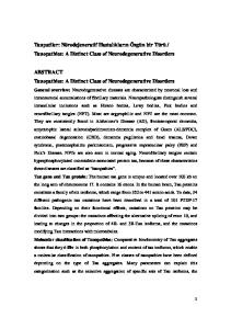
The human Tau gene is unique and located over 100 kb on the long arm of chromosome 17 at band position 17q21 [7], and contains 16 exons [8] (Fig. 1). The restriction analysis and sequencing of the gene shows that it contains two CpG islands, one associated with the promoter region, the other with exon 9 [8]. The CpG island in the putative Tau promoter region resembles previously described neuron-specific promoters. Two regions homologous to the mouse Alu-like sequence are present. The sequence of the promoter region also reveals a TATA-less sequence that is likely to be related to the presence of multiple initiation sites, typical of housekeeping genes. Three SP1-binding sites that are important in directing transcription initiation in other TATA-less promoters are also found in the proximity of the first transcription initiation site [9]. The SP1-binding
* Corresponding author. Tel.: +33 320 622074; fax: +33 320 622079. E-mail address:
[email protected] (L. Bue´e). URL: http://www.lille.inserm.fr/u422/. 0925-4439/$ - see front matter D 2004 Elsevier B.V. All rights reserved. doi:10.1016/j.bbadis.2004.06.020
180
N. Sergeant et al. / Biochimica et Biophysica Acta 1739 (2005) 179–197
sites are suggested to control neuronal specific expression of Tau [10]. The Tau primary transcript contains 16 exons (Fig. 1A). However, two of them (exons 4A, and 8) are skipped in human brain. They are specific to peripheral Tau proteins. Exon 4A is found in bovine, human and rodent peripheral tissues with a high degree of homology. Cryptic splicing sites are described in exon 6 that would generate Tau mRNA lacking the remaining 3V exon cassettes [11]. Recently, expression of Tau isoform including exons 2 and 3 plus exon 6 in neuroblastoma cells was shown to inhibit neurite outgrowth [12]. Exon 1 is part of the promoter, and is transcribed but not translated. Exons 1, 4, 5, 7, 9, 11, 12 and 13 are constitutive exons. Exon 14 is part of the 3V untranslated region of Tau mRNA [13,14]. Exons 2, 3 and 10 are alternatively spliced and are adult brain-specific [8]. Exon 3 never appears independently of exon 2 [15]. Thus,
alternative splicing of these three exons allows for six combinations (2 3 10 ; 2+3 10 ; 2+3+10 ; 2 3 10+; 2+3 10+; 2+3+10+). In the human brain, the Tau primary transcript gives rise to six mRNAs [13,14] (Fig. 1B).
3. Tau structure and functions In the human brain, Tau proteins constitute a family of six isoforms, which range from 352 to 441 amino acids (Fig. 1C). Their molecular weight is ranging from 45 to 65 kDa when resolved on polyacrylamide gel electrophoresis in the presence of sodium dodecyl sulfate (SDSPAGE). The Tau isoforms differ from each other by the presence of either three- (3R) or four-repeat regions (4R) in the carboxy-terminal (C-terminal) part of the molecule
Fig. 1. Tau gene structure, pre-mRNA alternative splicing in the central nervous system (CNS) and protein isoforms translated from alternative tau mRNAs. (A) The human tau gene, located on chromosome 17 at position q21, spans over 110 kb. It is composed of 16 exons numbered from 1 to 14. The start codon is located in exon 1 and two codons are described: one in the intron between exons 13 and 14 and the second in exon 14. The 3V ending of exon 14 is not completely characterized in human. The initiation of transcription is indicated by +1. (B) Schematic representation of Tau mRNAs. In the CNS, exons 4A, 6 and 8 are constitutively skipped. A small proportion of tau mRNA might arise from the use of cryptic sites in exon 6 and would give tau proteins lacking the microtubule-binding domain. This potential use of cryptic splicing site is not detailed in the figure. Exons 1 and 14 correspond to the 5V and 3V untranslated regions of Tau mRNA, respectively. Two cleavage-polyadenylation sites are described: one in intron 13/14 and one in exon 14. The polyadenylated Tau mRNA including exon 14 is less represented in human. In the CNS, the alternative splicing of exons 2, 3 and 10 generates six tau proteins. Inclusion of exon 3 is associated with that of exon 2, whereas exon 2 can be included alone. C. The six human brain tau isoforms are represented as they are resolved by polyacrylamide gel electrophoresis. The amino acid sequences corresponding respectively to exons 2, 3 and 10 are detailed. The tau isoform lacking the alternative exons 2, 3 and 10 is the only tau isoform to be expressed in fetal CNS.
N. Sergeant et al. / Biochimica et Biophysica Acta 1739 (2005) 179–197
and the absence or presence of one or two inserts (29 or 58 amino acids) in the amino-terminal (N-terminal) part [13,14,16]. Each of these isoforms is likely to have particular physiological roles since they are differentially expressed during development. For instance, only one Tau isoform, characterized by 3R and no N-terminal inserts, is present during fetal stages, while the six isoforms (with one or two N-terminal inserts and 3- or 4R) are expressed during adulthood [17,18]. Thus, Tau isoforms are likely to have specific functions related to the absence or presence of regions encoded by the cassette exons 2, 3 and 10. Furthermore, the six Tau isoforms may not be equally expressed in neurons. For example, Tau mRNAs containing exon 10 are not found in granular cells of the dentate gyrus [19]. Thus, Tau isoforms may be differentially distributed in neuronal subpopulations.
4. The projection domain The two 29-amino-acid sequences encoded by exons 2 and 3 give different lengths to the N-terminal part of Tau proteins. These two additional inserts are highly acidic, and are followed by a basic proline-rich region. The Nterminal part is referred to as the projection domain since it projects from the microtubule surface where it may interact with other cytoskeletal elements and plasma membrane [20,21]. In mice lacking the Tau gene, an increase in microtubuleassociated protein 1A, which may compensate for the functions of Tau proteins, has been observed [22]. In contrast to this report, a more recent study has shown that embryonic hippocampal cultures from tau deficient mice show a significant delay in maturation as measured by axonal and neuritic extensions [23]. However, in both studies, axonal growth and axonal diameter are particularly affected. This may be related to the particular length of the N-terminal domain (with or without sequences encoded by exons 2 and 3) of Tau proteins in specific axons. In fact, projection domains of Tau determine spacings between microtubule in axon and may increase axonal diameter [24]. It should be noted that in peripheral neurons which often project a very long axon with large diameter, an additional N-terminal Tau sequence encoded by exon 4A is present, generating a specific Tau isoform called Tbig Taur [8,25]. These results strongly suggest that N-terminal regions of Tau proteins are crucial in the stabilization and organization of certain types of axons. Tau proteins bind to spectrin and actin filaments [26–29]. Through these interactions, Tau proteins may allow microtubules to interconnect with other cytoskeletal components such as neurofilaments [30] and may restrict the flexibility of the microtubules [31]. There is also evidence that Tau proteins interact with cytoplasmic organelles. Such interactions may allow for binding between microtubules and mitochondria [32]. The Tau N-terminal projection domain
181
also permits interactions with the neural plasma membrane [20]. Thus, Tau may act as a mediator between microtubules and plasma membrane. More recently, this interaction has been defined as involving a binding between the prolinerich sequence in the N-terminal part of Tau proteins and the SH3 domains of src-family non-receptor tyrosine kinases, such as fyn. Recent studies determined that human tau Tyr18 and 29 are phosphorylated by the src family tyrosine kinase fyn [33,34]. The same proline-rich region of Tau proteins is likely involved in the interaction with phospholipase C-g (PLC-g) isozymes [35,36]. Hwang and colleagues have demonstrated in vitro that Tau proteins complex specifically with the SH3 domain of PLC-g, and enhance its activity in the presence of unsaturated fatty acids such as arachidonic acid. These results suggest that in cells that express Tau proteins, receptors coupled to cytosolic phospholipase A2 may activate PLC-g indirectly, in the absence of the usual tyrosine phosphorylation, through the hydrolysis of phosphatidylcholine to generate arachidonic acid [35,36]. Altogether, these data indicate that Tau proteins may also play a role in the signal transduction pathway involving PLC-g.
5. The microtubule assembly domain Tau proteins bind microtubules through repetitive regions in their C-terminal part. These repetitive regions are the repeat domains (R1–R4) encoded by exons 9–12 [37] (Fig. 1C). The three (3R) or four copies (4R) are made of a highly conserved 18-amino-acid repeat [14,16,37] separated from each other by less conserved 13- or 14-amino-acid interrepeat domains. Tau proteins are known to act as promoter of tubulin polymerization in vitro, and are involved in axonal transport [38–42]. They have been shown to increase the rate of microtubule polymerization and to inhibit the rate of depolymerization [43]. The 18-amino-acid repeats bind to microtubules through a flexible array of distributed weak sites [37,44]. It has been demonstrated that adult Tau isoforms with 4R (R1–R4) are more efficient at promoting microtubule assembly than the fetal isoform with 3R (R1, R3, R4) [18,44,45]. Interestingly, the most potent part to induce microtubule polymerization is the inter-region between repeats 1 and 2 (R1–R2 inter-region) and more specifically peptide 274KVQIINKK281 within this sequence. This R1–R2 inter-region is unique to 4R-Tau, adult-specific and responsible for difference in the binding affinities between 3R and 4R-Tau [46]. Recent data indicated that during elongation of microtubules, no difference in binding to microtubules between 3R and 4R could be observed, suggesting that Tau proteins are also able to bind the inner part of microtubules [47,48]. Recent evidences support a role for the microtubulebinding domain in the modulation of the phosphorylation state of Tau proteins. A direct and competitive binding has been demonstrated between this part (residues 224–236
182
N. Sergeant et al. / Biochimica et Biophysica Acta 1739 (2005) 179–197
according to the numbering of the longest isoform) and microtubules on one hand, and this part and protein phosphatase 2A (PP2A) on the other hand [49]. As a consequence, microtubules could inhibit PP2A activity by competing for binding to Tau at the microtubule-binding domains.
6. Posttranslational modifications 6.1. O-glycosylation O-glycosylation is a dynamic and abundant posttranslational modification that is characterized by the addition of a O-linked N-acetylglucosamine (O-GlcNAc) residue on Ser or Thr in the proximity of Pro residues [50]. The O-GlcNAc transferase was recently identified [51]. Although the functional significance of O-GlcNAc modification is not yet fully understood, it is implicated in transcriptional regulation, protein degradation, cell activation, cell cycle regulation and the proper assembly of multimeric protein complexes [52]. This modification is often reciprocal to phosphorylation (for review, see Ref. [53]). It occurs in neurofilaments [54], microtubule-associated proteins including MAP2 and Tau proteins [55,56]. The number of OGlcNAcylated sites on Tau proteins is lower than the number of phosphorylation sites. Site-specific or stoichiometric changes in O-GlcNAcylation may modulate Tau function. In fact, phosphorylation and O-GlcNacylation may have opposite effects (see below for the role of Tau phosphorylation). For instance, O-GlcNacylation of Tau proteins and other microtubule-associated proteins suggests a role for O-GlcNac in mediating their interactions with tubulin. O-GlcNacylation may also play a role in subcellular localization and degradation of Tau proteins [53,55,56].
7. Phosphorylation 7.1. Sites of phosphorylation There are 80 putative Ser or Thr phosphorylation sites on the longest brain Tau isoform (441 amino acids). Using phosphorylation-dependent monoclonal antibodies against Tau, mass spectrometry and sequencing, at least 30 phosphorylation sites have been described, including Thr39, Ser46Pro, Thr50Pro, Thr69Pro, Thr153Pro, Thr175Pro, Thr 181Pro, Ser198, Ser199Pro, Ser202Pro, Thr205Pro, Ser208, Ser210, Thr212Pro, Ser214, Thr217Pro, Thr231Pro, Ser235Pro, Ser237, Ser241, Ser262, Ser285, Ser305, Ser324, Ser352, Ser356, Ser396Pro, Ser400, Thr403, Ser404Pro, Ser409, Ser412, Ser413, Ser416 and Ser422Pro [57–63]. All of these sites are localized outside the microtubule-binding domains with the exception of Ser 262 (R1), Ser285 (R1–R2 inter-repeat), Ser305 (R2–R3 inter-repeat), Ser324 (R3), Ser352 (R4) and
Ser356 (R4) [64,65]. Both Ser/Thr-Pro and non-Ser/Thr-Pro sites have been identified [61,62]. The different states of Tau phosphorylation result from the activity of specific kinases and phosphatases towards these sites. 7.2. Kinases Most of the kinases involved in Tau phosphorylation are part of the proline-directed protein kinases (PDPK), which include mitogen activated protein kinase (MAP) [66–69], Tau-tubulin kinase [70,71] and cyclin-dependent kinases including cdc2 and cdk5 [72,73]. Stress-activated protein kinases (SAP kinases) have been recently involved in Tau phosphorylation [67,74,75]. Non Ser/Thr-Pro sites can be phosphorylated by many other protein kinases, including microtubule-affinity regulating kinase (MARK) [76], Ca2+/ calmodulin-dependent protein kinase II (CaMPK II) [77,78], cyclic-AMP-dependent kinase (PKA) [79,80] and casein kinase I and II [81,82]. Glycogen synthase kinase 3 (GSK3) is a Tau kinase able to phosphorylate both non-Ser/Thr-Pro sites and Ser/ThrPro sites (for review, see Ref. [83]). Numerous kinases, proline-directed and non-proline-directed, have to be used in tandem in order to observe a complete phosphorylation of recombinant Tau, and may be positively modulated at the substrate level by non-PDPK-catalyzed phosphorylations [84]. A recent example is DYRK and GSK3h [85]. 7.3. Phosphatases Tau proteins from brain tissue or neuroblastoma cells are rapidly dephosphorylated by endogenous phosphatases [86– 89]. Ser/Thr phosphatase proteins 1, 2A, 2B (calcineurin) and 2C are present in the brain [90,91] and are developmentally regulated [92]. Like kinases, phosphatases have many direct or indirect physiological effects, and counterbalance the action of kinases. They are associated directly or indirectly with microtubules [49,92,93]. Thus, Tau proteins have been demonstrated to act as a link between PP1 and the tubulin [93], whereas PP2A is directly linked to the microtubules by ionic interactions [49]. Purified phosphatase proteins 1, 2A and 2B can dephosphorylate Tau proteins in vitro [94–97]. For instance, in fetal rat primary cultured neurons, the use of phosphatase 2A inhibitors induces phosphorylation of Tau proteins on some sites, whereas phosphatase 2B inhibitors allow phosphorylation on other sites [98,99], suggesting that phosphatases 2A and 2B are involved in dephosphorylation of different sites on Tau proteins in neurons. Protein phosphatase 5 (PP5) is a 58-kDa novel phosphoseryl/phosphothreonyl protein phosphatase. It is ubiquitously expressed in all mammalian tissues examined, with a high level in the brain, but little is known about its physiological substrates. This phosphatase dephosphorylated recombinant tau phosphorylated with cAMP-dependent protein kinase and glycogen synthase kinase-3beta. The
N. Sergeant et al. / Biochimica et Biophysica Acta 1739 (2005) 179–197
specific activity of PP5 toward tau was comparable to those reported with other protein substrates examined to date. Immunostaining demonstrated that PP5 was primarily cytoplasmic in PC12 cells. A small pool of PP5 associated with microtubules. Expression of active PP5 in PC12 cells resulted in reduced phosphorylation of tau, suggesting that PP5 can also dephosphorylate tau in cells [100]. 7.4. Tau phosphorylation and microtubule assembly Tau proteins bind microtubules through the microtubulebinding domains. However, microtubule assembly depends partially upon the phosphorylation state since phosphorylated Tau proteins are less effective than non-phosphorylated Tau proteins on microtubule polymerization [40,41,96,101– 104]. Phosphorylation of Ser262 dramatically reduces the affinity of Tau for microtubules in vitro [101]. Nevertheless, this site alone, which is present in fetal Tau, adult Tau as well as in hyperphosphorylated Tau proteins found in NFT, is insufficient to eliminate Tau binding to microtubules [64]. The heptapeptide 224KKVAVVR230 located in the prolinerich region has a high microtubule binding activity in combination with the repeats regions [105], suggesting intramolecular interactions between the both regions. Thus, phosphorylation outside the microtubule-binding domains can strongly influence tubulin assembly by modifying the affinity between Tau and microtubules. 7.5. Hyper- and abnormal phosphorylation and Tau aggregation In numerous neurodegenerative disorders, tau proteins aggregate into intraneuronal filamentous inclusions. In AD, these filaments are named paired helical filaments (PHF), and their constitutive proteins are referred to as PHF-tau proteins. Despite the fact that many phosphorylation sites are common to PHF-tau proteins and native tau, there are biochemical characteristics that differentiate them and support the concept of pathological tau proteins. Twodimensional immunoblot analysis reveals that PHF-tau proteins are more acidic than normal tau from biopsyderived samples [87]. Using an immunological probe against an unphosphorylated epitope, biopsy-derived tau protein are visualized whereas PHF-tau are not, showing hyperphosphorylation of PHF-tau at physiologically regulated sites [87]. Insoluble polymers of tau are present exclusively in AD brain extracts, where they are visualized as bsmearsQ on Western blots. Therefore, the main difference between biopsy and postmortem tissues is that PHF-tau are aggregated, while tau from biopsies are not. Few phosphorylation-dependent antibodies such as AT100 [88], AP422 [106], 988 [107], PHF-27 [108] or the TG/MC antibodies (i.e., TG3) [109] only detected PHF-tau, demonstrating the presence of abnormal phosphorylated sites. With the exception of Ser422, these phosphorylated sites found in PHF-tau are in addition conformation-dependent epitopes.
183
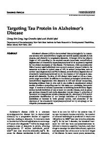
Overall, there is a direct relationship between hyperphosphorylation, abnormal phosphorylation and Tau aggregation but it remains to be determined whether phosphorylation is a cause or a consequence in the aggregation process. 7.6. Classification of tau aggregation in tauopathies In neurodegenerative disorders, other than Alzheimer’s disease, and referred to as tauopathies, abnormally and hyperphosphorylated tau proteins aggregate in the absence of amyloid deposits. Comparative biochemistry of Tau aggregates shows that they differ in both phosphorylation and content of tau isoforms, which enable a molecular classification of tauopathies. Five classes of tauopathies have been defined depending on the type of Tau aggregates that constitute the bBar CodeQ for neurodegenerative disorders (Fig. 2). 7.7. Class 0: frontal lobe degeneration non-Alzheimer non-Pick Frontal lobe degeneration is a neurological disorder, which has been recently characterized, despite the fact that it is the most common presenile dementing disorder in Europe, after AD. As Pick’s disease, it has a bfrontalQ pathology. However, while Pick’s disease is neuropathologically characterized by Pick bodies, frontal lobe degeneration has no specific neuropathologic hallmarks. Morphological changes comprise a neuronal cell loss, spongiosis and gliosis mainly in the superficial cortical layers of the frontal and temporal cortex. No tau aggregates are observed although a loss of tau protein expression is observed in this disorder, also named DLDH (Dementia Lacking Distinctive Histopathological features) [110–112]. 7.8. Class I: a major Tau triplet at 60, 64, 69 Class I is characterized by a pathological tau triplet at 60, 64 and 69 kDa and a minor pathological tau at 72/74 kDa (Fig. 2B). It is now well established that this pathological tau triplet corresponds to the aggregation of the six tau isoforms [113,114]. The pathological tau 60 is composed of shortest tau isoform. The tau 64 and 69 are a mix of tau isoforms with exon 10 or exon 2 alone, and exon 2+10 or exon 2+3, respectively. The longest tau isoform including exon 2+3+10 constitutes the 72/74 kDa pathological component. The prototypical neurological disorder that characterizes this class is Alzheimer’s disease, but includes nine additional neurological disorders such as hippocampal tauopathy in cerebral aging, amyotrophic lateral sclerosis parkinsonism-dementia complex of Guam, Parkinsonism with dementia of Guadeloupe, postencephalitic parkinsonism, dementia pugislistica, Down’s syndrome, Niemann– Pick disease of type C, Familial British dementia as well as Fronto-temporal dementia with parkinsonism linked to
184
N. Sergeant et al. / Biochimica et Biophysica Acta 1739 (2005) 179–197
Fig. 2. The bar code of tauopathies and their classification. (A) Human brain tissue from patients affected by different tauopathies, which are separated by polyacrylamide gel electrophoresis, and pathological tau proteins revealed by Western blotting using phospho-dependent tau antibodies (e.g., AD2 monoclonal antibody recognizing the phosphorylated Ser396/404 of tau, numbering according to the longest tau isoform). Four different electrophoretic patterns of pathological tau proteins are illustrated. These are composed of pathological tau bands at 60, 64, 69 and 74 kDa, which correspond to pathological tau that are found in aggregates. Type I of aggregate is characterized by the presence of the four pathological tau components, whereas types II and III include two major pathological tau components at 64 and 69 kDa, or 60 and 64 kDa, respectively. Finally, the fourth aggregate type, characterized by a strong pathological tau component at 60, 64 and 69 kDa components, is often observed depending on the severity of the affected region analyzed. These four main patterns of pathological tau thus represent a bar code of tauopathies. (B) Neurological disorders for which a pathological tau pattern has been defined are classified according to the bbar codeQ. Five classes are defined including a unique or multiple neurological disorders. The four aggregate types are detailed as well as the pathological tau pattern identified. Class 0 includes a unique neurological disorder characterized by the loss of expression of tau protein and lacking distinctive neuropathological features, which is Frontal dementia of non-AD, non-Pick type.
chromosome 17 (FTDP-17) ([115,116] Hof, 1994 #120, [117–125]). 7.9. Class II: a major Tau doublet at 64 and 69 kDa and the concept of 4R tauopathies The class II profile is characterized essentially by the aggregation of 4R-Tau isoforms. This pathological tau profile is observed in progressive supranuclear palsy, corticobasal degeneration, Argyrophylic grain and FTDP17 [126–129]. Progressive supranuclear palsy (PSP) is a late-onset atypical parkinsonism disorder described by Steele, Richardson, and Olszewski in 1964 [130,131]. Dementia is also a common feature at the end-stage of the disease [132,133]. Neuropathologically, PSP is characterized by neuronal loss, gliosis and NFT formation. Neurofibrillary tangles were first described in basal ganglia, brain stem, and cerebellum [130]. More later, the degenerating process has been described in the perirhinal, inferior
temporal and prefrontal cortex, with the same features as subcortical NFT [134,135]. Glial fibrillary tangles are also described [136–140]. Corticobasal degeneration (CBD) was first described in 1967 and referred to as corticodentatonigral degeneration with neuronal achromasia [141,142]. It is a rare, sporadic and slowly progressive late-onset neurodegenerative disorder. It is clinically characterized by cognitive disturbances and extrapyramidal motor dysfunction. Moderate dementia emerges sometimes late in the course of the disease [143]. There is a clinical and pathological overlap between PSP and corticobasal degeneration [133,144,145]. Neuropathological examination reveals severe glial and neuronal abnormalities. The glial pathology is constituted of astrocytic plaques and numerous tau-immunoreactive inclusions in the white matter while the one in PSP is characterized by btuftedQ plaques. Achromatic ballooned neurons are detected in cortex, brainstem and subcortical structures, and in neuritic changes and NFT. In both PSP and CBD, the pathological tau profile consists
N. Sergeant et al. / Biochimica et Biophysica Acta 1739 (2005) 179–197
essentially of the aggregation of 4R-Tau isoforms, although a recent study of a large series of PSP patients suggests that the pathological tau profile is heterogeneous and includes variable amounts of 3R-Tau isoforms as well. Thus, an increased ratio of 4R/3R pathological tau isoform is rather considered to define the class II of tauopathies. In 1987, Braak and Braak [146] reported a series of eight patients with a non-Alzheimer, late onset dementia. Clinically, Argyrophilic grain dementia (AGD) fit with the clinical spectrum that is reported: behavioural disturbances such as personality change and emotional imbalance, memory and cognitive impairment [147]. At the neuropathological level, AGD is characterized by the occurrence of argyrophilic grains (ArG) on light microscopy of the brain tissue, and therefore referred to as Argyrophilic Grain Dementia (AGD). ArGs are neuronal inclusions stained by silver dyes [146,148]. The diagnosis of Dementia with ArGs is based on widespread occurrence of minute, spindle or comma-shaped, argyrophilic, tau-immunoreactive structures distinct from neuropile threads and predominantly located in the hippocampus and related limbic areas [149]. The 4R/3R ratio has been shown to be increased in AGD, thus demonstrating that AGD is most likely a 4R-Tau pathology [128,150]. The tau pathology affects essentially the limbic system but, more recently, the tau pathology has been shown to extend toward all cerebral cortex, very distant from the limbic temporal region, in brain areas that are considered to be spared in AGD [151]. The neocortical extension of tau pathology in AGD would be a further feature common to PSP and CBD, and might be a clue to a pathological continuum between PSP, CBD, limbic AGD and diffuse AGD [151]. Many features are shared by all four repeat tauopathies. For example, like in PSP and CBD, the subthalamic nuclei are selectively involved by tau aggregates in AGD patients [152]. The H1/H1 haplotype is more frequent in PSP/CBD patients than in controls or other tauopathies [153–155]. H1/H1 may be more frequent in AGD patients than in controls, though recent studies have failed to establish statistically a significant difference [156,157]. 7.10. Class III: a major Tau doublet at 60 and 64 kDa and the concept of 3R tauopathies This class of tauopathy includes a single neurological disorder that is Pick’s disease (PiD). Pick’s disease is a rare form of neurodegenerative disorder characterized by a progressive dementing process. Early in the clinical course, patients show signs of frontal disinhibition [158]. Neuropathologically, Pick’s disease is characterized by prominent fronto-temporal lobar atrophy, gliosis, severe neuronal loss, ballooned neurons and the presence of neuronal inclusions called Pick bodies [159,160]. Pick bodies are labeled by tau antibodies, with a higher density in the hippocampus than in the neocortex [159–161]. The laminar distribution of Pick bodies is clearly different from other tauopathies such as
185
PSP and CBD. In the hippocampus, Pick bodies are numerous in granular cell neurons of the dentate gyrus, in CA1, subiculum and entorhinal cortex, whereas in the neocortex, they are mainly found in layers II and VI of the temporal and frontal lobes. Ultrastructurally, Pick bodies consist of accumulation of both random coiled and straight filaments. The biochemical analysis using a quantitative Western blot approach with phosphorylation-dependent anti-tau antibodies has revealed that in all cases of PiD studied, a major 60- and 64-kDa pathological Tau doublet is observed in the isocortex, in the limbic areas and in subcortical nuclei [160]. A faint pathological tau band is observed at 69 kDa [162,163]. The pathological tau profile of PiD is opposed to that of class II tauopathies. Indeed, the pathological tau isoforms is shown to consist essentially of the 3R-Tau isoforms [162]. In addition, aggregated tau proteins in Pick’s disease are not detected by the monoclonal antibody 12E8 raised against the phosphorylated residue Ser262/Ser356 whereas in other neurodegenerative disorders, this phosphorylation site is detected [160,164]. The lack of phosphorylation at Ser262 and -356 sites is likely to be related to either a kinase inhibition in neurons that degenerate in Pick’s disease or an absence of these kinases within degenerating neurons [165]. 7.11. Class IV: a major Tau 60 Class IV is also represented by a single neurological disorder: Myotonic dystrophy of type I. Myotonic dystrophy (DM) is the commonest form of adult-onset muscular dystrophy. It is a multisystemic disease affecting many systems as the central nervous system (cognitive and neuropsychiatric impairments), the heart (cardiac conduction defects), the genital tract (testicular atrophy), the eyes (cataracts), the ears (deafness), gastrointestinal tract (smooth muscle), endocrine system (insulin resistance), thus leading to a wide and variable complex panel of symptoms [166– 169]. At the clinical level, DM includes two entities designated as myotonic dystrophy of type I (DM1) and myotonic dystrophy of type II (DM2), as recommended by the international Myotonic Dystrophy Consortium (2000), as well as the European NeuroMuscular Centre (ENMC) DM2/PROMM Workshop (2003) [170]. Although the latter is also referred to as proximal myotonic myopathy (PROMM), because of subtle clinical differences [171,172], DM2 now refers to the recently discovered gene locus and mutation [173]. DM1, the commonest form of DM, is an inherited autosomal dominant disorder caused by a single gene mutation consisting of expansion of a CTG trinucleotide motif in the 3V untranslated of the myotonic dystrophy protein kinase gene (dmpk), located on chromosome 19q [174]. The mutation that causes DM2 corresponds also of an expansion of a CCTG tetranucleotide in the first intron
186
N. Sergeant et al. / Biochimica et Biophysica Acta 1739 (2005) 179–197
(untranslated sequence) of ZNF9 gene, located on chromosome 3q, that encodes a nuclear protein [173,175]. Both mutations are very unstable. The length changes from one generation to another as well as in somatic cells of an individual. Thus, DM1 and DM2 mutations share many pathogenic similarities (for review, see Ref. [176]). Cognitive impairment, as memory, visuo-spatial recall and verbal scale, cortical atrophy essentially of the frontal and the temporal lobe and white matter lesions are often described in both DM1 and DM2 [177,178]. Neuropathological lesions, as neurofibrillary tangles, have been observed in adult DM1 individuals aged over 50 years [179–181]. The pathological tau profile of DM1 is characterized by a strong pathological tau band at 60 kDa and, to a lesser extent, a pathological tau component at 64 and 69 kDa. This typical pathological tau profile is reflected by a reduced number of tau isoforms expressed in the brain of individuals with DM1, both at the protein and mRNA levels [182]. In addition, tau protein expression is also demonstrated to be altered in transgenic with human DM1 locus [183]. The analysis of multiple brain regions of one genetically confirmed DM2 patient aged of 71 years showed some neurofibrillary degenerating processes. Using specific immunological probes against exon 2 and exon 3 corresponding amino acid sequences, the neurofibrillary lesions were shown to be devoid of tau isoforms with N-terminal inserts [170]. An altered splicing of tau characterized by a reduced expression of tau isoforms containing the Nterminal inserts characterizes both DM1 and DM2. Overall, it demonstrates that the central nervous system is affected and that DM are real tauopathies. The direct relationship between the altered splicing of Tau and neurofibrillary degeneration in DM remains to be established. Indeed, such an altered splicing of Tau is commonly observed in FTDP17 and considered as reminiscent to neurofibrillary degeneration and tauopathies. 7.12. Tau mutations and familial frontotemporal dementia and chromosome 17-linked pathologies Historically, frontotemporal dementia (FTD) was often classified as a form of Pick’s disease, even when Pick cells or Pick bodies were not found [184]. However, this denomination may involve different subgroups of pathologies, and the Lund and Manchester Groups [185] published in 1994 a consensus on Clinical and Neuropathological Criteria for Frontotemporal Dementia. This publication clarified the position of Pick’s disease within FTD, and several of the reported cases of familial Pick’s disease were probably cases of familial FTD. Indeed, it is difficult to ascertain families, which have the classic pathological features of Pick’s disease from the literature [186], because they often have unusual clinical features. In 1994, Wilhelmsen and colleagues have described an autosomal dominantly inherited disease related to familial
FTD, characterized by adult-onset behavioral disturbances, frontal lobe dementia, parkinsonism and amyotrophy [187]. They demonstrated a genetic linkage between this pathology, denominated disinhibition-dementia-parkinsonismamyotrophy complex (DDPAC), and chromosome 17q21– 22 [187]. Since then, several families sharing strong clinical and pathological features and for which there is a linkage with chromosome 17q22–22 have been described [188– 191]. They have been included in a group of pathologies referred to as frontotemporal dementia with parkinsonism linked to chromosome 17 (FTDP-17) [192]. Although a clinical heterogeneity could be described between and within the families with FTDP-17, usual symptoms include behavioural changes, loss of frontal executive functions, language deficit and hyperorality. Parkinsonism and amyotrophy are described in some families, but are not consistent features. Neuropathologically, brains of FTD patients exhibit an atrophy of frontal and temporal lobes, a severe neuronal cell loss, a grey and white matter gliosis, and a superficial laminar spongiosis. One of the main important characteristic is the filamentous pathology affecting the neuronal cells, or the both neuronal and glial cells in some cases. The absence of amyloid aggregates is usually established [192–194]. FTDP-17 has been related to mutations on the Tau gene [195–198]. Tau mutations always segregate with the pathology and are not found in the control subjects, suggesting their pathogenic role. To date, 17 mutations have been described in the Tau gene among the different families with cases diagnosed as FTDP-17 (Table 1). Twenty missense mutations in coding regions R5H [199], R5L [200], K257T [201,202], I260V [203], L266V [204,205], G272V [195,198,206], N279K [207–210], N296H [211,212], P301L [189,206,213,214], P301S [215,216], S305N [208,217], L315R [206], S320F [206], Q336R [218], V337M [219], E342V, S352V, K369I [220], G389R [221], R406W [195,206], three silent mutation L284L [222], N296N [223], S305S [224], two single amino acid deletions DK280 and DN296 [206,212,222,225,226], and nine intronic mutations in the splicing region following exon 10 at position +3 [198], +11, +12, +13, +14 [195], +16 [227], +19, +29 and +33 after exon 9 [225] have been reported (Table 1). 7.13. Mutations affecting Tau splicing Depending on their functional effects, mutations on Tau proteins may be divided into two groups: the mutations affecting the alternative splicing of exon 10, and leading to changes in the proportion of 4R- and 3R-Tau isoforms, and the mutations modifying Tau interactions with microtubules. The first group includes intronic mutations (+3, +13, +14, +16) and some missense mutations. Intronic mutations disturb a stem loop structure in the 5V splice site of exon 10 that stabilizes this region of the pre-mRNA [195,196, 198,228]. Sequence analysis of this splicing region in
N. Sergeant et al. / Biochimica et Biophysica Acta 1739 (2005) 179–197 Table 1 FTDP-17: effects of tau mutations
7.14. Tau missense mutations and Tau aggregation
Mutation
Localization
Aggregates
Isoforms
R5H R5L K257T L266V G272V I260V N279K DK280 L284L N296H N296N DN296 P301L P301S S305N S305S +3, +11, +12, +13, +14, +16 +19, +29 +33 L315R S320F V337M E342V
Exon 1 Exon 1 Exon 9 Exon 9 Exon 9 Exon 9 Exon 10 Exon 10 Exon 10 Exon 10 Exon 10 Exon 10 Exon 10 Exon 10 Exon 10 Exon 10 Intron 10
Glial Neu(DNF) Neu(PiD) Neu/Glial Neu(PiD) ND Neu/Glial ND Neu Neu/Glial Neu/Glial ND Neu Neu Neu Neu Neu/Glial
4R 4R+1N3R 3RN4R 3R+4R 3R+4R ND 4R ND 4R ? 4R 4R ND 4R 4R 4R 4R 4R
Intron 10 Intron 10 Exon 11 Exon 11 Exon 12 Exon 12
ND ND ND Neu(PiD) Neu Neu
S352V K369I G389R R406W
Exon Exon Exon Exon
Neu Neu(PiD)/Glial Neu(PiD) Neu
3RJ4R ND ND ND 3R+4R 4R (0N4RN 1N4RN2N4R) ND 3R+4R 4RN3R 3R+4R
12 12 13 13
187
Neuronal aggregates are indicated by Neu. Neuronal inclusions corresponding to Pick bodies are precised (PiD). The cellular types of aggregates or pathological tau isoform profiles which have not been determined are indicated by ND.
different animals indicates that the lack of the stem loop structure is associated with an increase in Tau mRNAs containing exon 10 [196]. Indeed, without this stem loop, access of U1snRNP to this site may be facilitated, increasing the formation of exon 10+Tau mRNAs and thus the 4R-Tau isoform [196,222,228]. Interestingly, in these families, abnormally phosphorylated 4R-Tau isoforms aggregate into filaments and display a Tau electrophoretic profile similar to the major Tau doublet at 64 and 69 kDa found in PSP and CBD [126,159,194,198]. Some missense mutations (N279K and S305N) also modify the splicing of exon 10 [222]. For instance, the change in nucleotide for N279K and S305N mutations also creates an exon-splicing enhancer sequence [222]. The silent mutation L284L increases the formation of Tau mRNAs containing exon 10, presumably by destroying an exon splicing silencing element [222]. Families with one of these three missense mutations display the same electrophoretic Tau pattern than those having intronic mutations, namely a Tau doublet at 64 and 69 kDa [222,229,230]. Finally, the twisted ribbon filaments described in neurons and glial cells are a common neuropathological feature in all of the neurodegenerative disorders belonging to this first group.
The second group of Tau mutations found in FTDP-17 includes several missense mutations. The effects of mutations G272V, P301L, P301S, V337M and R406W in an in vitro system of microtubule assembly were reported by Hasegawa et al. [219]. These authors showed that mutated Tau isoforms bind microtubules to a lesser extent than wildtype isoforms. They suggest that the mutated isoforms may induce microtubule disassembly [219,227]. These data are now confirmed by a number of studies [222,229,231,232]. When missense mutations are located in Tau regions common to all isoforms, outside exon 10 (V272G, V337M, G389R, R406W), the six Tau isoforms do not bind properly to microtubules. These proteins aggregate into PHF and straight filaments similar to those described in AD, and are present in neuronal cells. Their biochemical characterization shows a Tau electrophoretic profile similar to the AD Tau-triplet. Conversely, when missense mutations are located in exon 10 (P301L, P301S), 4R-Tau isoforms are affected, do not bind to microtubules, and aggregate into twisted ribbon filaments. This type of filamentous inclusions is described in both neurons and glial cells. The biochemical characterization shows a Tau electrophoretic profile similar to the major Tau doublet encountered in PSP and CBD. The DK280 mutation is particular, since it modifies the ratio 4R-Tau/3R-Tau and could also affect the interaction between Tau and microtubules. This mutation may decrease the formation of Tau mRNAs containing exon 10 and thus enhancing the formation of 3R-Tau isoforms. Moreover, this deletion mutation is also responsible for a considerably reduced ability of Tau to promote microtubule assembly, and is stronger than the effect of the P301L mutation. No data are currently available on the biochemistry of Tau aggregates in this family [222,225]. Mutations of the Tau gene and their involvement in FTDP-17 emphasize the fact that abnormal Tau proteins may play a central role in the etiopathogenesis of neurodegenerative disorders, without any implication of the amyloid cascade. The functional effects of the mutations suggest that a reduced ability of Tau to interact with microtubules may be upstream of hyperphosphorylation and aggregation. These mutations may also lead to an increase in free cytoplasmic Tau (especially 4R-Tau isoforms), facilitating their aggregation into filaments [231]. Finally, some mutations may have a direct effect on Tau fibrillogenesis [233]. 7.15. Tau missense mutations and in vivo microtubule stabilization Regarding the functional effects of Tau mutants, it was shown that mutations G272V, DK280, P301L, P301S and V337M cause a decreased ability of Tau to promote microtubule assembly in vitro [219]. However, the magni-
188
N. Sergeant et al. / Biochimica et Biophysica Acta 1739 (2005) 179–197
tude of the observed effects and the reported rank order of potency of individual mutations have been variable. Overexpression of mutant Tau in transfected cells has given inconsistent results as far as effects on microtubule binding and stability are concerned [208,225,231,232]. Such studies are confounded by the problem that high expression of mutant Tau may override any effects that are present at more physiological levels. For in vivo investigation, recombinant tau 3R, 4R or mutated tau are injected in Xenopus oocytes and maturation of oocytes is used as an indicator of microtubule function [234]. Normal oocyte maturation, as visualized by the appearance of a white spot, indicates that oocyte meiosis driven by microtubule is normal. Wild-type 4R-Tau inhibits maturation of oocytes in a concentration-dependent manner, whereas 3R-Tau has no effect. These data suggest that there is a direct interaction of 4R-Tau and microtubules that interferes with oocyte maturation. Whatever the concentration of the following 4R mutant Tau (G272V, P301L, P301S and V337M) injected in oocyte, it fails to affect oocyte maturation although few changes in the organization of meiotic spindles are observed. In contrast to wild-type 4R-Tau, these mutations are likely to reduce the interaction of tau with microtubules. Two additional mutations (R406W and S305N) are analyzed and they are found to perturb oocyte maturation. Differential phosphorylation cannot explain the data obtained for the first group of Tau mutants since wild-type 3R- and 4R-Tau are found to be phosphorylated at the same extent as the G272V, P301L, P301S and V337M 4R-Tau mutants. Therefore, these mutations strongly reduce the ability of Tau to interact with microtubules [234]. Regarding the R406W mutation, there are some controversial effects: state of phosphorylation and reduced microtubule binding. At low concentrations, R406W Tau mutant strongly interferes with oocyte maturation even when no effect was observed with wild-type 4R-Tau. Conversely, at high concentrations similar to those of injected wild-type 4R-Tau that block maturation, oocyte maturation is never completely abolished. Altogether, these data suggest that at low concentrations, R406W mutant Tau is less phosphorylated and thus shows a better microtubule binding, and at high concentrations, the phosphorylation state is not sufficient to thwart the reduction of microtubule binding of this Tau mutant [234]. The most pronounced inhibitory effect on oocyte maturation is observed with S305N Tau, even following microinjections of small amounts of Tau protein. This effect is independent of phosphorylation. It was shown that S305N Tau has a slightly increased ability to promote microtubule assembly in vitro when compared with wild-type protein [208]. Altogether, our data demonstrate that in vivo Tau missense mutations either strongly reduce interactions with microtubules or increase these interactions. The observed phenotype is dependent on the combined effect of Tau
phosphorylation and concentration. It also demonstrates that the Xenopus oocyte is an interesting heterologous model system for following perturbations in microtubule function. 7.16. Familial versus sporadic tauopathies The mapping of the spatiotemporal distribution of tau pathology in the different brain areas is important to understand how the disease spreads in the brain. While tau gene mutations simultaneously affect different brain areas, many Tsporadicr tauopathies affect first a precise vulnerable area: the entorhinal and hippocampal area in Alzheimer’s disease, the brain stem in PSP and CBD. Indeed, there is a precise biochemical pathway of tau pathology in aging and in AD. The progression of tau pathology is sequential, invariable, hierarchical, and predictable. Ten stages (S1 to S10) were defined, corresponding to 10 brain areas sequentially affected [235]. The extension of the tauopathy fits well with the evolution of cognitive deficits, from memory disorders when the hippocampal formation is affected (stages 1 to 3) to language impairment (temporal areas affected at stages 4 to 6) and then to apraxia, and agnosia when frontal and parietal areas are involved (stage 7 to 10). It is interesting to note that the pathway of tau pathology in PSP in CBD is quite different from the one in AD, and roughly the opposite, emerging from subcortical nuclei toward the neocortex, and especially the frontal motor cortex. In most sporadic neurodegenerative disorders, the spreading of tau pathology follows specific neuronal connections, like a precise neuronal chain reaction. This pathway starts in the vulnerable brain area characteristic of the disease to specific neocortical brain areas. At that level of tau extension, a dementing process is occurring. These observations in the human brain demonstrate the progressive collapse of neuronal populations, along the corticocortical of subcortico-cortical connections. 7.17. Tau pathology is fueled by APP in Alzheimer’s disease The initiating cause of AD is currently based on the discovery of mutations in APP and presenilin genes in Familial Alzheimer’s disease (FAD). However, no mutations and even polymorphisms on tau, APP or presenilin genes have been associated with Sporadic Alzheimer’s disease (SAD) [119,236–241]. The major risk factor is aging, closely followed by ApoE gene polymorphisms [242,243]. Therefore, the initiating events in SAD remain a matter of debate. The APP metabolism has been poorly investigated in SAD. Thus, a better understanding of the precise and detailed progression of tau pathology in aged controls and SAD and its correlation with APP expression and with all APP catabolic products is of major importance.
N. Sergeant et al. / Biochimica et Biophysica Acta 1739 (2005) 179–197
7.18. Reduced expression of carboxy-terminal fragments of APP is correlated to tau pathology staging in AD Upstream catabolic products of APP, the so-called carboxy-terminal fragments (CTF) or stubs, result from proteases activities named secretases (for review see, Ref. [244]). Three secretases activities have been identified, that of the a, h and g-secretases. The a-secretases cleave APP in the middle of the Ah sequence (at position 17) and generate the a-stub. The CTF precursor for Ah results from the hsecretase activity, liberating the amino-terminus of Ah. An alternate cleavage by the h-secretase occurs at position 11 of Ah leading to hV-stub. APP-CTF h is further processed through the g-secretase producing the Ah peptide and the cytosolic C57 fragments. Being distinguished from the gsecretase activity that leads to the production of Ah, qsecretase, which can obviously process all APP-CTFs, liberates the cytosolic domain of APP, also called AICD or C51 (amyloid precursor protein intracellular domain) [244]. In AD, Ah-42 deposition strongly suggests that APP metabolism is altered and APP-CTFs should be modified. We have studied the APP-CTF expression in both nondemented and AD patients [245]. APP-CTFs were shown to be significantly diminished during the course of AD and, moreover, the decrease of APP-CTFs was well correlated with the progression of tau pathology. In addition, the brain tissue of individuals having an inherited form of AD linked to mutations of presenilin 1 also showed a decreased amount of APP-CTFs. Our results thus showed for the first time a relationship between Tau and APP pathologies, which is, in addition, observed at the earliest stages of AD [246]. 7.19. Amyloid deposition is correlated to tau pathology but is lacking the spatiotemporal overlap The decrease of APP-CTFs should be associated with modification of the downstream products that are the Ah peptides. Thus, Ah peptides were quantitatively and qualitatively investigated, using an extraction in pure formic acid and applying them on the brain tissue of all cases from our brain bank. Multiple immunological tools associated with a proteomic analysis were performed. Insoluble Ah-42 and -40 species were fully solubilized and quantified in the main neocortical areas [246,247]. The quantities of both species Ah were compared to the extent of tau pathology, as well as to cognitive impairment. In AD, there is a constellation of amyloid phenotypes, extending from cases with exclusively aggregated Ah-42 to cases with, in addition, large quantities of insoluble Ah-40 species. Nonetheless, insoluble Ah-40 detection was often observed late in the amyloid deposition process (starting at Tau pathology stages 4–5). We observed that there was no obvious spatial and temporal overlap in the distribution of these two insoluble Ah species in cortical brain areas. Thus, brain areas with Ah-42 were not systematically associated with Ah-40 deposition and, moreover, the quantities of Ah-
189
40 measured were not directly related to that of Ah-42. Physical properties were also different. Formic acid-solubilized Ah-40 aggregates were composed essentially of monomers and dimers, while solubilized Ah-42 was essentially observed as monomers, dimers, and oligomers. More importantly, Ah-42 aggregates were observed at the early stages of tau pathology in non-demented patients. All together, it was interesting to note that during the progression of the disease, Ah aggregates increase in quantity and heterogeneity, in close parallel to the extension of tau pathology. But unexpectedly, there was no spatial overlap between Ah aggregation that is widespread and heterogeneously distributed in cortical areas and tau pathology that is progressing sequentially, stereotypically, and hierarchically [246,247]. Therefore, these observations demonstrate that Ah-42 aggregation, and not Ah-40, is the marker that is close to Alzheimer aetiology. It should be the main target for the early biological diagnosis of AD and modelling. Furthermore, the spatial mismatch between Ah and tau pathologies in cortical areas was obvious. First of all, the neocortical areas that contain first or more Ah are not always the same. Generally, but not always, the occipital cortex is more prone to develop amyloid deposits [247]. These Ah-42 amyloid deposits observed at the neuropathological level were not related to amyloid angiopathy. Remarkably enough, this brain region is the last to develop tau pathology [235,247]. Conversely, the hippocampal region early affected by tau pathology is not especially affected by amyloid deposition. These observations confirm the findings of Braak and Braak [248,249]. Together, this Ah/Tau mismatch demonstrates that neurodegeneration is not a direct consequence of extracellular Ah neurotoxicity (considering that toxicity is mediated through the cell body). Hence, there is a synergetic effect of APP dysfunction on the neuron-to-neuron propagation of tau pathology. Indeed, tau pathology can be found in the hippocampal area without Ah deposits [121]. Nonetheless, decrease of APP-CTFs is likely to be more widely distributed over the brain regions, as established in the temporal cortex and occipital, and hence further supports the relationship between APP dysfunction and tau pathology [245]. In contrast, the extension of tau pathology in the polymodal association areas is always found in the presence of Ah deposits, as if these species, directly or indirectly, were necessary to stimulate the progression of tau pathology [121,246].
8. Conclusion Aggregation of tau proteins in filamentous inclusions is a common feature of more than 20 neurodegenerative disorders. The laminar and regional distributions of NFT or other inclusions are different among dementing conditions. Likewise, a bbar codeQ of pathological tau protein electrophoretic profiles permitted a classification of these disorders sharing similar biochemical signatures. Many
190
N. Sergeant et al. / Biochimica et Biophysica Acta 1739 (2005) 179–197
parameters can explain this classification such as the selective aggregation of specific sets of tau isoforms, the differential vulnerability of subneuronal population, in addition to possibly variable sets of enzymes (e.g. kinases, phosphatases). Notwithstanding the regional or laminar distribution or the electrophoretic pattern of pathological tau proteins, their aggregation is always correlated to dementia when association neocortical areas are involved. The recent discovery of mutations on the tau gene, resulting in an abnormal aggregation of tau isoforms into filamentous inclusions in FTDP-17, demonstrates that abnormal tau metabolism is sufficient to induce nerve cell degeneration, in relationship with the property of mutant tau to regulate the microtubule dynamic. Altogether, these data indicate that numerous mechanisms including cell vulnerability, dysregulation of many enzymes, and tau mutations could interact to disturb tau metabolism, and result in the disorganization of the cystoskeleton, including the microtubule network, commonly observed in all of these neurodegenerative illnesses. But the key event is always the disorganization of the cytoskeleton leading to nerve cell degeneration. Acknowledgments This work was supported by the Institut de la Sante´ et de la Recherche Me´dicale, the Centre National de la Recherche Scientifique, University of Lille II, AFM, Aventis Pharma, FEDER, IMPRT of Lille and the Conseil Re´gional Nord Pas-de-Calais (Ge´nopole de Lille). References [1] T.A. Schoenfeld, R.A. Obar, Diverse distribution and function of fibrous microtubule-associated proteins in the nervous system, Int. Rev. Cytol. 151 (1994) 67 – 137. [2] R.P. Tucker, The roles of microtubule-associated proteins in brain morphogenesis: a review, Brain Res. Brain Res. Rev. 15 (1990) 101 – 120. [3] S.S. Chin, J.E. Goldman, Glial inclusions in CNS degenerative diseases, J. Neuropathol. Exp. Neurol. 55 (1996) 499 – 508. [4] Y. Gu, F. Oyama, Y. Ihara, Tau is widely expressed in rat tissues, J. Neurochem. 67 (1996) 1235 – 1244. [5] M. Ingelson, E. Vanmechelen, L. Lannfelt, Microtubule-associated protein tau in human fibroblasts with the Swedish Alzheimer mutation, Neurosci. Lett. 220 (1996) 9 – 12. [6] M.T. Vanier, P. Neuville, L. Michalik, J.F. Launay, Expression of specific tau exons in normal and tumoral pancreatic acinar cells, J. Cell. Sci. 111 (Pt 10) (1998) 1419 – 1432. [7] R.L. Neve, P. Harris, K.S. Kosik, D.M. Kurnit, T.A. Donlon, Identification of cDNA clones for the human microtubule-associated protein tau and chromosomal localization of the genes for tau and microtubule-associated protein 2, Brain Res. 387 (1986) 271 – 280. [8] A. Andreadis, J.A. Broderick, K.S. Kosik, Relative exon affinities and suboptimal splice site signals lead to non-equivalence of two cassette exons, Nucleic Acids Res. 23 (1995) 3585 – 3593. [9] A. Andreadis, B.K. Wagner, J.A. Broderick, K.S. Kosik, A tau promoter region without neuronal specificity, J. Neurochem. 66 (1996) 2257 – 2263.
[10] A. Heicklen-Klein, I. Ginzburg, Tau promoter confers neuronal specificity and binds Sp1 and AP-2, J. Neurochem. 75 (2000) 1408 – 1418. [11] M.L. Wei, A. Andreadis, Splicing of a regulated exon reveals additional complexity in the axonal microtubule-associated protein tau, J. Neurochem. 70 (1998) 1346 – 1356. [12] M.H. Luo, M.L. Leski, A. Andreadis, Tau isoforms which contain the domain encoded by exon 6 and their role in neurite elongation, J. Cell. Biochem. 91 (2004) 880 – 895. [13] M. Goedert, M.G. Spillantini, R. Jakes, D. Rutherford, R.A. Crowther, Multiple isoforms of human microtubule-associated protein tau: sequences and localization in neurofibrillary tangles of Alzheimer’s disease, Neuron 3 (1989) 519 – 526. [14] M. Goedert, M.G. Spillantini, M.C. Potier, J. Ulrich, R.A. Crowther, Cloning and sequencing of the cDNA encoding an isoform of microtubule-associated protein tau containing four tandem repeats: differential expression of tau protein mRNAs in human brain, EMBO J. 8 (1989) 393 – 399. [15] A. Andreadis, W.M. Brown, K.S. Kosik, Structure and novel exons of the human tau gene, Biochemistry 31 (1992) 10626 – 10633. [16] A. Himmler, D. Drechsel, M.W. Kirschner, D.W. Martin Jr., Tau consists of a set of proteins with repeated C-terminal microtubulebinding domains and variable N-terminal domains, Mol. Cell. Biol. 9 (1989) 1381 – 1388. [17] K.S. Kosik, L.D. Orecchio, S. Bakalis, R.L. Neve, Developmentally regulated expression of specific tau sequences, Neuron 2 (1989) 1389 – 1397. [18] M. Goedert, R. Jakes, Expression of separate isoforms of human tau protein: correlation with the tau pattern in brain and effects on tubulin polymerization, EMBO J. 9 (1990) 4225 – 4230. [19] M. Goedert, M.G. Spillantini, Molecular neuropathology of Alzheimer’s disease: in situ hybridization studies, Cell. Mol. Neurobiol. 10 (1990) 159 – 174. [20] R. Brandt, J. Leger, G. Lee, Interaction of tau with the neural plasma membrane mediated by tau’s amino-terminal projection domain, J. Cell. Biol. 131 (1995) 1327 – 1340. [21] N. Hirokawa, Y. Shiomura, S. Okabe, Tau proteins: the molecular structure and mode of binding on microtubules, J. Cell. Biol. 107 (1988) 1449 – 1459. [22] A. Harada, K. Oguchi, S. Okabe, J. Kuno, S. Terada, T. Ohshima, R. Sato-Yoshitake, Y. Takei, T. Noda, N. Hirokawa, Altered microtubule organization in small-calibre axons of mice lacking tau protein, Nature 369 (1994) 488 – 491. [23] H.N. Dawson, A. Ferreira, M.V. Eyster, N. Ghoshal, L.I. Binder, M.P. Vitek, Inhibition of neuronal maturation in primary hippocampal neurons from tau deficient mice, J. Cell Sci. 114 (2001) 1179 – 1187. [24] J. Chen, Y. Kanai, N.J. Cowan, N. Hirokawa, Projection domains of MAP2 and tau determine spacings between microtubules in dendrites and axons, Nature 360 (1992) 674 – 677. [25] I.S. Georgieff, R.K. Liem, D. Couchie, C. Mavilia, J. Nunez, M.L. Shelanski, Expression of high molecular weight tau in the central and peripheral nervous systems, J. Cell. Sci. 105 (Pt 3) (1993) 729 – 737. [26] M.F. Carlier, C. Simon, R. Cassoly, L.A. Pradel, Interaction between microtubule-associated protein tau and spectrin, Biochimie 66 (1984) 305 – 311. [27] I. Correas, R. Padilla, J. Avila, The tubulin-binding sequence of brain microtubule-associated proteins, tau and MAP-2, is also involved in actin binding, Biochem. J. 269 (1990) 61 – 64. [28] J.P. Henriquez, D. Cross, C. Vial, R.B. Maccioni, Subpopulations of tau interact with microtubules and actin filaments in various cell types, Cell Biochem. Funct. 13 (1995) 239 – 250. [29] S.C. Selden, T.D. Pollard, Phosphorylation of microtubule-associated proteins regulates their interaction with actin filaments, J. Biol. Chem. 258 (1983) 7064 – 7071. [30] Y. Miyata, M. Hoshi, E. Nishida, Y. Minami, H. Sakai, Binding of microtubule-associated protein 2 and tau to the intermediate filament
N. Sergeant et al. / Biochimica et Biophysica Acta 1739 (2005) 179–197
[31] [32]
[33]
[34]
[35] [36]
[37] [38]
[39]
[40]
[41]
[42]
[43]
[44]
[45]
[46]
[47]
[48]
[49]
[50]
reassembled from neurofilament 70-kDa subunit protein. Its regulation by calmodulin, J. Biol. Chem. 261 (1986) 13026 – 13030. A. Matus, Microtubule-associated proteins and the determination of neuronal form, J. Physiol. (Paris) 84 (1990) 134 – 137. D. Jung, D. Filliol, M. Miehe, A. Rendon, Interaction of brain mitochondria with microtubules reconstituted from brain tubulin and MAP2 or TAU, Cell Motil. Cytoskelet. 24 (1993) 245 – 255. G. Lee, R. Thangavel, V.M. Sharma, J.M. Litersky, K. Bhaskar, S.M. Fang, L.H. Do, A. Andreadis, G. Van Hoesen, H. Ksiezak-Reding, Phosphorylation of tau by fyn: implications for Alzheimer’s disease, J. Neurosci. 24 (2004) 2304 – 2312. R. Williamson, T. Scales, B.R. Clark, G. Gibb, C.H. Reynolds, S. Kellie, I.N. Bird, I.M. Varndell, P.W. Sheppard, I. Everall, B.H. Anderton, Rapid tyrosine phosphorylation of neuronal proteins including tau and focal adhesion kinase in response to amyloid-beta peptide exposure: involvement of Src family protein kinases, J. Neurosci. 22 (2002) 10 – 20. S.M. Jenkins, G.V. Johnson, Tau complexes with phospholipase Cgamma in situ, NeuroReport 9 (1998) 67 – 71. S.C. Hwang, D.Y. Jhon, Y.S. Bae, J.H. Kim, S.G. Rhee, Activation of phospholipase C-gamma by the concerted action of tau proteins and arachidonic acid, J. Biol. Chem. 271 (1996) 18342 – 18349. G. Lee, R.L. Neve, K.S. Kosik, The microtubule binding domain of tau protein, Neuron 2 (1989) 1615 – 1624. R. Brandt, G. Lee, The balance between tau protein’s microtubule growth and nucleation activities: implications for the formation of axonal microtubules, J. Neurochem. 61 (1993) 997 – 1005. R. Brandt, G. Lee, Functional organization of microtubule-associated protein tau. Identification of regions which affect microtubule growth, nucleation, and bundle formation in vitro, J. Biol. Chem. 268 (1993) 3414 – 3419. D.W. Cleveland, S.Y. Hwo, M.W. Kirschner, Physical and chemical properties of purified tau factor and the role of tau in microtubule assembly, J. Mol. Biol. 116 (1977) 227 – 247. D.W. Cleveland, S.Y. Hwo, M.W. Kirschner, Purification of tau, a microtubule-associated protein that induces assembly of microtubules from purified tubulin, J. Mol. Biol. 116 (1977) 207 – 225. M.D. Weingarten, A.H. Lockwood, S.Y. Hwo, M.W. Kirschner, A protein factor essential for microtubule assembly, Proc. Natl. Acad. Sci. U. S. A. 72 (1975) 1858 – 1862. D.N. Drechsel, A.A. Hyman, M.H. Cobb, M.W. Kirschner, Modulation of the dynamic instability of tubulin assembly by the microtubule-associated protein tau, Mol. Biol. Cell. 3 (1992) 1141 – 1154. K.A. Butner, M.W. Kirschner, Tau protein binds to microtubules through a flexible array of distributed weak sites, J. Cell. Biol. 115 (1991) 717 – 730. N. Gustke, B. Trinczek, J. Biernat, E.M. Mandelkow, E. Mandelkow, Domains of tau protein and interactions with microtubules, Biochemistry 33 (1994) 9511 – 9522. B.L. Goode, S.C. Feinstein, Identification of a novel microtubule binding and assembly domain in the developmentally regulated interrepeat region of tau, J. Cell Biol. 124 (1994) 769 – 782. S. Kar, G.J. Florence, I. Paterson, L.A. Amos, Discodermolide interferes with the binding of tau protein to microtubules, FEBS Lett. 539 (2003) 34 – 36. S. Kar, J. Fan, M.J. Smith, M. Goedert, L.A. Amos, Repeat motifs of tau bind to the insides of microtubules in the absence of taxol, EMBO J. 22 (2003) 70 – 77. E. Sontag, V. Nunbhakdi-Craig, G. Lee, R. Brandt, C. Kamibayashi, J. Kuret, C.L. White III, M.C. Mumby, G.S. Bloom, Molecular interactions among protein phosphatase 2A, tau, and microtubules. Implications for the regulation of tau phosphorylation and the development of tauopathies, J. Biol. Chem. 274 (1999) 25490 – 25498. R.S. Haltiwanger, S. Busby, K. Grove, S. Li, D. Mason, L. Medina, D. Moloney, G. Philipsberg, R. Scartozzi, O-glycosylation of nuclear
[51]
[52]
[53]
[54]
[55]
[56]
[57]
[58] [59]
[60]
[61]
[62]
[63]
[64]
[65]
[66]
[67]
191
and cytoplasmic proteins: regulation analogous to phosphorylation? Biochem. Biophys. Res. Commun. 231 (1997) 237 – 242. L.K. Kreppel, M.A. Blomberg, G.W. Hart, Dynamic glycosylation of nuclear and cytosolic proteins. Cloning and characterization of a unique O-GlcNAc transferase with multiple tetratricopeptide repeats, J. Biol. Chem. 272 (1997) 9308 – 9315. G.W. Hart, L.K. Kreppel, F.I. Comer, C.S. Arnold, D.M. Snow, Z. Ye , X. Cheng , D. DellaManna , D.S. Caine , B.J. Earles, Y. Akimoto, R.N. Cole, B.K. Hayes, O-GlcNAcylation of key nuclear and cytoskeletal proteins: reciprocity with O-phosphorylation and putative roles in protein multimerization, Glycobiology 6 (1996) 711 – 716. K. Kamemura, G.W. Hart, Dynamic interplay between O-glycosylation and O-phosphorylation of nucleocytoplasmic proteins: a new paradigm for metabolic control of signal transduction and transcription, Prog. Nucleic. Acid Res. Mol. Biol. 73 (2003) 107 – 136. D.L. Dong, Z.S. Xu, G.W. Hart, D.W. Cleveland, Cytoplasmic OGlcNAc modification of the head domain and the KSP repeat motif of the neurofilament protein neurofilament-H, J. Biol. Chem. 271 (1996) 20845 – 20852. T. Lefebvre, M.L. Caillet-Boudin, L. Buee, A. Delacourte, J.C. Michalski, O-GlcNAc glycosylation and neurological disorders, Adv. Exp. Med. Biol. 535 (2003) 189 – 202. T. Lefebvre, S. Ferreira, L. Dupont-Wallois, T. Bussiere, M.J. Dupire, A. Delacourte, J.C. Michalski, M.L. Caillet-Boudin, Evidence of a balance between phosphorylation and O-GlcNAc glycosylation of Tau proteins—a role in nuclear localization, Biochim. Biophys. Acta 1619 (2003) 167 – 176. M. Hasegawa, M. Morishima-Kawashima, K. Takio, M. Suzuki, K. Titani, Y. Ihara, Protein sequence and mass spectrometric analyses of tau in the Alzheimer’s disease brain, J. Biol. Chem. 267 (1992) 17047 – 17054. M. Hasegawa, Phosphorylation in tau protein, Seikagaku 65 (1993) 469 – 473. M. Hasegawa, A. Watanabe, K. Takio, M. Suzuki, T. Arai, K. Titani, Y. Ihara, Characterization of two distinct monoclonal antibodies to paired helical filaments: further evidence for fetal-type phosphorylation of the tau in paired helical filaments, J. Neurochem. 60 (1993) 2068 – 2077. S. Lovestone, C.H. Reynolds, The phosphorylation of tau: a critical stage in neurodevelopment and neurodegenerative processes, Neuroscience 78 (1997) 309 – 324. M. Morishima-Kawashima, M. Hasegawa, K. Takio, M. Suzuki, H. Yoshida, A. Watanabe, K. Titani, Y. Ihara, Hyperphosphorylation of tau in PHF, Neurobiol. Aging 16 (1995) 365 – 371. M. Morishima-Kawashima, M. Hasegawa, K. Takio, M. Suzuki, H. Yoshida, K. Titani, Y. Ihara, Proline-directed and non-proline-directed phosphorylation of PHF-tau, J. Biol. Chem. 270 (1995) 823 – 829. L. Buee, T. Bussiere, V. Buee-Scherrer, A. Delacourte, P.R. Hof, Tau protein isoforms, phosphorylation and role in neurodegenerative disorders, Brain Res. Brain Res. Rev. 33 (2000) 95 – 130. P. Seubert, M. Mawal-Dewan, R. Barbour, R. Jakes, M. Goedert, G.V. Johnson, J.M. Litersky, D. Schenk, I. Lieberburg, J.Q. Trojanowski, et al., Detection of phosphorylated Ser262 in fetal tau, adult tau, and paired helical filament tau, J. Biol. Chem. 270 (1995) 18917 – 18922. H.M. Roder, R.P. Fracasso, F.J. Hoffman, J.A. Witowsky, G. Davis, C.B. Pellegrino, Phosphorylation-dependent monoclonal Tau antibodies do not reliably report phosphorylation by extracellular signal-regulated kinase 2 at specific sites, J. Biol. Chem. 272 (1997) 4509 – 4515. G. Drewes, B. Lichtenberg-Kraag, F. Doring, E.M. Mandelkow, J. Biernat, J. Goris, M. Doree, E. Mandelkow, Mitogen activated protein (MAP) kinase transforms tau protein into an Alzheimer-like state, EMBO J. 11 (1992) 2131 – 2138. M. Goedert, M. Hasegawa, R. Jakes, S. Lawler, A. Cuenda, P. Cohen, Phosphorylation of microtubule-associated protein tau
192
[68]
[69]
[70] [71]
[72]
[73]
[74]
[75]
[76]
[77]
[78]
[79]
[80]
[81]
[82]
[83] [84]
[85]
N. Sergeant et al. / Biochimica et Biophysica Acta 1739 (2005) 179–197 by stress-activated protein kinases, FEBS Lett. 409 (1997) 57 – 62. C.H. Reynolds, M.A. Utton, G.M. Gibb, A. Yates, B.H. Anderton, Stress-activated protein kinase/c-jun N-terminal kinase phosphorylates tau protein, J. Neurochem. 68 (1997) 1736 – 1744. R. Vulliet, S.M. Halloran, R.K. Braun, A.J. Smith, G. Lee, Prolinedirected phosphorylation of human Tau protein, J. Biol. Chem. 267 (1992) 22570 – 22574. M. Takahashi, K. Tomizawa, K. Sato, A. Ohtake, A. Omori, A novel tau-tubulin kinase from bovine brain, FEBS Lett. 372 (1995) 59 – 64. M. Takahashi, K. Tomizawa, K. Ishiguro, M. Takamatsu, S.C. Fujita, K. Imahori, Involvement of tau protein kinase I in paired helical filament-like phosphorylation of the juvenile tau in rat brain, J. Neurochem. 64 (1995) 1759 – 1768. K. Baumann, E.M. Mandelkow, J. Biernat, H. Piwnica-Worms, E. Mandelkow, Abnormal Alzheimer-like phosphorylation of tauprotein by cyclin-dependent kinases cdk2 and cdk5, FEBS Lett. 336 (1993) 417 – 424. W.K. Liu, R.T. Williams, F.L. Hall, D.W. Dickson, S.H. Yen, Detection of a Cdc2-related kinase associated with Alzheimer paired helical filaments, Am. J. Pathol. 146 (1995) 228 – 238. S.M. Jenkins, M. Zinnerman, C. Garner, G.V. Johnson, Modulation of tau phosphorylation and intracellular localization by cellular stress, Biochem. J. 345 (Pt 2) (2000) 263 – 270. V. Buee-Scherrer, M. Goedert, Phosphorylation of microtubuleassociated protein tau by stress-activated protein kinases in intact cells, FEBS Lett. 515 (2002) 151 – 154. G. Drewes, A. Ebneth, U. Preuss, E.M. Mandelkow, E. Mandelkow, MARK, a novel family of protein kinases that phosphorylate microtubule-associated proteins and trigger microtubule disruption, Cell 89 (1997) 297 – 308. J. Baudier, R.D. Cole, Interactions between the microtubuleassociated tau proteins and S100b regulate tau phosphorylation by the Ca2+/calmodulin-dependent protein kinase II, J. Biol. Chem. 263 (1988) 5876 – 5883. B. Steiner, E.M. Mandelkow, J. Biernat, N. Gustke, H.E. Meyer, B. Schmidt, G. Mieskes, H.D. Soling, D. Drechsel, M.W. Kirschner, et al., Phosphorylation of microtubule-associated protein tau: identification of the site for Ca2(+)-calmodulin dependent kinase and relationship with tau phosphorylation in Alzheimer tangles, EMBO J. 9 (1990) 3539 – 3544. G.A. Jicha, C. Weaver, E. Lane, C. Vianna, Y. Kress, J. Rockwood, P. Davies, cAMP-dependent protein kinase phosphorylations on tau in Alzheimer’s disease, J. Neurosci. 19 (1999) 7486 – 7494. J.M. Litersky, G.V. Johnson, Phosphorylation by cAMP-dependent protein kinase inhibits the degradation of tau by calpain, J. Biol. Chem. 267 (1992) 1563 – 1568. M. Pierre, J. Nunez, Multisite phosphorylation of tau proteins from rat brain, Biochem. Biophys. Res. Commun. 115 (1983) 212 – 219. J.A. Greenwood, C.W. Scott, R.C. Spreen, C.B. Caputo, G.V. Johnson, Casein kinase II preferentially phosphorylates human tau isoforms containing an amino-terminal insert. Identification of threonine 39 as the primary phosphate acceptor, J. Biol. Chem. 269 (1994) 4373 – 4380. E. Planel, X. Sun, A. Takashima, Role of GSK-3beta in Alzheimer’s disease pathology, Drug Dev. Res. 56 (2002) 491 – 510. T.J. Singh, T. Zaidi, I. Grundke-Iqbal, K. Iqbal, Non-prolinedependent protein kinases phosphorylate several sites found in tau from Alzheimer disease brain, Mol. Cell. Biochem. 154 (1996) 143 – 151. Y.L. Woods, P. Cohen, W. Becker, R. Jakes, M. Goedert, X. Wang, C.G. Proud, The kinase DYRK phosphorylates protein-synthesis initiation factor eIF2Bepsilon at Ser539 and the microtubuleassociated protein tau at Thr212: potential role for DYRK as a glycogen synthase kinase 3-priming kinase, Biochem. J. 355 (2001) 609 – 615.
[86] V. Buee-Scherrer, O. Condamines, C. Mourton-Gilles, R. Jakes, M. Goedert, B. Pau, A. Delacourte, AD2, a phosphorylationdependent monoclonal antibody directed against tau proteins found in Alzheimer’s disease, Brain Res. Mol. Brain Res. 39 (1996) 79 – 88. [87] N. Sergeant, T. Bussiere, P. Vermersch, J.P. Lejeune, A. Delacourte, Isoelectric point differentiates PHF-tau from biopsy-derived human brain tau proteins, NeuroReport 6 (1995) 2217 – 2220. [88] E.S. Matsuo, R.W. Shin, M.L. Billingsley, A. Van deVoorde, M. O’Connor, J.Q. Trojanowski, V.M. Lee, Biopsy-derived adult human brain tau is phosphorylated at many of the same sites as Alzheimer’s disease paired helical filament tau, Neuron 13 (1994) 989 – 1002. [89] C. Soulie, J. Lepagnol, A. Delacourte, M.L. Caillet-Boudin, Dephosphorylation studies of SKNSH-SY 5Y cell Tau proteins by endogenous phosphatase activity, Neurosci. Lett. 206 (1996) 189 – 192. [90] P. Cohen, The structure and regulation of protein phosphatases, Annu. Rev. Biochem. 58 (1989) 453 – 508. [91] T.S. Ingebritsen, P. Cohen, The protein phosphatases involved in cellular regulation: 1. Classification and substrate specificities, Eur. J. Biochem. 132 (1983) 255 – 261. [92] S.M. Dudek, G.V. Johnson, Postnatal changes in serine/threonine protein phosphatases and their association with the microtubules, Brain Res. Dev. Brain Res. 90 (1995) 54 – 61. [93] H. Liao, Y. Li, D.L. Brautigan, G.G. Gundersen, Protein phosphatase 1 is targeted to microtubules by the microtubule-associated protein Tau, J. Biol. Chem. 273 (1998) 21901 – 21908. [94] M. Goedert, R. Jakes, Z. Qi, J.H. Wang, P. Cohen, Protein phosphatase 2A is the major enzyme in brain that dephosphorylates tau protein phosphorylated by proline-directed protein kinases or cyclic AMP-dependent protein kinase, J. Neurochem. 65 (1995) 2804 – 2807. [95] S. Goto, H. Yamamoto, K. Fukunaga, T. Iwasa, Y. Matsukado, E. Miyamoto, Dephosphorylation of microtubule-associated protein 2, tau factor, and tubulin by calcineurin, J. Neurochem. 45 (1985) 276 – 283. [96] H. Yamamoto, Y. Saitoh, K. Fukunaga, H. Nishimura, E. Miyamoto, Dephosphorylation of microtubule proteins by brain protein phosphatases 1 and 2A, and its effect on microtubule assembly, J. Neurochem. 50 (1988) 1614 – 1623. [97] H. Yamamoto, Y. Saitoh, S. Yasugawa, E. Miyamoto, Dephosphorylation of tau factor by protein phosphatase 2A in synaptosomal cytosol fractions, and inhibition by aluminum, J. Neurochem. 55 (1990) 683 – 690. [98] T. Ono, H. Yamamoto, K. Tashima, H. Nakashima, E. Okumura, K. Yamada, S. Hisanaga, T. Kishimoto, T. Miyakawa, E. Miyamoto, Dephosphorylation of abnormal sites of tau factor by protein phosphatases and its implication for Alzheimer’s disease, Neurochem. Int. 26 (1995) 205 – 215. [99] T. Saito, K. Ishiguro, T. Uchida, E. Miyamoto, T. Kishimoto, S. Hisanaga, In situ dephosphorylation of tau by protein phosphatase 2A and 2B in fetal rat primary cultured neurons, FEBS Lett. 376 (1995) 238 – 242. [100] C.X. Gong, F. Liu, G. Wu, S. Rossie, J. Wegiel, L. Li, I. GrundkeIqbal, K. Iqbal, Dephosphorylation of microtubule-associated protein tau by protein phosphatase 5, J. Neurochem. 88 (2004) 298 – 310. [101] J. Biernat, N. Gustke, G. Drewes, E.M. Mandelkow, E. Mandelkow, Phosphorylation of Ser262 strongly reduces binding of tau to microtubules: distinction between PHF-like immunoreactivity and microtubule binding, Neuron 11 (1993) 153 – 163. [102] G.T. Bramblett, M. Goedert, R. Jakes, S.E. Merrick, J.Q. Trojanowski, V.M. Lee, Abnormal tau phosphorylation at Ser396 in Alzheimer’s disease recapitulates development and contributes to reduced microtubule binding, Neuron 10 (1993) 1089 – 1099. [103] D. Drubin, S. Kobayashi, M. Kirschner, Association of tau protein with microtubules in living cells, Ann. N. Y. Acad. Sci. 466 (1986) 257 – 268.
N. Sergeant et al. / Biochimica et Biophysica Acta 1739 (2005) 179–197 [104] G. Lindwall, R.D. Cole, Phosphorylation affects the ability of tau protein to promote microtubule assembly, J. Biol. Chem. 259 (1984) 5301 – 5305. [105] B.L. Goode, P.E. Denis, D. Panda, M.J. Radeke, H.P. Miller, L. Wilson, S.C. Feinstein, Functional interactions between the prolinerich and repeat regions of tau enhance microtubule binding and assembly, Mol. Biol. Cell. 8 (1997) 353 – 365. [106] M. Hasegawa, R. Jakes, R.A. Crowther, V.M. Lee, Y. Ihara, M. Goedert, Characterization of mAb AP422, a novel phosphorylationdependent monoclonal antibody against tau protein, FEBS Lett. 384 (1996) 25 – 30. [107] T. Bussiere, P.R. Hof, C. Mailliot, C.D. Brown, M.L. Caillet-Boudin, D.P. Perl, L. Buee, A. Delacourte, Phosphorylated serine422 on tau proteins is a pathological epitope found in several diseases with neurofibrillary degeneration, Acta Neuropathol. (Berl.) 97 (1999) 221 – 230. [108] R. Hoffmann, V.M. Lee, S. Leight, I. Varga, L. Otvos Jr., Unique Alzheimer’s disease paired helical filament specific epitopes involve double phosphorylation at specific sites, Biochemistry 36 (1997) 8114 – 8124. [109] I. Vincent, M. Rosado, P. Davies, Mitotic mechanisms in Alzheimer’s disease? J. Cell Biol. 132 (1996) 413 – 425. [110] A. Delacourte, L. Buee, Normal and pathological Tau proteins as factors for microtubule assembly, Int. Rev. Cytol. 171 (1997) 167 – 224. [111] V. Zhukareva, V. Vogelsberg-Ragaglia, V.M. Van Deerlin, J. Bruce, T. Shuck, M. Grossman, C.M. Clark, S.E. Arnold, E. Masliah, D. Galasko, J.Q. Trojanowski, V.M. Lee, Loss of brain tau defines novel sporadic and familial tauopathies with frontotemporal dementia, Ann. Neurol. 49 (2001) 165 – 175. [112] V. Zhukareva, S. Sundarraj, D. Mann, M. Sjogren, K. Blenow, C.M. Clark, D.W. McKeel, A. Goate, C.F. Lippa, J.P. Vonsattel, J.H. Growdon, J.Q. Trojanowski, V.M. Lee, Selective reduction of soluble tau proteins in sporadic and familial frontotemporal dementias: an international follow-up study, Acta Neuropathol. (Berl.) 105 (2003) 469 – 476. [113] N. Sergeant, J.P. David, M. Goedert, R. Jakes, P. Vermersch, L. Buee, D. Lefranc, A. Wattez, A. Delacourte, Two-dimensional characterization of paired helical filament-tau from Alzheimer’s disease: demonstration of an additional 74-kDa component and age-related biochemical modifications, J. Neurochem. 69 (1997) 834 – 844. [114] M. Goedert, M.G. Spillantini, N.J. Cairns, R.A. Crowther, Tau proteins of Alzheimer paired helical filaments: abnormal phosphorylation of all six brain isoforms, Neuron 8 (1992) 159 – 168. [115] P.R. Hof, C. Bouras, L. Buee, A. Delacourte, D.P. Perl, J.H. Morrison, Differential distribution of neurofibrillary tangles in the cerebral cortex of dementia pugilistica and Alzheimer’s disease cases, Acta Neuropathol. (Berl.) 85 (1992) 23 – 30. [116] P.R. Hof, A. Charpiot, A. Delacourte, L. Buee, D. Purohit, D.P. Perl, C. Bouras, Distribution of neurofibrillary tangles and senile plaques in the cerebral cortex in postencephalitic parkinsonism, Neurosci. Lett. 139 (1992) 10 – 14. [117] V. Buee-Scherrer, L. Buee, P.R. Hof, B. Leveugle, C. Gilles, A.J. Loerzel, D.P. Perl, A. Delacourte, Neurofibrillary degeneration in amyotrophic lateral sclerosis/parkinsonism–dementia complex of Guam. Immunochemical characterization of tau proteins, Am. J. Pathol. 146 (1995) 924 – 932. [118] V. Buee-Scherrer, L. Buee, B. Leveugle, D.P. Perl, P. Vermersch, P.R. Hof, A. Delacourte, Pathological tau proteins in postencephalitic parkinsonism: comparison with Alzheimer’s disease and other neurodegenerative disorders, Ann. Neurol. 42 (1997) 356 – 359. [119] A. Delacourte, L. Buee, Tau pathology: a marker of neurodegenerative disorders, Curr. Opin. Neurol. 13 (2000) 371 – 376. [120] D. Caparros-Lefebvre, N. Sergeant, A. Lees, A. Camuzat, S. Daniel, A. Lannuzel, A. Brice, E. Tolosa, A. Delacourte, C. Duyckaerts,
[121]
[122] [123]
[124]
[125]
[126]
[127]
[128]
[129]
[130]
[131]
[132]
[133] [134]
[135]
[136]
[137]
193
Guadeloupean parkinsonism: a cluster of progressive supranuclear palsy-like tauopathy, Brain 125 (2002) 801 – 811. A. Delacourte, N. Sergeant, A. Wattez, C.A. Maurage, F. Lebert, F. Pasquier, J.P. David, Tau aggregation in the hippocampal formation: an ageing or a pathological process? Exp. Gerontol. 37 (2002) 1291 – 1296. S. Love, L.R. Bridges, C.P. Case, Neurofibrillary tangles in Niemann–Pick disease type C, Brain 118 (Pt 1) (1995) 119 – 129. M.G. Spillantini, M. Tolnay, S. Love, M. Goedert, Microtubuleassociated protein tau, heparan sulphate and alpha-synuclein in several neurodegenerative diseases with dementia, Acta Neuropathol. (Berl) 97 (1999) 585 – 594. T. Revesz, J.L. Holton, B. Doshi, B.H. Anderton, F. Scaravilli, G.T. Plant, Cytoskeletal pathology in familial cerebral amyloid angiopathy (British type) with non-neuritic amyloid plaque formation, Acta Neuropathol. (Berl). 97 (1999) 170 – 176. J.L. Holton, J. Ghiso, T. Lashley, A. Rostagno, C.J. Guerin, G. Gibb, H. Houlden, H. Ayling, L. Martinian, B.H. Anderton, N.W. Wood, R. Vidal, G. Plant, B. Frangione, T. Revesz, Regional distribution of amyloid-Bri deposition and its association with neurofibrillary degeneration in familial British dementia, Am. J. Pathol. 158 (2001) 515 – 526. N. Sergeant, A. Wattez, A. Delacourte, Neurofibrillary degeneration in progressive supranuclear palsy and corticobasal degeneration: tau pathologies with exclusively bexon 10Q isoforms, J. Neurochem. 72 (1999) 1243 – 1249. S. Flament, A. Delacourte, M. Verny, J.J. Hauw, F. JavoyAgid, Abnormal Tau proteins in progressive supranuclear palsy. Similarities and differences with the neurofibrillary degeneration of the Alzheimer type, Acta Neuropathol. (Berl.) 81 (1991) 591 – 596. M. Tolnay, N. Sergeant, A. Ghestem, S. Chalbot, R.A. De Vos, E.N. Jansen Steur, A. Probst, A. Delacourte, Argyrophilic grain disease and Alzheimer’s disease are distinguished by their different distribution of tau protein isoforms, Acta Neuropathol. (Berl). 104 (2002) 425 – 434. M. Tolnay, M.G. Spillantini, M. Goedert, J. Ulrich, D. Langui, A. Probst, Argyrophilic grain disease: widespread hyperphosphorylation of tau protein in limbic neurons, Acta Neuropathol. (Berl.) 93 (1997) 477 – 484. J.C. Steele, J.C. Richardson, J. Olszewski, Progressive supranuclear palsy. A heterogeneous degeneration involving the brain stem, basal ganglia and cerebellum with vertical gaze and pseudobulbar palsy, nuchal dystonia and dementia, Arch. Neurol. 10 (1964) 333 – 359. J.C. Richardson, J. Steele, J. Olszewski, Supranuclear ophthalmoplegia, pseudobulbar palsy, nuchal dystonia and dementia. A clinical report on eight cases of bheterogeneous system degenerationQ, Trans. Am. Neurol. Assoc. 88 (1963) 25 – 29. I. Litvan, M. Hutton, Clinical and genetic aspects of progressive supranuclear palsy, J. Geriatr. Psychiatry Neurol. 11 (1998) 107 – 114. I. Litvan, Diagnosis and management of progressive supranuclear palsy, Semin. Neurol. 21 (2001) 41 – 48. J.J. Hauw, M. Verny, P. Delaere, P. Cervera, Y. He, C. Duyckaerts, Constant neurofibrillary changes in the neocortex in progressive supranuclear palsy. Basic differences with Alzheimer’s disease and aging, Neurosci. Lett. 119 (1990) 182 – 186. P.R. Hof, A. Delacourte, C. Bouras, Distribution of cortical neurofibrillary tangles in progressive supranuclear palsy: a quantitative analysis of six cases, Acta Neuropathol. (Berl.) 84 (1992) 45 – 51. C. Bergeron, M.S. Pollanen, L. Weyer, A.E. Lang, Cortical degeneration in progressive supranuclear palsy. A comparison with cortical-basal ganglionic degeneration, J. Neuropathol. Exp. Neurol. 56 (1997) 726 – 734. M. Verny, K.A. Jellinger, J.J. Hauw, C. Bancher, I. Litvan, Y. Agid, Progressive supranuclear palsy: a clinicopathological study of 21 cases, Acta Neuropathol. (Berl.) 91 (1996) 427 – 431.
194
N. Sergeant et al. / Biochimica et Biophysica Acta 1739 (2005) 179–197
[138] M. Verny, C. Duyckaerts, Y. Agid, J.J. Hauw, The significance of cortical pathology in progressive supranuclear palsy. Clinico-pathological data in 10 cases, Brain 119 (Pt 4) (1996) 1123 – 1136. [139] T. Komori, Tau-positive glial inclusions in progressive supranuclear palsy, corticobasal degeneration and Pick’s disease, Brain Pathol. 9 (1999) 663 – 679. [140] T. Komori, N. Arai, M. Oda, H. Nakayama, H. Mori, S. Yagishita, T. Takahashi, N. Amano, S. Murayama, S. Murakami, N. Shibata, M. Kobayashi, S. Sasaki, M. Iwata, Astrocytic plaques and tufts of abnormal fibers do not coexist in corticobasal degeneration and progressive supranuclear palsy, Acta Neuropathol. (Berl.) 96 (1998) 401 – 408. [141] J.J. Rebeiz, E.H. Kolodny, E.P. Richardson Jr., Corticodentatonigral degeneration with neuronal achromasia, Arch. Neurol. 18 (1968) 20 – 33. [142] J.J. Rebeiz, E.H. Kolodny, E.P. Richardson Jr., Corticodentatonigral degeneration with neuronal achromasia: a progressive disorder of late adult life, Trans. Am. Neurol. Assoc. 92 (1967) 23 – 26. [143] J.O. Rinne, M.S. Lee, P.D. Thompson, C.D. Marsden, Corticobasal degeneration. A clinical study of 36 cases, Brain 117 (Pt 5) (1994) 1183 – 1196. [144] I. Litvan, Y. Agid, C. Goetz, J. Jankovic, G.K. Wenning, J.P. Brandel, E.C. Lai, M. Verny, K. Ray-Chaudhuri, A. McKee, K. Jellinger, R.K. Pearce, J.J. Bartko, Accuracy of the clinical diagnosis of corticobasal degeneration: a clinicopathologic study, Neurology 48 (1997) 119 – 125. [145] I. Litvan, J.L. Cummings, M. Mega, Neuropsychiatric features of corticobasal degeneration, J. Neurol. Neurosurg. Psychiatry 65 (1998) 717 – 721. [146] H. Braak, E. Braak, Argyrophilic grains: characteristic pathology of cerebral cortex in cases of adult onset dementia without Alzheimer changes, Neurosci. Lett. 76 (1987) 124 – 127. [147] H. Braak, E. Braak, Argyrophilic grain disease: frequency of occurrence in different age categories and neuropathological diagnostic criteria, J. Neural Transm. 105 (1998) 801 – 819. [148] H. Braak, E. Braak, Cortical and subcortical argyrophilic grains characterize a disease associated with adult onset dementia, Neuropathol. Appl. Neurobiol. 15 (1989) 13 – 26. [149] M. Tolnay, M. Schwietert, A.U. Monsch, H.B. Staehelin, D. Langui, A. Probst, Argyrophilic grain disease: distribution of grains in patients with and without dementia, Acta Neuropathol. (Berl.) 94 (1997) 353 – 358. [150] T. Togo, N. Sahara, S.H. Yen, N. Cookson, T. Ishizawa, M. Hutton, R. de Silva, A. Lees, D.W. Dickson, Argyrophilic grain disease is a sporadic 4-repeat tauopathy, J. Neuropathol. Exp. Neurol. 61 (2002) 547 – 556. [151] C.A. Maurage, N. Sergeant, S. Schraen-Maschke, F. Lebert, M.M. Ruchoux, B. Sablonniere, F. Pasquier, A. Delacourte, Diffuse form of argyrophilic grain disease: a new variant of four-repeat tauopathy different from limbic argyrophilic grain disease, Acta Neuropathol. (Berl.) 106 (2003) 575 – 583. [152] P. Mattila, T. Togo, D.W. Dickson, The subthalamic nucleus has neurofibrillary tangles in argyrophilic grain disease and advanced Alzheimer’s disease, Neurosci. Lett. 320 (2002) 81 – 85. [153] M. Hutton, Molecular genetics of chromosome 17 tauopathies, Ann. N. Y. Acad. Sci. 920 (2000) 63 – 73. [154] M. Baker, I. Litvan, H. Houlden, J. Adamson, D. Dickson, J. PerezTur, J. Hardy, T. Lynch, E. Bigio, M. Hutton, Association of an extended haplotype in the tau gene with progressive supranuclear palsy, Hum. Mol. Genet. 8 (1999) 711 – 715. [155] H. Houlden, M. Baker, H.R. Morris, N. MacDonald, S. Pickering-Brown, J. Adamson, A.J. Lees, M.N. Rossor, N.P. Quinn, A. Kertesz, M.N. Khan, J. Hardy, P.L. Lantos, P. St George-Hyslop, D.G. Munoz, D. Mann, A.E. Lang, C. Bergeron, E.H. Bigio, I. Litvan, K.P. Bhatia, D. Dickson, N.W. Wood, M. Hutton, Corticobasal degeneration and progressive supranuclear
[156]
[157]
[158] [159]
[160]
[161]
[162]
[163]
[164]
[165]
[166] [167]
[168] [169] [170]
[171]
[172]
[173]
palsy share a common tau haplotype, Neurology 56 (2001) 1702 – 1706. T. Ishizawa, L.W. Ko, N. Cookson, P. Davias, M. Espinoza, D.W. Dickson, Selective neurofibrillary degeneration of the hippocampal CA2 sector is associated with four-repeat tauopathies, J. Neuropathol. Exp. Neurol. 61 (2002) 1040 – 1047. A.R. Miserez, F. Clavaguera, A.U. Monsch, A. Probst, M. Tolnay, Argyrophilic grain disease: molecular genetic difference to other four-repeat tauopathies, Acta Neuropathol. (Berl.) 106 (2003) 363 – 366. S. Brion, J. Plas, A. Jeanneau, Pick’s disease. Anatomo-clinical point of view, Rev. Neurol. (Paris) 147 (1991) 693 – 704. V. Buee Scherrer, P.R. Hof, L. Buee, B. Leveugle, P. Vermersch, D.P. Perl, C.W. Olanow, A. Delacourte, Hyperphosphorylated tau proteins differentiate corticobasal degeneration and Pick’s disease, Acta Neuropathol. (Berl.) 91 (1996) 351 – 359. A. Delacourte, Y. Robitaille, N. Sergeant, L. Buee, P.R. Hof, A. Wattez, A. Laroche-Cholette, J. Mathieu, P. Chagnon, D. Gauvreau, Specific pathological Tau protein variants characterize Pick’s disease, J. Neuropathol. Exp. Neurol. 55 (1996) 159 – 168. P.R. Hof, C. Bouras, D.P. Perl, J.H. Morrison, Quantitative neuropathologic analysis of Pick’s disease cases: cortical distribution of Pick bodies and coexistence with Alzheimer’s disease, Acta Neuropathol. (Berl.) 87 (1994) 115 – 124. N. Sergeant, J.P. David, D. Lefranc, P. Vermersch, A. Wattez, A. Delacourte, Different distribution of phosphorylated tau protein isoforms in Alzheimer’s and Pick’s diseases, FEBS Lett. 412 (1997) 578 – 582. A. Delacourte, N. Sergeant, A. Wattez, D. Gauvreau, Y. Robitaille, Vulnerable neuronal subsets in Alzheimer’s and Pick’s disease are distinguished by their tau isoform distribution and phosphorylation, Ann. Neurol. 43 (1998) 193 – 204. A. Probst, M. Tolnay, D. Langui, M. Goedert, M.G. Spillantini, Pick’s disease: hyperphosphorylated tau protein segregates to the somatoaxonal compartment, Acta Neuropathol. (Berl.) 92 (1996) 588 – 596. C. Mailliot, N. Sergeant, T. Bussiere, M.L. Caillet-Boudin, A. Delacourte, L. Buee, Phosphorylation of specific sets of tau isoforms reflects different neurofibrillary degeneration processes, FEBS Lett. 433 (1998) 201 – 204. P.S. Harper, W. Reardon, Heart disease in myotonic dystrophy, Lancet 339 (1992) 939. W. Reardon, P.S. Harper, Advances in myotonic dystrophy: a clinical and genetic perspective, Curr. Opin. Neurol. Neurosurg. 5 (1992) 605 – 609. G. Meola, Clinical and genetic heterogeneity in myotonic dystrophies, Muscle Nerve 23 (2000) 1789 – 1799. G. Meola, Myotonic dystrophies, Curr. Opin. Neurol. 13 (2000) 519 – 525. B. Udd, G. Meola, R. Krahe, C. Thornton, L. Ranum, J. Day, G. Bassez, K. Ricker, Report of the 115th ENMC workshop: DM2/ PROMM and other myotonic dystrophies, 3rd Workshop, 14–16 February 2003, Naarden, The Netherlands, Neuromuscul. Disord., vol. 13, 2003, pp. 589 – 596. K. Ricker, M.C. Koch, F. Lehmann-Horn, D. Pongratz, M. Otto, R. Heine, R.T. Moxley III, Proximal myotonic myopathy: a new dominant disorder with myotonia, muscle weakness, and cataracts, Neurology 44 (1994) 1448 – 1452. G. Meola, V. Sansone, S. Radice, S. Skradski, L. Ptacek, A family with an unusual myotonic and myopathic phenotype and no CTG expansion (proximal myotonic myopathy syndrome): a challenge for future molecular studies, Neuromuscul. Disord. 6 (1996) 143 – 150. C.L. Liquori, K. Ricker, M.L. Moseley, J.F. Jacobsen, W. Kress, S.L. Naylor, J.W. Day, L.P. Ranum, Myotonic dystrophy type 2 caused by a CCTG expansion in intron 1 of ZNF9, Science 293 (2001) 864 – 867.
N. Sergeant et al. / Biochimica et Biophysica Acta 1739 (2005) 179–197 [174] J.D. Brook, M.E. McCurrach, H.G. Harley, A.J. Buckler, D. Church, H. Aburatani, K. Hunter, V.P. Stanton, J.P. Thirion, T. Hudson, et al., Molecular basis of myotonic dystrophy: expansion of a trinucleotide (CTG) repeat at the 3V end of a transcript encoding a protein kinase family member, Cell 69 (1992) 385. [175] L.P. Ranum, P.F. Rasmussen, K.A. Benzow, M.D. Koob, J.W. Day, Genetic mapping of a second myotonic dystrophy locus, Nat. Genet. 19 (1998) 196 – 198. [176] A. Mankodi, C.A. Thornton, Myotonic syndromes, Curr. Opin. Neurol. 15 (2002) 545 – 552. [177] G. Meola, V. Sansone, D. Perani, A. Colleluori, S. Cappa, M. Cotelli, F. Fazio, C.A. Thornton, R.T. Moxley, Reduced cerebral blood flow and impaired visual-spatial function in proximal myotonic myopathy, Neurology 53 (1999) 1042 – 1050. [178] G. Meola, V. Sansone, D. Perani, S. Scarone, S. Cappa, C. Dragoni, E. Cattaneo, M. Cotelli, C. Gobbo, F. Fazio, G. Siciliano, M. Mancuso, E. Vitelli, S. Zhang, R. Krahe, R.T. Moxley, Executive dysfunction and avoidant personality trait in myotonic dystrophy type 1 (DM-1) and in proximal myotonic myopathy (PROMM/DM2), Neuromuscul. Disord. 13 (2003) 813 – 821. [179] P. Vermersch, N. Sergeant, M.M. Ruchoux, H. Hofmann-Radvanyi, A. Wattez, H. Petit, P. Dwailly, A. Delacourte, Specific tau variants in the brains of patients with myotonic dystrophy, Neurology 47 (1996) 711 – 717. [180] N. Yoshimura, Alzheimer’s neurofibrillary changes in the olfactory bulb in myotonic dystrophy, Clin. Neuropathol. 9 (1990) 240 – 243. [181] N. Yoshimura, M. Otake, K. Igarashi, M. Matsunaga, K. Takebe, H. Kudo, Topography of Alzheimer’s neurofibrillary change distribution in myotonic dystrophy, Clin. Neuropathol. 9 (1990) 234 – 239. [182] N. Sergeant, B. Sablonniere, S. Schraen-Maschke, A. Ghestem, C.A. Maurage, A. Wattez, P. Vermersch, A. Delacourte, Dysregulation of human brain microtubule-associated tau mRNA maturation in myotonic dystrophy type 1, Hum. Mol. Genet. 10 (2001) 2143 – 2155. [183] H. Seznec, O. Agbulut, N. Sergeant, C. Savouret, A. Ghestem, N. Tabti, J.C. Willer, L. Ourth, C. Duros, E. Brisson, C. Fouquet, G. Butler-Browne, A. Delacourte, C. Junien, G. Gourdon, Mice transgenic for the human myotonic dystrophy region with expanded CTG repeats display muscular and brain abnormalities, Hum. Mol. Genet. 10 (2001) 2717 – 2726. [184] J. Constantinidis, J. Richard, R. Tissot, Pick’s disease. Histological and clinical correlations, Eur. Neurol. 11 (1974) 208 – 217. [185] The Lund and Manchester Groups, Clinical and neuropathological criteria for frontotemporal dementia, J. Neurol. Neurosurg. Psychiatry 57 (1994) 416 – 418. [186] J. Brown, Pick’s disease, Baillieres Clin. Neurol. 1 (1992) 535 – 557. [187] K.C. Wilhelmsen, T. Lynch, E. Pavlou, M. Higgins, T.G. Nygaard, Localization of disinhibition-dementia-parkinsonismamyotrophy complex to 17q21–22, Am. J. Hum. Genet. 55 (1994) 1159 – 1165. [188] T.D. Bird, E.M. Wijsman, D. Nochlin, M. Leehey, S.M. Sumi, H. Payami, P. Poorkaj, E. Nemens, M. Rafkind, G.D. Schellenberg, Chromosome 17 and hereditary dementia: linkage studies in three non-Alzheimer families and kindreds with late-onset FAD, Neurology 48 (1997) 949 – 954. [189] P. Heutink, M. Stevens, P. Rizzu, E. Bakker, J.M. Kros, A. Tibben, M.F. Niermeijer, C.M. van Duijn, B.A. Oostra, J.C. van Swieten, Hereditary frontotemporal dementia is linked to chromosome 17q21–q22: a genetic and clinicopathological study of three Dutch families, Ann. Neurol. 41 (1997) 150 – 159. [190] J.R. Murrell, D. Koller, T. Foroud, M. Goedert, M.G. Spillantini, H.J. Edenberg, M.R. Farlow, B. Ghetti, Familial multiple-system tauopathy with presenile dementia is localized to chromosome 17, Am. J. Hum. Genet. 61 (1997) 1131 – 1138. [191] M. Wijker, Z.K. Wszolek, E.C. Wolters, M.A. Rooimans, G. Pals, R.F. Pfeiffer, T. Lynch, R.L. Rodnitzky, K.C. Wilhelmsen, F. Arwert, Localization of the gene for rapidly progressive autosomal dominant
[192]
[193]
[194]
[195]
[196]
[197]
[198]
[199]
[200]
[201]
[202]
[203]
[204]
[205]
195
parkinsonism and dementia with pallido-ponto-nigral degeneration to chromosome 17q21, Hum. Mol. Genet. 5 (1996) 151 – 154. N.L. Foster, K. Wilhelmsen, A.A. Sima, M.Z. Jones, C.J. D’Amato, S. Gilman, Frontotemporal dementia and parkinsonism linked to chromosome 17: a consensus conference, Conf. Particip., Ann. Neurol. 41 (1997) 706 – 715. M.G. Spillantini, M. Goedert, R.A. Crowther, J.R. Murrell, M.R. Farlow, B. Ghetti, Familial multiple system tauopathy with presenile dementia: a disease with abundant neuronal and glial tau filaments, Proc. Natl. Acad. Sci. U. S. A. 94 (1997) 4113 – 4118. M.G. Spillantini, T.D. Bird, B. Ghetti, Frontotemporal dementia and Parkinsonism linked to chromosome 17: a new group of tauopathies, Brain Pathol. 8 (1998) 387 – 402. M. Hutton, C.L. Lendon, P. Rizzu, M. Baker, S. Froelich, H. Houlden, S. Pickering-Brown, S. Chakraverty, A. Isaacs, A. Grover, J. Hackett, J. Adamson, S. Lincoln, D. Dickson, P. Davies, R.C. Petersen, M. Stevens, E. de Graaff, E. Wauters, J. van Baren, M. Hillebrand, M. Joosse, J.M. Kwon, P. Nowotny, P. Heutink, et al., Association of missense and 5V-splice-site mutations in tau with the inherited dementia FTDP-17, Nature 393 (1998) 702 – 705. A. Grover, H. Houlden, M. Baker, J. Adamson, J. Lewis, G. Prihar, S. Pickering-Brown, K. Duff, M. Hutton, 5V Splice site mutations in tau associated with the inherited dementia FTDP-17 affect a stem– loop structure that regulates alternative splicing of exon 10, J. Biol. Chem. 274 (1999) 15134 – 15143. P. Poorkaj, T.D. Bird, E. Wijsman, E. Nemens, R.M. Garruto, L. Anderson, A. Andreadis, W.C. Wiederholt, M. Raskind, G.D. Schellenberg, Tau is a candidate gene for chromosome 17 frontotemporal dementia, Ann. Neurol. 43 (1998) 815 – 825. M.G. Spillantini, J.R. Murrell, M. Goedert, M.R. Farlow, A. Klug, B. Ghetti, Mutation in the tau gene in familial multiple system tauopathy with presenile dementia, Proc. Natl. Acad. Sci U. S. A. 95 (1998) 7737 – 7741. S. Hayashi, Y. Toyoshima, M. Hasegawa, Y. Umeda, K. Wakabayashi, S. Tokiguchi, T. Iwatsubo, H. Takahashi, Late-onset frontotemporal dementia with a novel exon 1 (Arg5His) tau gene mutation, Ann. Neurol. 51 (2002) 525 – 530. P. Poorkaj, N.A. Muma, V. Zhukareva, E.J. Cochran, K.M. Shannon, H. Hurtig, W.C. Koller, T.D. Bird, J.Q. Trojanowski, V.M. Lee, G.D. Schellenberg, An R5L tau mutation in a subject with a progressive supranuclear palsy phenotype, Ann. Neurol. 52 (2002) 511 – 516. S. Pickering-Brown, M. Baker, S.H. Yen, W.K. Liu, M. Hasegawa, N. Cairns, P.L. Lantos, M. Rossor, T. Iwatsubo, Y. Davies, D. Allsop, R. Furlong, F. Owen, J. Hardy, D. Mann, M. Hutton, Pick’s disease is associated with mutations in the tau gene, Ann. Neurol. 48 (2000) 859 – 867. C. Rizzini, M. Goedert, J.R. Hodges, M.J. Smith, R. Jakes, R. Hills, J.H. Xuereb, R.A. Crowther, M.G. Spillantini, Tau gene mutation K257T causes a tauopathy similar to Pick’s disease, J. Neuropathol. Exp. Neurol. 59 (2000) 990 – 1001. A. Grover, E. England, M. Baker, N. Sahara, J. Adamson, B. Granger, H. Houlden, U. Passant, S.H. Yen, M. DeTure, M. Hutton, A novel tau mutation in exon 9 (1260V) causes a four-repeat tauopathy, Exp. Neurol. 184 (2003) 131 – 140. T. Kobayashi, S. Ota, K. Tanaka, Y. Ito, M. Hasegawa, Y. Umeda, Y. Motoi, M. Takanashi, M. Yasuhara, M. Anno, Y. Mizuno, H. Mori, A novel L266V mutation of the tau gene causes frontotemporal dementia with a unique tau pathology, Ann. Neurol. 53 (2003) 133 – 137. M. Hogg, Z.M. Grujic, M. Baker, S. Demirci, A.L. Guillozet, A.P. Sweet, L.L. Herzog, S. Weintraub, M.M. Mesulam, N.E. LaPointe, T.C. Gamblin, R.W. Berry, L.I. Binder, R. de Silva, A. Lees, M. Espinoza, P. Davies, A. Grover, N. Sahara, T. Ishizawa, D. Dickson, S.H. Yen, M. Hutton, E.H. Bigio, The L266V tau mutation is associated with frontotemporal dementia and Pick-like 3R and 4R tauopathy, Acta Neuropathol. (Berl.) 106 (2003) 323 – 336.
196
N. Sergeant et al. / Biochimica et Biophysica Acta 1739 (2005) 179–197
[206] S.M. Rosso, L. Donker Kaat, T. Baks, M. Joosse, I. de Koning, Y. Pijnenburg, D. de Jong, D. Dooijes, W. Kamphorst, R. Ravid, M.F. Niermeijer, F. Verheij, H.P. Kremer, P. Scheltens, C.M. van Duijn, P. Heutink, J.C. van Swieten, Frontotemporal dementia in The Netherlands: patient characteristics and prevalence estimates from a population-based study, Brain 126 (2003) 2016 – 2022. [207] M.B. Delisle, J.R. Murrell, R. Richardson, J.A. Trofatter, O. Rascol, X. Soulages, M. Mohr, P. Calvas, B. Ghetti, A mutation at codon 279 (N279K) in exon 10 of the Tau gene causes a tauopathy with dementia and supranuclear palsy, Acta Neuropathol. (Berl.) 98 (1999) 62 – 77. [208] M. Hasegawa, M.J. Smith, M. Iijima, T. Tabira, M. Goedert, FTDP17 mutations N279K and S305N in tau produce increased splicing of exon 10, FEBS Lett. 443 (1999) 93 – 96. [209] M. Yasuda, T. Kawamata, O. Komure, S. Kuno, I. D’Souza, P. Poorkaj, J. Kawai, S. Tanimukai, Y. Yamamoto, H. Hasegawa, M. Sasahara, F. Hazama, G.D. Schellenberg, C. Tanaka, A mutation in the microtubule-associated protein tau in pallido-nigro-luysian degeneration, Neurology 53 (1999) 864 – 868. [210] Z.K. Wszolek, Y. Tsuboi, R.J. Uitti, L. Reed, Two brothers with frontotemporal dementia and parkinsonism with an N279K mutation of the tau gene, Neurology 55 (2000) 1939. [211] E. Iseki, T. Matsumura, W. Marui, H. Hino, T. Odawara, N. Sugiyama, K. Suzuki, H. Sawada, T. Arai, K. Kosaka, Familial frontotemporal dementia and parkinsonism with a novel N296H mutation in exon 10 of the tau gene and a widespread tau accumulation in the glial cells, Acta Neuropathol. (Berl.) 102 (2001) 285 – 292. [212] H. Yoshida, R.A. Crowther, M. Goedert, Functional effects of tau gene mutations deltaN296 and N296H, J. Neurochem. 80 (2002) 548 – 551. [213] C. Dumanchin, A. Camuzat, D. Campion, P. Verpillat, D. Hannequin, B. Dubois, P. Saugier-Veber, C. Martin, C. Penet, F. Charbonnier, Y. Agid, T. Frebourg, A. Brice, Segregation of a missense mutation in the microtubule-associated protein tau gene with familial frontotemporal dementia and parkinsonism, Hum. Mol. Genet. 7 (1998) 1825 – 1829. [214] P. Poorkaj, M. Grossman, E. Steinbart, H. Payami, A. Sadovnick, D. Nochlin, T. Tabira, J.Q. Trojanowski, S. Borson, D. Galasko, S. Reich, B. Quinn, G. Schellenberg, T.D. Bird, Frequency of tau gene mutations in familial and sporadic cases of non-Alzheimer dementia, Arch. Neurol. 58 (2001) 383 – 387. [215] O. Bugiani, J.R. Murrell, G. Giaccone, M. Hasegawa, G. Ghigo, M. Tabaton, M. Morbin, A. Primavera, F. Carella, C. Solaro, M. Grisoli, M. Savoiardo, M.G. Spillantini, F. Tagliavini, M. Goedert, B. Ghetti, Frontotemporal dementia and corticobasal degeneration in a family with a P301S mutation in tau, J. Neuropathol. Exp. Neurol. 58 (1999) 667 – 677. [216] A.D. Sperfeld, M.B. Collatz, H. Baier, M. Palmbach, A. Storch, J. Schwarz, K. Tatsch, S. Reske, M. Joosse, P. Heutink, A.C. Ludolph, FTDP-17: an early-onset phenotype with parkinsonism and epileptic seizures caused by a novel mutation, Ann. Neurol. 46 (1999) 708 – 715. [217] M. Iijima, T. Tabira, P. Poorkaj, G.D. Schellenberg, J.Q. Trojanowski, V.M. Lee, M.L. Schmidt, K. Takahashi, T. Nabika, T. Matsumoto, Y. Yamashita, S. Yoshioka, H. Ishino, A distinct familial presenile dementia with a novel missense mutation in the tau gene, NeuroReport 10 (1999) 497 – 501. [218] S.M. Pickering-Brown, M. Baker, T. Nonaka, K. Ikeda, S. Sharma, J. Mackenzie, S.A. Simpson, J.W. Moore, J.S. Snowden, R. Silva Rd, T. Revesz, M. Hasegawa, M. Hutton, D.M. Mann, Frontotemporal dementia with Pick-type histology associated with Q336R mutation in the tau gene, Brain (2004). [219] M. Hasegawa, M.J. Smith, M. Goedert, Tau proteins with FTDP-17 mutations have a reduced ability to promote microtubule assembly, FEBS Lett. 437 (1998) 207 – 210. [220] M. Neumann, W. Schulz-Schaeffer, R.A. Crowther, M.J. Smith, M.G. Spillantini, M. Goedert, H.A. Kretzschmar, Pick’s disease
[221]
[222]
[223]
[224]
[225]
[226]
[227]
[228]
[229]
[230]
[231]
[232]
[233]
[234]
[235]
associated with the novel Tau gene mutation K369I, Ann. Neurol. 50 (2001) 503 – 513. J.R. Murrell, M.G. Spillantini, P. Zolo, M. Guazzelli, M.J. Smith, M. Hasegawa, F. Redi, R.A. Crowther, P. Pietrini, B. Ghetti, M. Goedert, Tau gene mutation G389R causes a tauopathy with abundant Pick body-like inclusions and axonal deposits, J. Neuropathol. Exp. Neurol. 58 (1999) 1207 – 1226. I. D’Souza, P. Poorkaj, M. Hong, D. Nochlin, V.M. Lee, T.D. Bird, G.D. Schellenberg, Missense and silent tau gene mutations cause frontotemporal dementia with parkinsonism-chromosome 17 type, by affecting multiple alternative RNA splicing regulatory elements, Proc. Natl. Acad. Sci. U.S.A. 96 (1999) 5598 – 5603. M.G. Spillantini, J.C. Van Swieten, M. Goedert, Tau gene mutations in frontotemporal dementia and parkinsonism linked to chromosome 17 (FTDP-17), Neurogenetics 2 (2000) 193 – 205. P.M. Stanford, G.M. Halliday, W.S. Brooks, J.B. Kwok, C.E. Storey, H. Creasey, J.G. Morris, M.J. Fulham, P.R. Schofield, Progressive supranuclear palsy pathology caused by a novel silent mutation in exon 10 of the tau gene: expansion of the disease phenotype caused by tau gene mutations, Brain 123 (Pt 5) (2000) 880 – 893. P. Rizzu, J.C. Van Swieten, M. Joosse, M. Hasegawa, M. Stevens, A. Tibben, M.F. Niermeijer, M. Hillebrand, R. Ravid, B.A. Oostra, M. Goedert, C.M. van Duijn, P. Heutink, High prevalence of mutations in the microtubule-associated protein tau in a population study of frontotemporal dementia in the Netherlands, Am. J. Hum. Genet. 64 (1999) 414 – 421. R. Oliva, P. Pastor, Tau gene delN296 mutation, Parkinson’s disease, and atypical supranuclear palsy, Ann Neurol. 55 (2004) 448 – 449. M. Goedert, M.G. Spillantini, R.A. Crowther, S.G. Chen, P. Parchi, M. Tabaton, D.J. Lanska, W.R. Markesbery, K.C. Wilhelmsen, D.W. Dickson, R.B. Petersen, P. Gambetti, Tau gene mutation in familial progressive subcortical gliosis, Nat. Med. 5 (1999) 454 – 457. L. Varani, M. Hasegawa, M.G. Spillantini, M.J. Smith, J.R. Murrell, B. Ghetti, A. Klug, M. Goedert, G. Varani, Structure of tau exon 10 splicing regulatory element RNA and destabilization by mutations of frontotemporal dementia and parkinsonism linked to chromosome 17, Proc. Natl. Acad. Sci. U. S. A. 96 (1999) 8229 – 8234. M. Hong, V. Zhukareva, V. Vogelsberg-Ragaglia, Z. Wszolek, L. Reed, B.I. Miller, D.H. Geschwind, T.D. Bird, D. McKeel, A. Goate, J.C. Morris, K.C. Wilhelmsen, G.D. Schellenberg, J.Q. Trojanowski, V.M. Lee, Mutation-specific functional impairments in distinct tau isoforms of hereditary FTDP-17, Science 282 (1998) 1914 – 1917. L.A. Reed, M.L. Schmidt, Z.K. Wszolek, B.J. Balin, V. Soontornniyomkij, V.M. Lee, J.Q. Trojanowski, R.L. Schelper, The neuropathology of a chromosome 17-linked autosomal dominant parkinsonism and dementia (bpallido-ponto-nigral degenerationQ), J. Neuropathol. Exp. Neurol. 57 (1998) 588 – 601. S. Yen, C. Easson, P. Nacharaju, M. Hutton, S.H. Yen, FTDP-17 tau mutations decrease the susceptibility of tau to calpain I digestion, FEBS Lett. 461 (1999) 91 – 95. R. Dayanandan, M. Van Slegtenhorst, T.G. Mack, L. Ko, S.H. Yen, K. Leroy, J.P. Brion, B.H. Anderton, M. Hutton, S. Lovestone, Mutations in tau reduce its microtubule binding properties in intact cells and affect its phosphorylation, FEBS Lett. 446 (1999) 228 – 232. F. Chiti, M. Stefani, N. Taddei, G. Ramponi, C.M. Dobson, Rationalization of the effects of mutations on peptide and protein aggregation rates, Nature 424 (2003) 805 – 808. P. Delobel, S. Flament, M. Hamdane, R. Jakes, A. Rousseau, A. Delacourte, J.P. Vilain, M. Goedert, L. Buee, Functional characterization of FTDP-17 tau gene mutations through their effects on Xenopus oocyte maturation, J. Biol. Chem. 277 (2002) 9199 – 9205. A. Delacourte, J.P. David, N. Sergeant, L. Buee, A. Wattez, P. Vermersch, F. Ghozali, C. Fallet-Bianco, F. Pasquier, F. Lebert, H.
N. Sergeant et al. / Biochimica et Biophysica Acta 1739 (2005) 179–197
[236]
[237]
[238]
[239]
[240]
[241]
Petit, C. Di Menza, The biochemical pathway of neurofibrillary degeneration in aging and Alzheimer’s disease, Neurology 52 (1999) 1158 – 1165. G. Roks, B. Dermaut, P. Heutink, A. Julliams, H. Backhovens, M. Van de Broeck, S. Serneels, A. Hofman, C. Van Broeckhoven, C.M. van Duijn, M. Cruts, Mutation screening of the tau gene in patients with early-onset Alzheimer’s disease, Neurosci. Lett. 277 (1999) 137 – 139. F. Crawford, M. Freeman, T. Town, D. Fallin, M. Gold, R. Duara, M. Mullan, No genetic association between polymorphisms in the Tau gene and Alzheimer’s disease in clinic or population based samples, Neurosci. Lett. 266 (1999) 193 – 196. C. Russ, J.F. Powell, J. Zhao, M. Baker, M. Hutton, F. Crawford, M. Mullan, G. Roks, M. Cruts, S. Lovestone, The microtubule associated protein Tau gene and Alzheimer’s disease—an association study and meta-analysis, Neurosci. Lett. 314 (2001) 92 – 96. E.S. Athan, J.H. Lee, A. Arriaga, R.P. Mayeux, B. Tycko, Polymorphisms in the promoter of the human APP gene: functional evaluation and allele frequencies in Alzheimer disease, Arch. Neurol. 59 (2002) 1793 – 1799. A. Rocchi, S. Pellegrini, G. Siciliano, L. Murri, Causative and susceptibility genes for Alzheimer’s disease: a review, Brain Res. Bull. 61 (2003) 1 – 24. S. Sorbi, B. Nacmias, A. Tedde, P. Forleo, S. Piacentini, S. Latorraca, L. Amaducci, Presenilin-1 gene intronic polymorphism in sporadic and familial Alzheimer’s disease, Neurosci. Lett. 222 (1997) 132 – 134.
197
[242] J.C. Lambert, J. Perez-Tur, M.J. Dupire, D. Galasko, D. Mann, P. Amouyel, J. Hardy, A. Delacourte, M.C. Chartier-Harlin, Distortion of allelic expression of apolipoprotein E in Alzheimer’s disease, Hum. Mol. Genet. 6 (1997) 2151 – 2154. [243] M.C. Chartier-Harlin, L. Araria-Goumidi, J.C. Lambert, Genetic complexity of Alzheimer’s disease, Rev. Neurol. (Paris) 160 (2004) 251 – 255. [244] Y.H. Suh, F. Checler, Amyloid precursor protein, presenilins, and alpha-synuclein: molecular pathogenesis and pharmacological applications in Alzheimer’s disease, Pharmacol. Rev. 54 (2002) 469 – 525. [245] N. Sergeant, J.P. David, D. Champain, A. Ghestem, A. Wattez, A. Delacourte, Progressive decrease of amyloid precursor protein carboxy terminal fragments (APP-CTFs), associated with tau pathology stages, in Alzheimer’s disease, J. Neurochem. 81 (2002) 663 – 672. [246] N. Sergeant, A. Delacourte, Alzheimer’s disease: a true tauopathy fueled by APP dysfunction, Brain Aging 4 (2004) 15 – 21. [247] A. Delacourte, N. Sergeant, D. Champain, A. Wattez, C.A. Maurage, F. Lebert, F. Pasquier, J.P. David, Nonoverlapping but synergetic tau and APP pathologies in sporadic Alzheimer’s disease, Neurology 59 (2002) 398 – 407. [248] H. Braak, E. Braak, Neuropathological stageing of Alzheimer-related changes, Acta Neuropathol. (Berl.) 82 (1991) 239 – 259. [249] H. Braak, E. Braak, Staging of Alzheimer’s disease-related neurofibrillary changes, Neurobiol. Aging 16 (1995) 271 – 282.