Comp. by: GVasenthan Date:7/11/07 Time:15:17:31 Stage:First Proof File Path://spiina1001z/ womat/production/PRODENV/0000000001/0000006536/0000000016/0000747757.3D Proof by: QC by: ProjectAcronym:bs:DUY Volume:01209
Handbook of Clinical Neurology, Vol. 89 (3rd series) Dementias C. Duyckaerts, I. Litvan, Editors # 2008 Elsevier B.V. All rights reserved
Chapter 9
Tau as a biological marker of Alzheimer’s disease ANDRE DELACOURTE * INSERM, Lille, France
9.1. Introduction Alzheimer’s disease (AD) physiopathology appears today far more complicated than initially thought when the first molecular and genetic (1984–1996) findings suggested simple mechanisms. The physiopathology implies two pathological processes, neurofibrillary degeneration and amyloidosis, that affect polymodal association cortices, a feature never observed in non-human primates and difficult to model. The molecular analyses of these lesions reveals that tau is the basic component of the tangles (Brion et al., 1986), while amyloid beta (Ab) peptides, catabolic fragments of AbPP (Ab protein precursor) are the basic components of amyloid plaques (Glenner and Wong, 1984). Genetic studies have shown that AbPP plays a central role in familial and sporadic AD (Hardy and Higgins, 1992; Hardy and Selkoe, 2002), but the role of tau has been for a long time understated. In fact, there is accumulating evidence that tau is not only a specific and differential marker of numerous degenerative disorders, but also a good biological marker as it correlates well with cognitive impairment and therapeutic targets.
9.2. Tau as a marker in the central nervous system 9.2.1. Tangles are found in numerous diseases Most neurodegenerative disorders are characterized by specific brain lesions. These lesions were demonstrated at the beginning of the 20th century at the histological level, using different staining methods. One of them, silver staining, used with different protocols (Bodian, Gallyas, Bielschowsky, Palmgrem) (Yamamoto and Hirano, 1986) has been an excellent tool to
reveal the so-called “tangles”, the markers of neurofibrillary degeneration. Using silver methods, neuropathologists have been able to detect tangles in different clinical entities that are now well defined, such as Alzheimer’s disease (AD), Down syndrome (DS) (Wisniewski et al., 1979), progressive supranuclear palsy (PSP) (Jellinger et al., 1980), corticobasal degeneration (CBD) (Ikeda et al., 1994), frontotemporal dementia with parkinsonism linked to chromosome 17 (FTDP-17), myotonic dystrophy, subacute sclerosing panencephalitis, post-encephalic parkinsonism, dementia pugilistica, Niemann-Pick disease type C (Love et al., 1995), and Hallervorden-Spatz disease (Mitsuyama and Seyama, 1981), to which Pick’s disease (PiD) may be added since Pick bodies resemble tangles in many aspects. 9.2.2. There are different types of tangles Since tangles are found in numerous and very different diseases, they appear as a stereotypic neuronal response to different types of aggression, and therefore a consequence, not a cause (Terry, 2000). However, electron microscope observations reveal that the tangles are not identical in all the disorders in which they are found. Indeed, they are composed of bundles of paired-helical filaments (PHF) in AD, while they are made up of straight filaments in PSP (Tomonaga, 1977; Ghatak et al., 1980) and of random filaments in PiD (Kato et al., 1989). These structural differences are associated with biochemical ones that we will consider later. The location of the tangles in the brain is also a distinctive feature. The brain areas affected and the most affected cortical layers are different in AD compared to the Guam syndrome or PSP (Hof et al., 1994).
*Correspondence to: Andre Delacourte, INSERM Director, INSERM UNIT 815, 1, Place de Verdun, 59045 Lille cedex, France. E-mail:
[email protected].
Comp. by: GVasenthan Date:7/11/07 Time:15:17:32 Stage:First Proof File Path://spiina1001z/ womat/production/PRODENV/0000000001/0000006536/0000000016/0000747757.3D Proof by: QC by: ProjectAcronym:bs:DUY Volume:01209
96
A. DELACOURTE
9.2.3. Tau is the basic component of tangles The molecular era of the study of neurodegenerative disorders started in 1984 and 1985, with the discovery of Ab peptide as the basic component of amyloid plaques (Wong et al., 1985) and microtubule-associated protein tau as the basic component of tangles (Brion et al., 1986; Delacourte and Defossez, 1986; GrundkeIqbal et al., 1986a; Kosik et al., 1986). Rapidly, it was demonstrated with antibodies against tau proteins that all types of tangles found in brain diseases are labeled by antibodies against PHF or tau proteins (Joachim et al., 1987). Why are tau proteins involved in such a large group of neurodegenerative diseases? 9.2.4. What do we know about tau proteins? Neurons, like all other cells, contain a cytoskeleton made up of different types of filaments: microtubules, neurofilaments, actin filaments. Microtubules are especially abundant in the axoplasm. They have a role in its three-dimensional organization as well as in the transport of cargo-vesicles from the cell body towards the nerve endings. Cargo-vesicles contain the biological material (proteins, enzymes) for the replacement of aging structures and for the neuronal activity of nerve endings. Microtubules are labile structures that are stabilized by tau–tubulin interactions. These interactions depend on the phosphorylation state and on the isoforms of the tau proteins: an increase in tau phosphorylation favors the polymerization of microtubules. In the normal brain there are six isoforms of tau due to an alternative splicing of exons 2, 3 and 10 (Sergeant et al., 2005). Three isoforms contain 3 repeated binding domains to microtubules (3R), and the other three isoforms contain 4 of them (4R) in the carboxy-terminal part of the protein. 4R tau isoforms stabilize the microtubules more efficiently. Altogether tau proteins play, indirectly, important physiological roles in controlling microtubule stability, and therefore the intraneuronal transport of cargo-vesicles as well as the organization of nerve endings. 9.2.5. Tau proteins are modified in AD It has been demonstrated, by immunohistochemical and biochemical means, that tau proteins are modified in AD and in many other degenerative disorders. Abnormal phosphorylation was suggested when it was demonstrated that tau1, a monoclonal antibody against tau, labeled tangles better after their dephosphorylation by alkaline phosphatase (Grundke-Iqbal et al., 1986b). This was further corroborated by Western blotting: a
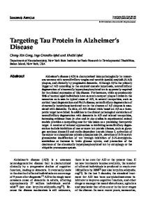
triplet of abnormal tau species, with a high molecular mass (tau 60, 64, 68 according to their molecular weight), were specifically detected in AD brains. After dephosphorylation, these abnormal tau species recovered their predicted mass (i.e. without phosphorylation) between 55 and 64 kDa (Flament et al., 1989; Goedert et al., 1995). Two-dimensional (2D) gels and mass spectrometry analyses confirmed that AD tau are more phosphorylated (Goedert et al., 1992; Hasegawa et al., 1992), and therefore more acidic on 2D gels, due to additional negative phosphate charges (Sergeant et al., 1995). A fourth abnormal tau component was discovered later on, of 72/74 kDa (Sergeant et al., 1997a). From these results, it was concluded that abnormal phosphorylation was responsible for the collapse of microtubules, and was the initiator of tau aggregation. The six tau isoforms are involved in the PHF structure (Sergeant et al., 1997a; Goedert and Klug, 1999). 9.2.6. From tau to tauopathies: the bar-code of tauopathies In neurodegenerative disorders, other than AD in which tangles are present, the comparison of tau aggregates shows that they differ in both phosphorylation and content of tau isoforms. These differences are the basis of the molecular classification of tauopathies (Pasquier and Delacourte, 1998). Five classes of tauopathies have been defined based on the type of tau aggregates that constitute the “barcode” of neurodegenerative disorders (Fig. 9.1) (Sergeant et al., 2005). 9.2.6.1. Class I: a major tau triplet at 60, 64, 68 Class I is characterized by a pathological tau triplet at 60, 64 and 68 kDa and a minor pathological tau at 72 kDa (molecular mass according to the literature) (Fig. 9.1). It is now well established that this pathological tau triplet corresponds to the aggregation of the six tau isoforms (Goedert et al., 1992; Sergeant et al., 1997a). The prototypical neurological disorder of this class is AD, but it includes additional neurological disorders such as amyotrophic lateral sclerosis parkinsonism-dementia complex of Guam , postencephalitic parkinsonism, dementia pugilistica, Down’s syndrome, Niemann-Pick disease type C, familial British dementia as well as some forms of fronto-temporal dementia with parkinsonism linked to chromosome 17 (FTDP-17) (reviewed in Buee and Delacourte, 1999). This type of tauopathy is also systematically found in the entorhinal formation of people aged over 75 years, and frequently found in addition in the hippocampal formation, with or without amyloid deposition (Delacourte et al., 1999).
Comp. by: GVasenthan Date:7/11/07 Time:15:17:32 Stage:First Proof File Path://spiina1001z/ womat/production/PRODENV/0000000001/0000006536/0000000016/0000747757.3D Proof by: QC by: ProjectAcronym:bs:DUY Volume:01209
TAU AS A BIOLOGICAL MARKER OF ALZHEIMER’S DISEASE
97
Bar code of Tauopathies τ72 τ 68 τ 64 τ 60
Aggregates A
Type I
Type II
Type III
Type IV
Tauopathies: 5 Classes Class 0
• Frontal lobe dementia non-AD, non-Pick (DLDH)
• • • • • • • • • •
Cerebral aging (indivuduals aged over 75 years) Alzheimer’s disease (sporadic and familial) ALS/parkinsonism-dementia complex of Guam Parkinson with dementia of Guadeloupe Niemann-Pick disease type C Postencephalitic parkinsonism Familial British dementia Dementia pugilistica Down’s syndrome FTDP-17
Class 2
• • • •
Corticobasal degeneration Progressive supranuclear palsy FTDP-17 Argyrophilic grain dementia
Class 3
Class 4
Class 1
Loss of tau protein expression Aggregated proteins:
Tau triplet: Type I Tau 60, 64 and 69
Type II
Upper tau doublet: Tau 64 and 69
• Pick’s disease • FTDP-17
Type III
Lower tau doublet: Tau 60 and 64
• Myotonic dystrophy of type I and II
Type IV
Tau 60
B Fig. 9.1 The biochemical signatures of the tauopathies. Direct immunostaining of tau proteins from brain homogenates by the phospho-dependent monoclonal antibody AD2, revealing the different patterns of immunopositive bands which can be compared to barcodes specific to or highly suggestive of specific disorders. Classification of the tauopathies by the patterns of tau Western blots (“barcodes”).
9.2.6.2. Class II: a major tau doublet at 64 and 69 kDa; 4R tauopathies The class II profile is specifically characterized by the presence of an upper doublet of aggregated tau proteins resulting from the aggregation of 4R tau isoforms (Fig. 9.1). This pathological tau profile is observed in progressive supranuclear palsy (PSP) (Flament et al., 1991; Vermersch et al., 1994), corticobasal degeneration (CBD) (Sergeant et al., 1999), atypical parkinsonism from Guadaloupe, argyrophylic grain (AGD) (Maurage et al., 2003b) and some forms of frontotem-
poral dementia with parkinsonism linked to chromosome 17 (FTDP-17) (Goedert, 2005). 9.2.6.2.1. Progressive supranuclear palsy Progressive supranuclear palsy (PSP) is a late-onset atypical parkinsonism disorder described by Steele, Richardson, and Olszewski in 1964 (Steele, 1994), in which dementia is also a common feature (Litvan, 2001, 2004). Neuropathologically, PSP is characterized by neuronal loss, gliosis, and NFT formation. Neurofibrillary tangles were first described in the basal
Comp. by: GVasenthan Date:7/11/07 Time:15:17:33 Stage:First Proof File Path://spiina1001z/ womat/production/PRODENV/0000000001/0000006536/0000000016/0000747757.3D Proof by: QC by: ProjectAcronym:bs:DUY Volume:01209
98
A. DELACOURTE
ganglia, brain stem, and cerebellum (Jellinger et al., 1980). Later on, the degenerating process was also found in the prefrontal cortex, with the same features as subcortical NFT (Hauw et al., 1990; Hof et al., 1992). Glial fibrillary tangles, also called astrocytic tufts, are thought to be quite characteristic of the disease (Bergeron et al., 1997). Biochemical studies show that aggregated tau in PSP is widely distributed in subcortical and neocortical areas, but with a major involvement of the subcortico-frontal neuronal networks (Vermersch et al., 1994). 9.2.6.2.2. Corticobasal degeneration Corticobasal degeneration (CBD) was first described in 1967 and referred to as corticodentatonigral degeneration with neuronal achromasia (Rebeiz et al., 1968). It is a rare, sporadic and slowly progressive late-onset neurodegenerative disorder clinically characterized by cognitive disturbances and extrapyramidal motor dysfunction. Dementia may occur early or late in the course of the disease (Rinne et al., 1994). There is a clinical and pathological overlap between PSP and CBD (Ikeda et al., 1994; Litvan, 1997, 2001). Neuropathological examination reveals severe glial and neuronal abnormalities. The glial pathology consists of astrocytic plaques and numerous tauimmunoreactive inclusions in the white matter, while glial pathology in PSP is characterized by tufted astrocytes. Achromatic ballooned neurons are detected in the cortex, brainstem, and subcortical structures as well as neuritic changes and NFT. In both PSP and CBD, the pathological tau profile consists essentially of the aggregation of 4R tau isoforms (Sergeant et al., 1999). The neocortical distribution of tau pathology is similar in CBD and PSP (Delacourte, 2001). 9.2.6.2.3. Argyrophilic grain disease In 1987, Braak and Braak (Braak and Braak, 1998) reported a series of eight patients with a non-AD, late-onset dementia. Clinically, argyrophilic grain disease (AGD) is associated with behavioral disturbances such as personality changes, memory and cognitive impairment (Braak and Braak, 1987) (see Ch. 48 by Ikeda in this volume). At the neuropathological level, AGD is characterized by the occurrence of grains stained by the Gallyas method, a silver technique – hence the term of argyrophilic grains (ArG). ArGs are minute, spindle or comma-shaped, argyrophilic, tau-immunoreactive structures distinct from neuropil threads and predominantly located in the hippocampus and related limbic areas (Braak and Braak, 1989). The 4R/3R ratio has been shown to be increased in AGD, thus demonstrating that AGD is mostly a 4R tau
pathology (Togo et al., 2002; Tolnay and Clavaguera, 2004). The tau pathology affects essentially the limbic system but, more recently, it has been shown to also involve, in some cases, all the cerebral cortex, even areas distant from the limbic temporal region, which are usually spared in AGD (Maurage et al., 2003a) – see Ch. 49 by Tolnay and Probst. As in PSP and CBD, the two other 4R tauopathies, the subthalamic nuclei are selectively involved by tau aggregates in AGD patients (Mattila et al., 2002). The H1/H1 haplotype is more prevalent in PSP/CBD patients than in controls or in patients with other tauopathies (Hutton, 2000). H1/H1 may also be more frequent in AGD patients than in controls, though recent studies have failed to establish a significant statistical difference (Ishizawa et al., 2002; Miserez et al., 2003). 9.2.6.3. Class III: a major tau doublet at 60 and 64 kDa; 3R tauopathies This class of tauopathy only includes Pick’s disease (PiD). It is a rare form of neurodegenerative disorder characterized by a progressive dementing process. Early in the clinical course, patients show signs of frontal disinhibition or severe apathy (Brion et al., 1991). Neuropathologically, Pick’s disease is characterized by prominent fronto-temporal lobar atrophy, gliosis, severe neuronal loss, ballooned neurons and the presence of neuronal inclusions called Pick bodies (Constantinidis et al., 1974) – see Ch. 35 by Uchihara and Tsuchiya. Pick bodies are labeled by tau antibodies, with a higher density in the hippocampus than in the neocortex (Hof et al., 1994). The laminar distribution of Pick bodies is clearly different from other tauopathies such as PSP and CBD. In the hippocampus, Pick bodies are numerous in granular cell neurons of the dentate gyrus, in CA1, subiculum and entorhinal cortex, whereas in the neocortex they are mainly found in layers II and VI of the temporal and frontal lobes. Ultrastructurally, Pick bodies consist of an accumulation of both random coiled and straight filaments. Biochemical analysis using a quantitative Western blot approach with phosphorylation-dependent anti-tau antibodies has revealed that, in all cases of PiD studied, a major 60- and 64-kDa pathological tau doublet is observed in the isocortex, in the limbic areas and in subcortical nuclei (Bue´e Scherrer et al., 1996; Delacourte et al., 1996). A faint pathological tau band is observed at 69 kDa (Sergeant et al., 1997b). The specific pathological tau profile of PiD is due to the aggregation of 3R tau isoforms (Delacourte et al., 1998; de Silva et al., 2006). In addition, aggregated tau proteins in PiD are not detected by the monoclonal antibody 12E8 raised against the phosphorylated resi-
Comp. by: GVasenthan Date:7/11/07 Time:15:17:33 Stage:First Proof File Path://spiina1001z/ womat/production/PRODENV/0000000001/0000006536/0000000016/0000747757.3D Proof by: QC by: ProjectAcronym:bs:DUY Volume:01209
TAU AS A BIOLOGICAL MARKER OF ALZHEIMER’S DISEASE due Ser262/Ser356, contrarily to what is observed in other neurodegenerative disorders (Delacourte et al., 1996; Probst et al., 1996). The lack of phosphorylation at Ser262 and -356 sites is likely to be related to an inhibition or to an absence of a particular kinase in the neurons that degenerate in PiD (Mailliot et al., 1998). The tauopathy in PiD, as studied by Western blotting of samples from multiple cortical areas, preferentially involves the fronto-temporal regions, in accordance with the clinical information and the neuropathological data. In some cases, there is also a strong involvement of the parietal cortex.
9.2.6.4. Class IV: a major tau 60 Class IV is represented by a single neurological disorder: type I myotonic dystrophy (MD), the commonest form of adult-onset muscular dystrophy. It is a multisystemic disease affecting many systems as well as the central nervous system (cognitive and neuropsychiatric impairment), thus leading to a wide and variable complex of symptoms (Harper and Reardon, 1992). At the clinical level, MD includes two entities designated type I (MD1) and type II (MD2) (Udd et al., 2003). MD1, the commonest form, is an inherited autosomal dominant disorder caused by a single gene mutation consisting of an expansion of a CTG trinucleotide motif in the 3V untranslated region of the myotonic dystrophy protein kinase gene (MDPK), located on chromosome 19q (Brook et al., 1992). Neuropathological lesions, such as neurofibrillary tangles, have been observed in adult MD1 individuals aged over 50 (Yoshimura et al., 1990; Vermersch et al., 1996). The tau profile of MD1 is characterized by a strong pathological tau band at 60 kDa and, to a lesser extent, pathological tau components at 64 and 69 kDa. This typical tau profile is associated with a reduced number of tau isoforms expressed in the brain of individuals with MD1, at both the protein and mRNA levels (Sergeant et al., 2001). In addition, tau protein expression is also demonstrated to be altered in transgenic mice with human MD1 locus (Seznec et al., 2001). The analysis of multiple brain regions of one genetically confirmed MD2 patient at the age of 71 years showed some neurofibrillary degenerating processes. Using specific immunological probes against the amino acid sequences corresponding to exon 2 and exon 3, the neurofibrillary lesions were shown to be devoid of tau isoforms with N-terminal inserts (Seznec et al., 2001). An altered splicing of tau characterized by a reduced expression of tau isoforms containing the N-terminal inserts characterizes both MD1 and MD2 (Maurage et al., 2005). These results show that MDs are also tauopathies and suggest that
99
tau alteration is involved in the cognitive deficit that had long been reported (Modoni et al., 2004). 9.2.6.5. A diversity of tauopathies in FTDP-17, a familial dementia In 1994, Wilhelmsen and colleagues described familial cases of fronto-temporal dementia (FTD), of autosomal dominant transmission, characterized by adultonset behavioral disturbances, frontal lobe dementia, parkinsonism and amyotrophy. They demonstrated a genetic linkage between this pathology, denominated disinhibition-dementia-parkinsonism amyotrophy complex (DDPAC), and chromosome 17q21–22 (Wilhelmsen et al., 1994). Since then, several families sharing strong clinical and pathological features and for which there is a linkage with chromosome 17q21–22 have been described (Wijker et al., 1996; Bird et al., 1997; Heutink et al., 1997; Murrell et al., 1997) and are referred to as having frontotemporal dementia with parkinsonism linked to chromosome 17 (FTDP-17) (Foster et al., 1997). Although there is clinical heterogeneity between and within the families with FTDP-17, the usual symptoms include behavioral changes, loss of frontal executive function, language disturbances and hyperorality. Parkinsonism and amyotrophy are described in some families, but are not consistent features. Neuropathologically, most brains of FTDP-17 patients exhibit an atrophy of frontal and temporal lobes, variable neuronal loss and gliosis, and a superficial laminar spongiosis. In many cases, tau proteins accumulate in neurons and glia (see Ch. 34 by Munoz and Ferrer). Amyloid deposits are usually absent (Foster et al., 1997; Spillantini et al., 1997, 1998). Finding tau gene mutations in affected individuals with FTDP-17 confirmed the role of tau aggregation in the pathogenesis of this disorder (Hutton et al., 1998; Spillantini et al., 1998; Grover et al., 1999).. To date, 35 mutations have been described in the tau gene in over 100 families (see Ch. 33 by Pickering-Brown and Hutton). Some mutations reduce the ability of tau protein to interact with microtubules and increase its propensity to assemble into abnormal filaments and others have their primary effect on the RNA by disturbing the normal ratio of 3- to 4-repeat tau isoforms. All the mutations and their potential pathological effects are described in Goedert (2005), Sergeant et al. (2005) and Trojanowski and Lee (2005). Transgenic models of FTDP-17 have been developed. Mice with a mutated tau gene reproduce some of the clinical and neuropathological features of these diseases, confirming the causal role of tau mutations (Klein et al., 2004; Zhang et al., 2004; Ikeda et al., 2005).
Comp. by: GVasenthan Date:7/11/07 Time:15:17:34 Stage:First Proof File Path://spiina1001z/ womat/production/PRODENV/0000000001/0000006536/0000000016/0000747757.3D Proof by: QC by: ProjectAcronym:bs:DUY Volume:01209
100
A. DELACOURTE
9.2.6.6. Tau disease without tangles: tau-less tauopathy or type 0 tauopathy FTD, as Pick complex (Kertesz and Munoz, 2002), is a clinical syndrome rather than a disease. The clinical phenotype resulting from frontotemporal degeneration is caused by different degenerating processes, PiD or CBD for instance. The neuropathology of some cases of FTD lacks distinctive histopathological hallmarks, such as NFT. In some of these cases, a huge decrease (from 60 to 90%) of normal tau expression may be shown by biochemical means (Zhukareva et al., 2001). This drop in tau expression has not yet been explained. Mutations in the progranulin gene are responsible for FTD with ubiquitin (tau-negative) inclusions – also called FTD-U (Baker et al., 2006; Cruts et al., 2006).
9.3. Progression of tau pathology in Alzheimer’s disease and aging 9.3.1. The Braak stages Using silver staining on large tissue sections, Heiko and Eva Braak were able to describe the progressive spreading of neurofibrillary degeneration, from the entorhinal and hippocampal areas towards plurimodal association cortices and finally primary areas (Braak and Braak, 1996). Dementia is usually apparent when a threshold of tangle density is reached in the plurimodal association cortical areas, corresponding to stages IV to VI (Braak and Braak, 1991). This staging was incorporated in the criteria for the definite diagnosis of AD in 1997 (Consensus_Report, 1997). 9.3.2. The spatio-temporal biochemical pathway of tau pathology in aging and sporadic AD The Braak stages, describing the histopathology of AD, were corroborated with biochemical means, using Western blot abnormal tau profile as a probe to quantify neurofibrillary degeneration. In a study involving 130 cases, including 60 non-demented patients, documented clinically and pathologically, we performed Western blots of various areas using the triplet of abnormal tau proteins as a marker. We found that the progression of tau pathology could be described in ten stages, corresponding to ten brain areas that are successively affected. Paired helical filaments (PHF)tau pathology was systematically found to be present in variable amounts in the entorhinal and hippocampal regions of non-demented patients aged over 75 years. When tau pathology was found in other brain areas, it appeared to progress along a sequential and hierarchical pathway in accordance with Braak stages: transentorhinal cortex (S1), entorhinal cortex (S2),
hippocampus (S3), anterior temporal cortex (S4), inferior temporal cortex (S5), mid temporal cortex (S6), polymodal association areas (prefrontal, parietal inferior, temporal superior) (S7), unimodal areas (S8), primary motor (S9a) or sensory (S9b, S9c) areas, and all neocortical areas (S10) (Delacourte et al., 1999). This progression pattern correlated with the sequence of cognitive impairment: from amnesia (involvement of the hippocampal formation) to aphasia (temporal cortex), apraxia and agnosia (other polymodal association areas). 9.3.3. Relationship with aging Abnormal tau may be detected in all patients over 75 years of age at least in the entorhinal area, and frequently in the hippocampal formation, suggesting that tau pathology is inevitable in the human. A high vulnerability of the same areas has also been found in a few non-human species, i.e., the baboon and rhesus monkey (Hartig et al., 2000; Schultz et al., 2000). In humans, neurofibrillary degeneration may be detected in young cases (Braak and Braak, 1997). Using the same histological approach and antibodies against specific tau phosphorylated sites such as Ser199P, we also observed that tau pathology could be present at a young age (Maurage et al., 2003a). Together, these studies show that a mild tauopathy can be observed in two out of ten patients at the second to third decade of age. At the age of 50, as many as half of the cases may have a small but significant entorhinal tau pathology. The prevalence of the lesions increases with age and they are constant at the age of 75 years, whatever the technique used to reveal them, either histochemical or biochemical. However, tau pathology may remain limited, even in centenarians (personal observation). Finding the explanation for the selective vulnerability of the entorhinal and hippocampal areas will certainly give clues for efficient neuroprotection (Morrison et al., 1998). 9.3.4. The burden of tau and Ab pathology in mild cognitive impairment There is no clinical method to determine whether a patient with mild cognitive impairment (MCI) has incipient AD. A variable percentage of MCI cases will convert to AD, but not all. We have prospectively collected data on the cognitive status as well as on the extent of tau and Ab pathology in patients with MCI (Delacourte et al., 1999, 2002). We observed that tau pathology, from tau stage S3 to S6, could be detected in all 13 MCI patients from our brain bank but that some were devoid of Ab pathology. Furthermore, all
Comp. by: GVasenthan Date:7/11/07 Time:15:17:34 Stage:First Proof File Path://spiina1001z/ womat/production/PRODENV/0000000001/0000006536/0000000016/0000747757.3D Proof by: QC by: ProjectAcronym:bs:DUY Volume:01209
TAU AS A BIOLOGICAL MARKER OF ALZHEIMER’S DISEASE patients from our prospective study with mild tau pathology did not have MCI, probably because the tau burden in these patients was compensated by neuronal plasticity. These results are in perfect agreement with those of several groups (Guillozet et al., 2003; Bennett et al., 2005; Markesbery et al., 2006) showing that tau pathology is more closely related to cognitive impairment than is Ab. These studies show that the substrate of MCI is a tau pathology in the limbic system. Since Ab aggregation is the inevitable correlate of AD, the presence of Ab deposits in the brain of MCI patients could reflect a more advanced stage and be related to prodromal AD. Interestingly, it has been shown that the simultaneous decrease of Ab x-42 and increase of tau and phospho-tau in the CSF are markers of incipient AD, in good agreement with the neuropathological features of MCI (Blennow, 2004). 9.3.5. The dementia threshold All our patients at stage S7 had different degrees of cognitive impairment and a significant vascular pathology was generally associated in those who were fully demented. Vascular lesions lower the threshold of AD pathology causing dementia, maybe because they have a deleterious effect on neuronal plasticity of the spared neurons. These observations strengthen the idea that clinical impairment results from an imbalance between a progressing degenerating process and decreasing compensatory effects from spared neurons. 9.3.6. The mechanism of progression of tau pathology The progression of tau pathology in the various areas of the cortex and in the subcortical nuclei seems to follow neuronal connections from the limbic cortex to the neocortical association areas in AD. A non-random progression of tau pathology is observed in other sporadic tauopathies, such as PSP, in which the brain stem, striatum, primary motor frontal neocortical area (Broadmann area 4), the unimodal frontal areas and finally all neocortical and limbic areas (Sergeant et al., 1999) are involved, although the exact sequence is unknown. These data are compatible with a “domino effect” in sporadic tauopathies: tau pathology first affects a specific vulnerable neuronal population such as layer II of the entorhinal formation in AD; or perhaps the oculomotor nuclei for PSP. This local tauopathy then destabilizes the connected neuronal populations which, in turn, develop tau pathology and destabilize the neurons to which they are connected. In this way, the pathology progresses from
101
neuron to neuron (Delacourte, 2000). A disorganization of the cortico-cortical circuitry has been shown in a transgenic AßPP mouse (Delatour et al., 2004). Improving our knowledge of the mechanism of propagation of tau pathology could probably open new therapeutic strategies for AD as well as for the other sporadic tauopathies. 9.3.7. The cause of the spreading of tauopathy in Alzheimer’s disease Tau pathology, being linked with neurodegeneration, is well correlated with cognitive impairment. However, in AD, tau pathology is probably secondary. The primary factors that generate tauopathy and promote its extension in brain areas are still unknown. AbPP dysfunction is certainly involved, since mutations of that protein are responsible (although rarely) for full blown familial AD. AbPP dysfunction could promote tau pathology either through Ab neurotoxicity or through AbPP loss of function. 9.3.7.1. Ab neurotoxicity We have shown that Ab x-42 aggregates increase in quantity and heterogeneity in close parallelism to the extension of tau pathology. But, unexpectedly, there was no spatial overlap between Ab aggregation that was widespread and heterogeneously distributed in cortical areas, and tau pathology progressing sequentially, stereotypically, and hierarchically (Delacourte et al., 2002). This observation suggests that APP dysfunction has a synergetic effect on the neuron-toneuron propagation of tau pathology. Indeed, tau pathology could be found in the hippocampal area without Ab deposits, as mentioned by Braak and Braak (1991), but when it is found in polymodal association areas it is systematically associated with Ab deposits, which appear, directly or indirectly, linked to the progression of tau pathology (Fig. 9.2). Altogether, our study demonstrated that amyloid deposits did not precede tau pathology in sporadic AD, as postulated in the amyloid cascade hypothesis (Hardy, 1992) which was developed from the observation of familial cases. 9.3.7.1. APP loss of function The parallelism and apparent synergy between tau pathology and Ab aggregation point to a molecular event linking the two degenerating processes. AbPP is a ubiquitous protein found in all species, suggesting a basic and important role that remains to be identified. A neurotrophic activity for AbPP and secreted sAbPP is often mentioned (Turner et al., 2003). A loss of function
Comp. by: GVasenthan Date:7/11/07 Time:15:17:35 Stage:First Proof File Path://spiina1001z/ womat/production/PRODENV/0000000001/0000006536/0000000016/0000747757.3D Proof by: QC by: ProjectAcronym:bs:DUY Volume:01209
102
A. DELACOURTE Pathway of tau pathology S0 Entorhinal cortex
S1 S2
Hippocampus
S3
Temporal pole
S4
APP*
S6
Inf temporal cortex
APP*
S5
Mid temporal cortex
Threshold for dementia
S7
APP*
Polymodal ass. ctx
S8 Unimodal areas
S9 APP*
S10 45 Biochemical staging of tauopathy in AD
55
65
75
Primary cortex 85
years
Fig. 9.2 Neocortical progression of tau pathology fueled by the weight of the APP/Ab burden.
of AbPP rather than a gain of toxic function of Ab could be an alternative hypothesis to explain the stimulation of tau pathology and neurodegeneration (Fig. 9.2). The concentration of C-terminal fragments of AbPP (AßCTFs) was found to be significantly decreased during the course of AD. This decrease was correlated with the progression of tau pathology (Sergeant et al., 2002). Alpha, beta, and gamma stubs (the carboxylterminal fragments of AßPP cleaved by the alpha, beta and gamma-secretases respectively). were also significantly decreased in the brain tissue of individuals having an inherited form of AD linked to mutations of presenilin 1, showing a general defect common to familial and sporadic forms of AD. The gamma stub, also named AICD (AbPP intracellular domain) could play a role as a gene regulator. Its deficit could have a deleterious effect on gene regulation (Cao and Sudhof, 2004). The concept of a loss of function of AßPP stimulating tau pathology led to new therapeutic strategies : “Ab may be a planet, but AbPP is central” (Neve and Robakis, 1998; Neve, 2001; Lee et al., 2004).
9.4. Tau as a biological marker Tau is detected in the CSF and its concentration may be evaluated by ELISA. Antibodies directed to phosphorylated epitopes may be used to assay a subset of tau (P-tau). This analysis increases the specificity and sensitivity of the diagnosis of AD in the range of 85% (Hampel et al., 2004). This biomarker is used in conjunction with Ab to increase the accuracy of the clinical diagnosis (Wiltfang et al., 2005; Maccioni et al., 2006). An index combining Ab, tau and phospho-tau levels in one single value has been shown to contribute significantly to the diagnosis accuracy of AD (Hulstaert et al.,
1999). The concentration of tau in the CSF is increased when tangles are present in the brain, but also in diseases which cause massive neuronal death (such as Creutzfeldt-Jakob disease) (Riemenschneider et al., 2003). On the other hand, the tau concentration is not increased in the CSF in PSP or CBD, in which the tauopathy may be severe (the interested reader is referred to Ch. 19, by Zetterberg and Blennow).
References Baker M, Mackenzie IR, Pickering-Brown SM, et al (2006). Mutations in progranulin cause tau-negative frontotemporal dementia linked to chromosome 17. Nature 442: 916–919. Bennett DA, Schneider JA, Bienias JL, et al (2005). Mild cognitive impairment is related to Alzheimer disease pathology and cerebral infarctions. Neurology 64: 834–841. Bergeron C, Pollanen MS, Weyer L, et al (1997). Cortical degeneration in progressive supranuclear palsy. A comparison with cortical-basal ganglionic degeneration. J Neuropathol Exp Neurol 56: 726–734. Bird TD, Wijsman EM, Nochlin D, et al (1997). Chromosome 17 and hereditary dementia: linkage studies in three non-Alzheimer families and kindreds with late-onset FAD. Neurology 48: 949–954. Blennow K (2004). CSF biomarkers for mild cognitive impairment. J Intern Med 256: 224–234. Braak H, Braak E (1987). Argyrophilic grains: characteristic pathology of cerebral cortex in cases of adult onset dementia without Alzheimer changes. Neurosci Lett 76: 124–127. Braak H, Braak E (1989). Cortical and subcortical argyrophilic grains characterize a disease associated with adult onset dementia. Neuropathol Appl Neurobiol 15: 13–26. Braak H, Braak E (1991). Neuropathological stageing of Alzheimer-related changes. Acta Neuropathol 82: 239–259. Braak H, Braak E (1996). Development of Alzheimerrelated neurofibrillary changes in the neocortex inversely
Comp. by: GVasenthan Date:7/11/07 Time:15:17:40 Stage:First Proof File Path://spiina1001z/ womat/production/PRODENV/0000000001/0000006536/0000000016/0000747757.3D Proof by: QC by: ProjectAcronym:bs:DUY Volume:01209
TAU AS A BIOLOGICAL MARKER OF ALZHEIMER’S DISEASE recapitulates cortical myelogenesis. Acta Neuropathol (Berl) 92: 197–201. Braak H, Braak E (1997). Frequency of stages of Alzheimerrelated lesions in different age categories. Neurobiol Aging 18: 351–357. Braak H, Braak E (1998). Argyrophilic grain disease: frequency of occurrence in different age categories and neuropathological diagnostic criteria. J Neural Transm 105: 801–819. Brion JP, Flament-Durand J, Dustin P (1986). Alzheimer’s disease and tau proteins. Lancet 2: 1098. Brion S, Plas J, Jeanneau A (1991). [Pick’s disease. Anatomo-clinical point of view]. Rev Neurol 147: 693–704. Brook JD, McCurrach ME, Harley HG, et al (1992). Molecular basis of myotonic dystrophy: expansion of a trinucleotide (CTG) repeat at the 30 end of a transcript encoding a protein kinase family member. Cell 69: 385. Buee L, Delacourte A (1999). Comparative biochemistry of tau in progressive supranuclear palsy, corticobasal degeneration, FTDP-17 and Pick’s disease. Brain Pathol 9: 681–693. Bue´e Scherrer V, Hof PR, Bue´e L, et al (1996). Hyperphosphorylated tau proteins differentiate corticobasal degeneration and Pick’s disease. Acta Neuropathol 91: 351–359. Cao X, Sudhof TC (2004). Dissection of amyloid-beta precursor protein-dependent transcriptional transactivation. J Biol Chem 279: 24601–24611. Consensus_Report (1997). Consensus recommendations for the postmortem diagnosis of Alzheimer’s disease. The National Institute on Aging, and Reagan Institute Working Group on Diagnostic Criteria for the Neuropathological Assessment of Alzheimer’s Disease. Neurobiol Aging 18: S1–2. Constantinidis J, Richard J, Tissot R (1974). Pick’s disease. Histological and clinical correlations. Eur Neurol 11: 208–217. Cruts M, Gijselinck I, van der Zee J, et al (2006). Null mutations in progranulin cause ubiquitin-positive frontotemporal dementia linked to chromosome 17q21. Nature 442: 920–924. Delacourte A (2000). The biochemical pathway of neurofibrillary degeneration in aging and Alzheimer’s disease. Neurology 54: 538. Delacourte A (2001). The molecular parameters of tau pathology. Tau as a killer and a witness. Adv Exp Med Biol 487: 5–19. Delacourte A, Defossez A (1986). Alzheimer’s disease: Tau proteins, the promoting factors of microtubule assembly, are major components of paired helical filaments. J Neurol Sci 76: 173–186. Delacourte A, Robitaille Y, Sergeant N, et al (1996). Specific pathological Tau protein variants characterize Pick’s disease. J Neuropathol Exp Neurol 55: 159–168. Delacourte A, Sergeant N, Wattez A, et al (1998). Vulnerable neuronal subsets in Alzheimer’s and Pick’s disease are distinguished by their tau isoform distribution and phosphorylation. Ann Neurol 43: 193–204.
103
Delacourte A, David JP, Sergeant N, et al (1999). The biochemical pathway of neurofibrillary degeneration in aging and Alzheimer’s disease. Neurology 52: 1158–1165. Delacourte A, Sergeant N, Champain D, et al (2002). Nonoverlapping but synergetic tau and APP pathologies in sporadic Alzheimer’s disease. Neurology 59: 398–407. Delatour B, Blanchard V, Pradier L, et al (2004). Alzheimer pathology disorganizes cortico-cortical circuitry: direct evidence from a transgenic animal model. Neurobiol Dis 16: 41–47. de Silva R, Lashley T, Strand C, et al (2006). An immunohistochemical study of cases of sporadic and inherited frontotemporal lobar degeneration using 3R- and 4Rspecific tau monoclonal antibodies. Acta Neuropathol (Berl) 111(4): 329–340. Flament S, Delacourte A, Hemon B, et al (1989). Characterization of two pathological tau protein variants in Alzheimer brain cortices. J Neurol Sci 92: 133–141. Flament S, Delacourte A, Verny M, et al (1991). Abnormal Tau proteins in progressive supranuclear palsy. Similarities and differences with the neurofibrillary degeneration of the Alzheimer type. Acta Neuropathol 81: 591–596. Foster NL, Wilhelmsen K, Sima AA, et al (1997). Frontotemporal dementia and parkinsonism linked to chromosome 17: a consensus conference. Conference Participants. Ann Neurol 41: 706–715. Ghatak NR, Nochlin D, Hadfield MG (1980). Neurofibrillary pathology in progressive supranuclear palsy. Acta Neuropathol (Berl) 52: 73–76. Glenner GG, Wong CW (1984). Alzheimer’s disease: initial report of the purification and characterization of a novel cerebrovascular amyloid protein. Biochem Biophys Res Commun 120: 885–890. Goedert M (2005). Tau gene mutations and their effects. Mov Disord 20(Suppl 12): S45–52. Goedert M, Klug A (1999). Tau protein and the paired helical filament of Alzheimer’s disease. Brain Res Bull 50: 469–470. Goedert M, Spillantini MG, Cairns NJ, et al (1992). Tau proteins of Alzheimer paired helical filaments: abnormal phosphorylation of all six brain isoforms. Neuron 8: 159–168. Goedert M, Jakes R, Qi Z, et al (1995). Protein phosphatase 2A is the major enzyme in brain that dephosphorylates tau protein phosphorylated by proline-directed protein kinases or cyclic AMP-dependent protein kinase. J Neurochem 65: 2804–2807. Grover A, Houlden H, Baker M, et al (1999). 50 splice site mutations in tau associated with the inherited dementia FTDP-17 affect a stem-loop structure that regulates alternative splicing of exon 10. J Biol Chem 274: 15134–15143. Grundke-Iqbal I, Iqbal K, Quinlan M, et al (1986a). Microtubule-associated protein tau. A component of Alzheimer paired helical filaments. J Biol Chem 261: 6084–6089. Grundke-Iqbal I, Iqbal K, Tung YC, et al (1986b). Abnormal phosphorylation of the microtubule-associated protein tau (tau) in Alzheimer cytoskeletal pathology. Proc Natl Acad Sci U S A 83: 4913–4917.
Comp. by: GVasenthan Date:7/11/07 Time:15:17:44 Stage:First Proof File Path://spiina1001z/ womat/production/PRODENV/0000000001/0000006536/0000000016/0000747757.3D Proof by: QC by: ProjectAcronym:bs:DUY Volume:01209
104
A. DELACOURTE
Guillozet AL, Weintraub S, Mash DC, et al (2003). Neurofibrillary tangles, amyloid, and memory in aging and mild cognitive impairment. Arch Neurol 60: 729–736. Hampel H, Teipel SJ, Fuchsberger T, et al (2004). Value of CSF beta-amyloid1–42 and tau as predictors of Alzheimer’s disease in patients with mild cognitive impairment. Mol Psychiatry 9: 705–710. Hardy J (1992). An ‘anatomical cascade hypothesis’ for Alzheimer’s disease. Trends Neurosci 15: 200–201. Hardy JA, Higgins GA (1992). Alzheimer’s disease: the amyloid cascade hypothesis. Science 256: 184–185. Hardy J, Selkoe DJ (2002). The amyloid hypothesis of Alzheimer’s disease: progress and problems on the road to therapeutics. Science 297: 353–356. Harper PS, Reardon W (1992). Heart disease in myotonic dystrophy. Lancet 339: 939. Hartig W, Klein C, Brauer K, et al (2000). Abnormally phosphorylated protein tau in the cortex of aged individuals of various mammalian orders. Acta Neuropathol (Berl) 100: 305–312. Hasegawa M, Morishima-Kawashima M, Takio K, et al (1992). Protein sequence and mass spectrometric analyses of tau in the Alzheimer’s disease brain. J Biol Chem 267: 17047–17054. Hauw JJ, Verny M, Delaere P, et al (1990). Constant neurofibrillary changes in the neocortex in progressive supranuclear palsy. Basic differences with Alzheimer’s disease and aging. Neurosci Lett 119: 182–186. Heutink P, Stevens M, Rizzu P, et al (1997). Hereditary frontotemporal dementia is linked to chromosome 17q21-q22: a genetic and clinicopathological study of three Dutch families. Ann Neurol 41: 150–159. Hof PR, Delacourte A, Bouras C (1992). Distribution of cortical neurofibrillary tangles in progressive supranuclear palsy: a quantitative analysis of six cases. Acta Neuropathol 84: 45–51. Hof PR, Bouras C, Perl DP, et al (1994). Quantitative neuropathologic analysis of Pick’s disease cases: cortical distribution of Pick bodies and coexistence with Alzheimer’s disease. Acta Neuropathol (Berl) 87: 115–124. Hof PR, Perl DP, Loerzel AJ, et al (1994). Amyotrophic lateral sclerosis and parkinsonism-dementia from Guam: differences in neurofibrillary tangle distribution and density in the hippocampal formation and neocortex. Brain Res 650: 107–116. Hulstaert F, Blennow K, Ivanoiu A, et al (1999). Improved discrimination of AD patients using beta-amyloid(1–42) and tau levels in CSF. Neurology 52: 1555–1562. Hutton M (2000). Molecular genetics of chromosome 17 tauopathies. Ann N Y Acad Sci 920: 63–73. Hutton M, Lendon CL, Rizzu P, et al (1998). Association of missense and 50 -splice-site mutations in tau with the inherited dementia FTDP-17. Nature 393: 702–705. Ikeda K, Akiyama H, Haga C, et al (1994). Argyrophilic thread-like structure in corticobasal degeneration and supranuclear palsy. Neurosci Lett 174: 157–159. Ikeda M, Shoji M, Kawarai T, et al (2005). Accumulation of filamentous tau in the cerebral cortex of human tau R406W transgenic mice. Am J Pathol 166: 521–531.
Ishizawa T, Ko LW, Cookson N, et al (2002). Selective neurofibrillary degeneration of the hippocampal CA2 sector is associated with four-repeat tauopathies. J Neuropathol Exp Neurol 61: 1040–1047. Jellinger K, Riederer P, Tomonaga M (1980). Progressive supranuclear palsy: clinico-pathological and biochemical studies. J Neural Transm Suppl 16: 111–128. Joachim CL, Morris JH, Kosik KS, et al (1987). Tau antisera recognize neurofibrillary tangles in a range of neurodegenerative disorders. Ann Neurol 22: 514–520. Kato S, Nakamura H, Otomo E (1989). Reappraisal of neurofibrillary tangles. Immunohistochemical, ultrastructural, and immunoelectron microscopical studies. Acta Neuropathol 77: 258–266. Kertesz A, Munoz DG (2002). Primary progressive aphasia: a review of the neurobiology of a common presentation of Pick complex. Am J Alzheimers Dis Other Demen 17: 30–36. Klein RL, Lin WL, Dickson DW, et al (2004). Rapid neurofibrillary tangle formation after localized gene transfer of mutated tau. Am J Pathol 164: 347–353. Kosik KS, Joachim CL, Selkoe DJ (1986). Microtubuleassociated protein tau (tau) is a major antigenic component of paired helical filaments in Alzheimer disease. Proc Natl Acad Sci USA 83: 4044–4048. Lee HG, Casadesus G, Zhu X, et al (2004). Challenging the amyloid cascade hypothesis: senile plaques and amyloidbeta as protective adaptations to Alzheimer disease. Ann N Y Acad Sci 1019: 1–4. Litvan I (2001). Diagnosis and management of progressive supranuclear palsy. Semin Neurol 21: 41–48. Litvan I (2004). Update on progressive supranuclear palsy. Curr Neurol Neurosci Rep 4: 296–302. Litvan I, Agid Y, Goetz C, et al (1997). Accuracy of the clinical diagnosis of corticobasal degeneration: a clinicopathologic study. Neurology 48: 119–125. Love S, Bridges LR, Case CP (1995). Neurofibrillary tangles in Niemann-Pick disease type C. Brain 118: 119–129. Maccioni RB, Lavados M, Guillon M, et al (2006). Anomalously phosphorylated tau and Abeta fragments in the CSF correlates with cognitive impairment in MCI subjects. Neurobiol Aging 27: 237–244. Mailliot C, Sergeant N, Bussiere T, et al (1998). Phosphorylation of specific sets of tau isoforms reflects different neurofibrillary degeneration processes. FEBS Lett 433: 201–204. Markesbery WR, Schmitt FA, Kryscio RJ, et al (2006). Neuropathologic substrate of mild cognitive impairment. Arch Neurol 63: 38–46. Mattila P, Togo T, Dickson DW (2002). The subthalamic nucleus has neurofibrillary tangles in argyrophilic grain disease and advanced Alzheimer’s disease. Neurosci Lett 320: 81–85. Maurage CA, Sergeant N, Ruchoux MM, et al (2003a). Phosphorylated serine 199 of microtubule-associated protein tau is a neuronal epitope abundantly expressed in youth and an early marker of tau pathology. Acta Neuropathol (Berl) 105: 89–97.
Comp. by: GVasenthan Date:7/11/07 Time:15:17:47 Stage:First Proof File Path://spiina1001z/ womat/production/PRODENV/0000000001/0000006536/0000000016/0000747757.3D Proof by: QC by: ProjectAcronym:bs:DUY Volume:01209
TAU AS A BIOLOGICAL MARKER OF ALZHEIMER’S DISEASE Maurage CA, Sergeant N, Schraen-Maschke S, et al (2003b). Diffuse form of argyrophilic grain disease: a new variant of four-repeat tauopathy different from limbic argyrophilic grain disease. Acta Neuropathol (Berl) 106: 575–583. Maurage CA, Udd B, Ruchoux MM, et al (2005). Similar brain tau pathology in DM2/PROMM and DM1/Steinert disease. Neurology 65: 1636–1638. Miserez AR, Clavaguera F, Monsch AU, et al (2003). Argyrophilic grain disease: molecular genetic difference to other four-repeat tauopathies. Acta Neuropathol (Berl) 106: 363–366. Mitsuyama Y, Seyama S (1981). Frequency of Alzheimer’s neurofibrillary tangle in the brains of progressive supranuclear palsy, postencephalitic parkinsonism, Alzheimer’s disease, senile dementia and non-demented elderly person. Folia Psychiatr Neurol Jpn 35: 189–204. Modoni A, Silvestri G, Pomponi MG, et al (2004). Characterization of the pattern of cognitive impairment in myotonic dystrophy type 1. Arch Neurol 61: 1943–1947. Morrison BM, Hof PR, Morrison JH (1998). Determinants of neuronal vulnerability in neurodegenerative diseases. Ann Neurol 44: S32–44. Murrell JR, Koller D, Foroud T, et al (1997). Familial multiple-system tauopathy with presenile dementia is localized to chromosome 17. Am J Hum Genet 61: 1131–1138. Neve RL (2001). A beta may be a planet, but APP is central. Neurobiol Aging 22: 151–154. Neve RL, Robakis NK (1998). Alzheimer’s disease: a reexamination of the amyloid hypothesis. Trends Neurosci 21: 15–19. Pasquier F, Delacourte A (1998). Non-Alzheimer degenerative dementias. Curr Opin Neurol 11: 417–427. Probst A, Tolnay M, Langui D, et al (1996). Pick’s disease: hyperphosphorylated tau protein segregates to the somatoaxonal compartment. Acta Neuropathol (Berl) 92: 588–596. Rebeiz JJ, Kolodny EH, Richardson EP Jr (1968). Corticodentatonigral degeneration with neuronal achromasia. Arch Neurol 18: 20–33. Riemenschneider M, Wagenpfeil S, Vanderstichele H, et al (2003). Phospho-tau/total tau ratio in cerebrospinal fluid discriminates Creutzfeldt-Jakob disease from other dementias. Mol Psychiatry 8: 343–347. Rinne JO, Lee MS, Thompson PD, et al (1994). Corticobasal degeneration. A clinical study of 36 cases. Brain 117: 1183–1196. Schultz C, Hubbard GB, Rub U, et al (2000). Age-related progression of tau pathology in brains of baboons. Neurobiol Aging 21: 905–912. Sergeant N, Bussiere T, Vermersch P, et al (1995). Isoelectric point differentiates PHF-tau from biopsy-derived human brain tau proteins. Neuroreport 6: 2217–2220. Sergeant N, David JP, Goedert M, et al (1997a). Twodimensional characterization of paired helical filament-tau from Alzheimer’s disease: demonstration of an additional 74-kda component and age-related biochemical modifications. J Neurochem 69: 834–844.
105
Sergeant N, David JP, Lefranc D, et al (1997b). Different distribution of phosphorylated tau protein isoforms in Alzheimer’s and Pick’s diseases. FEBS Lett 412: 578–582. Sergeant N, Wattez A, Delacourte A (1999). Neurofibrillary degeneration in progressive supranuclear palsy and corticobasal degeneration: tau pathologies with exclusively “exon 10” isoforms. J Neurochem 72: 1243–1249. Sergeant N, Sablonniere B, Schraen-Maschke S, et al (2001). Dysregulation of human brain microtubule-associated tau mrna maturation in myotonic dystrophy type 1. Hum Mol Genet 10: 2143–2155. Sergeant N, David JP, Champain D, et al (2002). Progressive decrease of amyloid precursor protein carboxy terminal fragments (APP-ctfs), associated with tau pathology stages, in Alzheimer’s disease. J Neurochem 81: 663–672. Sergeant N, Delacourte A, Buee L (2005). Tau protein as a differential biomarker of tauopathies. Biochim Biophys Acta 1739: 179–197. Seznec H, Agbulut O, Sergeant N, et al (2001). Mice transgenic for the human myotonic dystrophy region with expanded CTG repeats display muscular and brain abnormalities. Hum Mol Genet 10: 2717–2726. Spillantini MG, Goedert M, Crowther RA, et al (1997). Familial multiple system tauopathy with presenile dementia: a disease with abundant neuronal and glial tau filaments. Proc Natl Acad Sci USA 94: 4113–4118. Spillantini MG, Bird TD, Ghetti B (1998). Frontotemporal dementia and Parkinsonism linked to chromosome 17: a new group of tauopathies. Brain Pathol 8: 387–402. Steele JC (1994). Progressive supranuclear palsy. Historical notes. J Neural Transm Suppl 42: 3–14. Terry RD (2000). Do neuronal inclusions kill the cell? J Neural Transm (Suppl 59): 91–93. Togo T, Sahara N, Yen SH, et al (2002). Argyrophilic grain disease is a sporadic 4-repeat tauopathy. J Neuropathol Exp Neurol 61: 547–556. Tolnay M, Clavaguera F (2004). Argyrophilic grain disease: a late-onset dementia with distinctive features among tauopathies. Neuropathology 24: 269–283. Tomonaga M (1977). Ultrastructure of neurofibrillary tangles in progressive supranuclear palsy. Acta Neuropathol (Berl) 37: 177–181. Trojanowski JQ, Lee VM (2005). Pathological tau: a loss of normal function or a gain in toxicity? Nat Neurosci 8: 1136–1137. Turner PR, O’Connor K, Tate WP, et al (2003). Roles of amyloid precursor protein and its fragments in regulating neural activity, plasticity and memory. Prog Neurobiol 70: 1–32. Udd B, Meola G, Krahe R, et al (2003). Report of the 115th ENMC workshop: DM2/PROMM and other myotonic dystrophies. 3rd Workshop, 14–16 February 2003, Naarden, The Netherlands. Neuromuscul Disord 13: 589–596. Vermersch P, Robitaille Y, Bernier L, et al (1994). Biochemical mapping of neurofibrillary degeneration in a case of progressive supranuclear palsy: evidence for general cortical involvement. Acta Neuropathol 87: 572–577.
Comp. by: GVasenthan Date:7/11/07 Time:15:17:52 Stage:First Proof File Path://spiina1001z/ womat/production/PRODENV/0000000001/0000006536/0000000016/0000747757.3D Proof by: QC by: ProjectAcronym:bs:DUY Volume:01209
106
A. DELACOURTE
Vermersch P, Sergeant N, Ruchoux MM, et al (1996). Specific tau variants in the brains of patients with myotonic dystrophy. Neurology 47: 711–717. \Wijker M, Wszolek ZK, Wolters EC, et al (1996). Localization of the gene for rapidly progressive autosomal dominant parkinsonism and dementia with pallido-ponto-nigral degeneration to chromosome 17q21. Hum Mol Genet 5: 151–154. Wilhelmsen KC, Lynch T, Pavlou E, et al (1994). Localization of disinhibition-dementia-parkinsonism-amyotrophy complex to 17q21–22. Am J Hum Genet 55: 1159–1165. Wiltfang J, Lewczuk P, Riederer P, et al (2005). Consensus paper of the WFSBP Task Force on Biological Markers of Dementia: the role of CSF and blood analysis in the early and differential diagnosis of dementia. World J Biol Psychiatry 6: 69–84. Wisniewski K, Jervis GA, Moretz RC, et al (1979). Alzheimer neurofibrillary tangles in diseases other than senile and presenile dementia. Ann Neurol 5: 288–294.
Wong CW, Quaranta V, Glenner GG (1985). Neuritic plaques and cerebrovascular amyloid in Alzheimer disease are antigenically related. Proc Natl Acad Sci USA 82: 8729–8732. Yamamoto T, Hirano A (1986). A comparative study of modified Bielschowsky, Bodian and thioflavin S stains on Alzheimer’s neurofibrillary tangles. Neuropathol Appl Neurobiol 12: 3–9. Yoshimura N, Otake M, Igarashi K, et al (1990). Topography of Alzheimer’s neurofibrillary change distribution in myotonic dystrophy. Clin Neuropathol 9: 234–239. Zhang B, Higuchi M, Yoshiyama Y, et al (2004). Retarded axonal transport of R406W mutant tau in transgenic mice with a neurodegenerative tauopathy. J Neurosci 24: 4657–4667. Zhukareva V, Vogelsberg-Ragaglia V, Van Deerlin VM, et al (2001). Loss of brain tau defines novel sporadic and familial tauopathies with frontotemporal dementia. Ann Neurol 49: 165–175.