INT. J. BIOAUTOMATION, 2015, 19(2), 223-236
Tangential Volumetric Modulated Radiotherapy – A New Technique for Large Scalp Lesions with a Case Study in Lentigo Maligna E. Daniel Santos1,2*, Julia A. Green1,2, Nastik Bhandari1,2, Angela Hong3,4,5, Pascale Guitera3,4,5, Gerald B. Fogarty1,2,3,4,5 1
Genesis Cancer Care Department of Radiation Oncology and Brachytherapy Lower Ground Floor, 25 Rocklands Road, Mater Hospital Crow’s Nest, New South Wales 2065, Australia E-mails:
[email protected],
[email protected],
[email protected],
[email protected] 2
Mater Sydney Radiation Oncology, Sydney, Australia
3
Melanoma Institute Australia, Sydney, Australia E-mails:
[email protected],
[email protected] 4
Sydney Medical School, The University of Sydney, Sydney, Australia
5
Australia and New Zealand Melanoma Trials Group, North Sydney, Australia
*
Corresponding author
Received: October 31, 2014
Accepted: June 18, 2015 Published: June 30, 2015
Abstract: Introduction: Dose homogeneity within and dose conformity to the target volume can be a challenge to achieve when treating large area scalp lesions. Traditionally High Dose Rate (HDR) brachytherapy (BT) scalp moulds have been considered the ultimate conformal therapy. We have developed a new technique, Tangential Volumetric Modulated Arc Therapy (TVMAT) that treats with the beam tangential to the surface of the scalp. In the TVMAT plan the collimating jaws protect dose-sensitive tissue in close proximity to the planning target volume (PTV). Not all the PTV is within the beam aperture as defined by the jaws during all the beam-on time. We report the successful treatment of one patient. Methods: A patient with biopsy proven extensive lentigo maligna on the scalp was simulated and three plans were created; one using a HDR brachytherapy surface mould, another using a conventional VMAT technique and a third using our new TVMAT technique. The patient was prescribed 55 Gy in 25 fractions. Plans were optimised so that PTV V 100% = 100%. Plans were compared using Dose-Value Histogram (DVH) analysis, and homogeneity and conformity indices. Results: BT, VMAT and TVMAT PTV median coverage was 105.51%, 103.46% and 103.62%, with homogeneity index of 0.33, 0.07 and 0.07 and the conformity index of 0.30, 0.69 and 0.83 respectively. The median dose to the left hippocampus was 11.8 Gy, 9.0 Gy and 0.6 Gy and the median dose to the right hippocampus was 12.6 Gy, 9.4 Gy and 0.7 Gy for the BT, VMAT and TVMAT respectively. Overall TVMAT delivered the least doses to the surrounding organs, BT delivered the highest. Conclusions: TVMAT was superior to VMAT which was in turn superior to BT in PTV coverage, conformity and homogeneity and delivery of dose to the surrounding organs at risk. The patient was successfully treated to full dose with TVMAT. TVMAT was verified as being the best amongst the three techniques in a second patient. Keywords: Lentigo, Maligna, Volumetric, Modulated, Radiotherapy, Brachytherapy.
223
INT. J. BIOAUTOMATION, 2015, 19(2), 223-236
Introduction Scalp mould brachytherapy (BT) is usually regarded as the gold standard radiotherapy treatment for large scalp areas [3, 5, 17]. However, planning is labour intensive and there is a concern for dose delivered to the brain. Intensity Modulated Radiation Therapy (IMRT) technologies like Volumetric Modulated Arc Therapy (VMAT) have improved dosimetric coverage and conformity to the PTV compared to conformal treatments [16]. Conventional external beam radiotherapy traditionally has the planning target volume (PTV) within the beam aperture at all times during treatment. We developed a new method of treating large scalp areas with an evolution of VMAT called Tangential VMAT (TVMAT). This new technique does not have the PTV within the beam at all times during treatment. We have treated two patients with this technique. In the first patient this new treatment was superior to BT and conventional VMAT in terms of dosimetry and efficiency. The second patient validated the technique superiority.
Methods A patient had a large area of biopsy proven recurrent scalp lentigo maligna and was referred for definitive radiation [10]. The physician outlined the scalp lesion as the clinical target volume (CTV) defined using Reflectance Confocal Microscopy (RCM). A margin of 1 cm was added for the PTV. 55 Gy in 25 fractions was prescribed to a depth of 5 mm from the surface [6].
Brachytherapy surface mould We followed our usual practice in creating a scalp brachytherapy mould. The patient was seated upright and a mould of the scalp was created by placing a conventional IMRT immobilization mask (Klarity Medical Products USA, Newark, Ohio). 8 mm of water equivalent build-up material (Lordell Trading Pty. Ltd., Wetherill Park, NSW, Australia) was then placed on a 20 mm margin around the PTV area as stand off to maximise the dose coverage of the BT plan. 19 BT catheters running inferiorly above the bolus were placed with a 10 mm separation (Fig. 1) from each other. An extra 5 mm of wax was placed on top of the catheters to aid in anchoring the catheters and to act as back-scatter build-up. The PTV area was marked on the inner-surface of the mask and wired using CT skin markers (Beekley Corp., Bristol, CT). The mould was CT scanned with 1mm slice thickness with the catheters perpendicular to the scanning plane. The scan was exported to the Oncentra Bracytherapy planning system version 4.3 (Nucletron, Veenedaal, Netherlands). The PTV was contoured as a volume within the area of the CT wires and 5 mm below the surface mould into the skin as this depth is necessary for treatment [6]. The 19 catheters were reconstructed and dwell positions were activated using the auto-activate feature in Oncentra including a 20 mm margin on the target volume with a 5 mm dwell position spacing to allow for sufficient PTV coverage at depth. The dwell positions were 13 mm from the treatment depth. This 13 mm was made up of 8 mm water equivalent material on skin acting as stand-off with treatment prescribed to depth of 5 mm into skin. The plan was optimised so that the V100% = 100% and the 100% isodose did not extend beyond the PTV volume. The dose algorithm used in the Oncentra planning system is based on the AAPM TG 43 dose formalism. The plan contained 492 active dwell positions with a total reference air kerma of 41.11 mGy·m2.
224
INT. J. BIOAUTOMATION, 2015, 19(2), 223-236
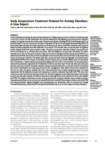
Fig. 1 Number 1: Brachytherapy surface mold: 1A – Lateral View, 1B – Superior View. With the patient sitting in an upright position, immobilization mask was placed over the scalp of the patient. 8 mm of water equivalent material (the red wax) was placed over the PTV area on the mask. 19 BT catheters were placed on the wax running inferiorly with a spacing approximately 1cm apart. A further 5 mm of wax was placed above the catheters to hold the catheters in place and to aid with back scatter. Number 2: VMAT and TVMAT immobilization mask with wax build-up: 2A – Lateral View, 2B – Superior View. With the patient supine, immobilization mask was placed over the scalp of the patient. 10 mm of water equivalent material (the red wax) was placed above the PTV area on the mask. A Nucletron MicroSelection HDR Brachytherapy Unit with an Iridium-192 source is used for treatment delivery. An additional 0.5cm of flexible bolus was placed above the mould as back-scatter material and quality assurance was then performed. Gafchromic EBT3 film (Ashland Inc., Covington, KY) was placed at various locations on the inner-surface of the mould. Phantom material (sandbags and rice) was placed into the mould and the treatment was delivered. The dose delivered to the Gafchromic film was obtained using a pre-calculated calibration curve and the measured results compared to the calculated doses from the plan.
VMAT An IMRT immobilization mask (Klarity Medical Products USA, Newark, Ohio) was created with the patient in the supine position. The PTV was outlined on the outer-surface and then on the inner-surface of the mask. A 10 mm water equivalent build-up material was moulded to the outside of the mask including a 40 mm lateral margin (Lordell Trading Pty. Ltd., Wetherill Park, NSW, Australia) (Fig. 1).
225
INT. J. BIOAUTOMATION, 2015, 19(2), 223-236
The patient was scanned (Scan 1) with the completed mask and bolus in the treatment position. The inner-surface of the mask was wired using CT skin markers (Beekley Corp., Bristol, CT) and scanned again (Scan 2) without the patient as per our usual protocol. Both scans were exported to Eclipse version 11 (Varian Medical Systems, Inc., Palo Alto, CA). The target volume, organs at risk and other parts of the patient’s anatomy were contoured on the Scan 1 CT scan and two VMAT plans were created. The first plan used conventional VMAT (cVMAT) treatment strategies for the head and neck region, which comprised of two complete arcs at the approximate geometric centre of the PTV which was used as the isocentre. Jaw positions for the two arcs were selected based on the size of the PTV in conjunction with the collimator rotations, of 20° and 340° for the two arcs respectively. No couch rotations or multiple isocentres were used to minimise the total treatment time. The plan was optimized using the Progressive Resolution Optimizer III in Eclipse (PROIII version 11.0) with the optimization target to achieve V100% = 100% PTV coverage and as low as achievable dose sparing to the organs at risk without sacrificing PTV coverage. At all times the full PTV was in the beam aperture as defined by the collimating jaws per conventional VMAT planning. Total MUs for the cVMAT plan was 1200 MUs.
TVMAT The second, tangential plan was created using 5 half-arcs around the patient’s head. The arcs were arranged so that all arcs would either start or complete with gantry at 0° and would rotate around either side of the patient. The field size set to an off-axis position such that at any one gantry angle during each half-arc could not treat the entire PTV. The beams eye view comparison can be seen in Fig. 3. Conventional VMAT is compared to TVMAT in Fig. 3. The jaws were also set based on which side of the patient the half-arc was. If the half-arc was on the patient’s right the jaws were set off-axis to the left; and to the right if the half-arc was on the patient’s left. The jaws were positioned so to minimize the dose to the organs at risk, particularly the brain. For arcs covering the forehead, the collimator was set very close to 0°. For arcs covering the crown of the head, the collimator was set to an angle closer to 45°. Thus the amount of time any single area is covered by the beam during an arc was controlled with the collimator rotation. No couch rotations or multiple isocentres were used to minimise the total treatment time. The plan was then optimized using the same conditions of V100% = 100% PTV coverage and as low as achievable dose sparing to the organs at risk without sacrificing PTV coverage. The Eclipse PROIII optimizer was used for plan optimisation. Total MUs for the TVMAT plan was 916 MUs.
Fig. 2 cVMAT (left) the beam center is perpendicular to the scalp and the jaws are opened to expose the entire PTV at its widest cross-section. TVMAT (right) the beam center is tangential to the scalp and the jaws are closed exposing only a small section of the PTV at any given time but exposes the entire PTV in an arc
226
INT. J. BIOAUTOMATION, 2015, 19(2), 223-236
Fig. 3 Beams Eye View of a conventional VMAT (top) versus a TVMAT (bottom) with the PTV (red) and Brain (blue) structures shown. The large yellow rectangles show the jaw position of each treatment, with the left image clearly irradiating more of the brain than the right image. Final dose calculations for both VMAT plans were done using the Anisotropic Analytical Algorithm (AAA version 11.0). The cVMAT plan was then normalized so that the PTV DVH matched the PTV dose value histogram (DVH) of the TVMAT plan. This was then also repeated for the BT plan. Plan quality assurance tests were performed using Gafchromic EBT3 film (Ashland Inc., Covington, KY) placed in-between two Perspex half-cylinder phantoms. The two VMAT plans were delivered to the films. An in-house film analysis program was used to analyse the films. Using gamma-value analysis the pass condition of 3 mm/3% was used for any singular point [4]. Treatment was delivered with Varian Unique linear accelerator mounted with a Millennium 120 leaf MLC.
Dose conformity and homogeneity The Conformity (CI) and the Homogeneity Index (HI) were used to compare the plans and were applied as defined by by ICRU Report 83.
Results The sagittal, coronal and axial views of the isodose plans for the three separate cases are shown in Fig. 4. DVH graphs for PTV, hippocampi, lenses and the lacrimal glands are shown in Fig. 5 and Fig. 6 respectively. The PTV DVH for the two VMAT plans was approximately identical with only the BT plan producing an inferior DVH for coverage. Table 1 outlines various dose outcomes for the PTV and shows that the TVMAT plan outperformed the other
227
INT. J. BIOAUTOMATION, 2015, 19(2), 223-236
two plans in all aspects. The TVMAT plan was empirically a more conformal plan compared to the other two plans as well as a more homogeneous plan compared to the BT plan.
Fig. 4 Isodose lines are shown as a percentage of prescribed dose: Purple = 120%, Red = 100%, Orange = 95%, Yellow = 90%, Green = 70%, Cyan = 50%, Brown = 25%, Pink = 10%. Number 1: Brachytherapy: 1A – sagittal, 1B – coronal, 1C – axial. Number 2: cVMAT 2A – sagittal, 2B – coronal, 2C – axial. Number 3: TVMAT 3A – sagittal, 3B – coronal, 3C – axial. Table 1 summarises the doses to various surrounding organs. Dose tolerances have been selected according to Marks et al. [13], Jeganathan et al. [12] and Henk et al. [11]. For the most part, each plan fell below the dose tolerance outlined by Marks et al. [13]. The exceptions were the dose to the lenses for the BT plan as well as the cVMAT plan. The BT plan delivered 31.8 Gy for both the left and right lens and the cVMAT plan which had lens doses at a maximum 6.2 Gy for the right lens. The TVMAT plan had a maximum lens dose of 3.3 Gy for the right lens. The dose to the lacrimal glands in the BT plan was also high at 39.4 Gy and 45.2 Gy for the left and right lacrimal glands respectively. The VMAT plans produced doses to the lacrimal glands within the specified tolerances. The median dose to the left hippocampi was 11.8 Gy, 9.0 Gy and 0.6 Gy for the BT, cVMAT and TVMAT respectively. The median dose to the right hippocampi was 12.6 Gy, 9.4 Gy and 0.7 Gy for
228
INT. J. BIOAUTOMATION, 2015, 19(2), 223-236
the BT, cVMAT and TVMAT respectively. The maximum dose to any of the hippocampi for the tangential plan was 1.5 Gy for right hippocampus. The maximum dose to either hippocampi the cVMAT and BT plan was 11.4 Gy and 16.8 Gy respectively. The TVMAT plan delivered less dose to every defined organ at risk and was well within all available tolerances.
Fig. 5 Dose value histogram comparisons for the PTV and the Hippocampi. Dashed lines represent the brachytherapy plan. Dotted lines represent the conventional VMAT plan. Unbroken lines represent the TVMAT plans.
Fig. 6 Dose value histogram comparison for the lenses and the lacrimal glands. Dashed lines represent the brachytherapy plan. Dotted lines represent the conventional VMAT plan. Unbroken lines represent the TVMAT plans.
229
INT. J. BIOAUTOMATION, 2015, 19(2), 223-236
Table 1. Various dose constraints for surrounding organs at risk and PTV coverage, homogeneity and conformity HDR BT cVMAT TVMAT % Gy % Gy % Gy D3 76.44 42.0 24.12 13.3 18.51 10.2 D5 72.53 39.9 22.85 12.6 17.3 9.5 Brain D10 64.11 35.3 21.09 11.6 14.6 8.0 47 25.9 25.61 14.1 2.77 1.5 Lt Optic Nerve D3 51.36 28.2 32.72 18.0 4.15 2.3 Rt Optic Nerve D3 D 33.99 18.7 18.86 10.4 1.55 0.9 Optic Chiasm 3 D100 60.62 33.3 21.28 11.7 90.4 49.7 D98 89.28 49.1 99.89 54.9 99.85 54.9 PTV D50 105.51 58.0 103.46 56.9 103.62 57.0 Coverage D2 124.32 68.4 107.13 58.9 107.13 58.9 HI 0.33 0.07 0.07 TVRI* 79.59 cm3 90.23 cm3 99.54 cm3 TV** 102.10 cm3 102.10 cm3 102.10 cm3 PTV Conformity VRI*** 210.23 cm3 115.70 cm3 116.67 cm3 CNCI 0.30 0.69 0.83 All values determined from the dose value histogram * Volume of the PTV covered by the 100% isodose ** Total volume of the PTV *** Volume covered of the 100% isodose within the patient The only incidence where the BT plan outperformed either of the two VMAT plans was a lower maximum dose to the brain compared to the cVMAT plan. In comparing the brain V5Gy the BT plan covers 90.6% while the cVMAT plan covers 98.57% of the brain with 5 Gy. The V5Gy for the TVMAT plan was18.37% of the brain. The film dosimetry results for the BT plan and both VMAT plans were all within a 5% tolerance of the expected dose obtained from the planning systems. Both VMAT planned dose distributions produced a gamma-value analysis pass rate of 100%. The clinical outcome for the TVMAT treatment can be seen in Fig. 7. The left figure shows the PTV mark-up, the right figure shows the acute skin reaction at the end of treatment. Note the sharp acute reaction edge of the treatment field. A second patient treated with TVMAT showed similar results thereby verifying the technique.
Discussion The geometry of large area scalp lesions, with a large area of high curvature, makes treatment challenging. We developed a new technique, TVMAT, to treat these lesions. This technique outperformed cVMAT and BT mould techniques. This challenges the accepted orthodoxy of radiotherapy which states that BT is the gold standard treatment in this scenario. It also challenges the accepted teaching that for external beam treatment the PTV must always be inside the beam aperture as defined by the collimating jaws during beam-on time.
230
INT. J. BIOAUTOMATION, 2015, 19(2), 223-236
Fig. 7 These figures show the excellent conformity of the TVMAT treatment (B figures) to the areas marked for treatment (A figures) at simulation: 1A – Mark-up (black lines) at simulation, lateral view; 1B – Mark-up (black lines) at simulation, anterior view; 2A – Post-treatment skin reaction, lateral view; 2B – Post-treatment skin reaction, anterior view. The TVMAT plan shields the brain with the jaws of the linear accelerator. Treatment is delivered tangentially to the PTV and thus the beam aperture only lines up with a small portion of the brain at any one gantry angle. An example of this can be seen in Fig. 3. The relative difference this makes can be seen in the results where there was no significant difference to the delivered dose to the PTV between the two types of VMAT plans, however the tangential plan significantly minimised the dose to the brain and optical structures. In particular, the BT plan underperformed for all organs at risk compared to the two VMAT plans. These findings are consistent with those found by Wojcika et al. [16] and Park et al. [15], and shows that this is a consistent effect for BT scalp mould treatments. Recent results from clinical trials show that even relatively small doses to the brain, specifically the hippocampus, will have an adverse effect on cognitive function [7-9]. The RTOG0933 study by Gondi et al. [7] limited the dose to the hippocampus to D100% ≤ 9 Gy and maximum dose ≤ 16 Gy. It is therefore imperative that the dose to the brain, especially the hippocampi, is minimised. The cVMAT plan significantly reduced the dose to the brain compared to the BT plan. However, the dose to the whole brain still remained high in the cVMAT plan. The TVMAT plan significantly reduced the dose to the brain and hippocampi even further. Most optical structures received dose lower than the tolerances outlined in Marks et al. [13]. The optical structures received significantly less dose with the VMAT plans compared to the BT plans. The lens doses in the BT plan were more than six times in excess than the tolerances in Henk et al. [11] increasing the risk of cataracts. For the lacrimal glands, Jeganathan et al. state that doses should not exceed a Dmax < 30 Gy for each gland [12]. For the BT treatment, the lacrimal glands received a greater dose, running the risk of chronic dry eyes.
231
INT. J. BIOAUTOMATION, 2015, 19(2), 223-236
BT has further disadvantages – it is both time and resource consuming. BT also relies heavily on the accurate construction of the surface mould. Realistically, significant gaps are produced in the construction of the BT mould and reproducibility of the mould’s position on the patient can be difficult. For BT there is less flexibility in the optimisation process compared with VMAT. The TPS for the BT plan also lacks accurate inhomegeneity correction. Although it is a small effect, particularly with a uniform medium such as the brain, there is increased uncertainty at the skin-skull and skull-brain interface due to photoelectric scattering effects in bone being more prominent for low energies [1, 14]. The difference treatment times between BT and both VMAT modalities are significant. The time taken from the moment the patient enters the treatment room to when they leave the treatment room for the BT treatment is on average 20 minutes for this type of treatment, while the TVMAT treatment and VMAT treatments in general took on average 10 minutes to complete the treatment. The advantages of IMRT/VMAT to treat large area scalp lesions are greater dose conformity and homogeneity compared to other treatment techniques and modalities [2, 15, 16]. The apparent weakness for IMRT/VMAT treatment is the increased percentage of the brain receiving a low dose. Even with this concern, the maximum dose to the brain from the cVMAT plan was significantly less than that of the BT plan. Furthermore, the cVMAT plan delivered significantly lower dose to the other critical organs, specifically the optical structures. The TVMAT plan was even better. A second patient diagnosed with scalp angio sarcoma was also treated with TVMAT. Planning, dosimetry and acute clinical results show that the TVMAT technique is better than cVMAT, verifying the technique in more than one patient. The new technique is therefore reproducible.
Conclusions TVMAT, a new technique to treat large scalp lesions, is described. Dose conformity and homogeneity was superior to cVMAT and BT and organ sparing was also improved. In essence, TVMAT treats with the beam tangential to the surface of the scalp. The collimating jaws protect dose-sensitive tissue in close proximity to the PTV. Not all the PTV is within the beam aperture as defined by the jaws during all the beam-on time. We report a successful treatment of one patient and reproduce and verify it in another.
List of abbreviations AAA BT CI CTV cVMAT DVH HDR HI IMRT PROIII PTV RCM TVMAT VMAT
– – – – – – – – – – – – – –
Anisotropic Analytical Algorithm Brachytherapy Conformity index Clinical Target Volume Conventional Volumetric Modulated Arc Therapy Dose Value Histogram High Dose Rate Homogeneity Index Intensity Modulated Arc Therapy Progressive Resolution Optimizer III Planning Target Volume Reflectance Confocal Microscopy Tangential Volumetric Modulated Arc Therapy Volumetric Modulated Arc Therapy
232
INT. J. BIOAUTOMATION, 2015, 19(2), 223-236
Competing interests The authors declare that they have no competing interests.
References 1. Beaulieu L., Å. C. Tedgren, J.-F. Carrier, S. D. Davis, F. Mourtada, M. J. Rivard, R. M. Thomson, F. Verhaegen, T. A. Wareing, J. F. Williamson (2012). Report of the Task Group 186 on Model-based Dose Calculation Methods in Brachytherapy Beyond the TG-43 Formalism: Current Status and Recommendations for Clinical Implementation, Medical Physics, 39(10), 6208-6236. 2. Bedford J. L., P. J. Childs, V. N. Hansen, A. P. Warrington, R. L. Mendes, J. P. Glees (2005). Treatment of Extensive Scalp Lesions with Segmental Intensity-modulated Photon Therapy, International Journal of Radiation Oncology • Biology • Physics, 62(5), 1549-1558. 3. Chan A., D. D’Souza, O. Vujovic, G. Rodrigues, J. T. F Whiston, J. Gilchrist (2003). High Dose Rate (HDR) Brachytherapy Treatment for Skin Cancer: A Novel Approach, International Journal of Radiation Oncology • Biology • Physics, 57(2), S450. 4. DeLuca P., D. Jones, R. Gahbauer, G. Whitmore, A. Wambersie (2010). Prescribing, Recording, and Reporting Photon-beam Intensity-modulated Radiation Therapy, ICRU Report No 83, Journal of the ICRU, 10(1). 5. Enis Özyar S. G. (2002). Mold Brachytherapy Can Be an Optional Technique for Total Scalp Irradiation, International Journal of Radiation Oncology • Biology • Physics, 54(4), 1286. 6. Fogarty G., A. Hong, R. Scolyer, E. Lin, L. Haydu, P. Guitera, J. Thompson (2014). Radiotherapy for Lentigo Maligna: A Literature Review and Recommendations for Treatment, British Journal of Dermatology, 170(1), 52-58. 7. Gondi V., M. Mehta, S. Pugh, W. Tome, A. Kanner, C. Caine, H. Rowley, V. Kundapur, J. Greenspoon, L. Kachnic (2013). Memory Preservation with Conformal Avoidance of the Hippocampus During Whole-brain Radiation Therapy for Patients with Brain Metastases: Primary Endpoint Results of RTOG 0933, International Journal of Radiation Oncology • Biology • Physics, 87(5), 1186. 8. Gondi V., W. A. Tome, J. Marsh, A. Struck, A. Ghia, J. V. Turian, S. M. Bentzen, J. S. Kuo, D. Khuntia, M. P. Mehta (2010). Estimated Risk of Perihippocampal Disease Progression after Hippocampal Avoidance during Whole-brain Radiotherapy: Safety Profile for RTOG 0933, Radiotherapy and Oncology, 95(3), 327-331. 9. Gondi V., W. A. Tomé, M. P. Mehta (2010). Why Avoid the Hippocampus? A Comprehensive Review, Radiotherapy and Oncology, 97(3), 370-376. 10. Guitera P., F. Moloney, S. Menzies, J. Stretch, M. Quinn, A. Hong, G. Fogarty, R. Scolyer (2013). Improving Management and Patient Care in Lentigo Maligna by Mapping with in vivo Confocal Microscopy, JAMA Dermatology, 149(6), 692-698. 11. Henk J., R. Whitelocke, A. Warrington, E. Bessell (1993). Radiation Dose to the Lens and Cataract Formation, International Journal of Radiation Oncology • Biology • Physics, 25(5), 815-820. 12. Jeganathan V. S. E., A. Wirth, M. P. MacManus (2011). Ocular Risks from Orbital and Periorbital Radiation Therapy: A Critical Review, International Journal of Radiation Oncology • Biology • Physics, 79(3), 650-659. 13. Marks L. B., E. D. Yorke, A. Jackson, R. K. T. Haken, L. S. Constine, A. Eisbruch, S. M. Bentzen, J. Nam, J. O. Deasy (2010). Use of Normal Tissue Complication Probability Models in the Clinic, International Journal of Radiation Oncology • Biology • Physics, 76(3), S10-S19.
233
INT. J. BIOAUTOMATION, 2015, 19(2), 223-236
14. Nath R., L. L. Anderson, G. Luxton, K. A. Weaver, J. F. Williamson, A. S. Meigooni (1995). Dosimetry of Interstitial Brachytherapy Sources: Recommendations of the AAPM Radiation Therapy Committee Task Group No. 43, Medical Physics, 22(2), 209-234. 15. Park S., M. Kamrava, O. Kayode, S. Lee, M. Steinberg (2012). Comparative Dosimetry of an Extensive Scalp Lesion: High-doserate Brachytherapy, Electronic Brachytherapy, and Volumetric Modulated Arc Therapy, International Journal of Radiation Oncology • Biology • Physics, 84(3S), S516-S517. 16. Wojcicka J. B., D. E. Lasher, S. S. McAfee, G. A. Fortier (2009). Dosimetric Comparison of Three Different Treatment Techniques in Extensive Scalp Lesion Irradiation, Radiotherapy and Oncology, 91, 255-260. 17. Woo V. H., C. G. Orton, G. Ezzell, A. T. Porter (1994). Whole Scalp Irradiation Using a High Dose Rate Brachytherapy Mold, International Journal of Radiation Oncology • Biology • Physics, 30(S1), 329. E. Daniel Santos, MMP E-mail:
[email protected],
[email protected] Daniel Santos is a Radiation Oncology Medical Physicist currently working for Genesis Cancer Care. He trained at the Mater Sydney Radiotherapy Centre with extended rotations through St Vincent’s Hospital, Sydney. He has gained extensive experience with Volumemetric Modulated Arc Therapy (VMAT), Intensity Modulated Radiotherapy (IMRT), Stereotactic Radiosurgary (SRS), Stereotactic Body Radiation Therapy (SBRT), high dose rate and low rate brachytherapy. He completed his Masters of Medical Physics at the University of Sydney on the optimization methods used in VMAT calculation. He recently received his certification in Radiation Oncology Medical Physics from the Australasian College of Physical Scientists and Engineers in Medicine (ACPSEM). He is currently working at the Waratah Private Hospital in Sydney’s south where is he working to implement an SRS and SBRT program. Julia A. Green, M.Sc. E-mail:
[email protected] Julia Green trained as a Radiation Oncology Medical Physicist at the Mater Sydney Radiotherapy Centre with extended rotations through St Vincent’s and Macquarie University Hospitals, Sydney. She has extensive experience in the quality assurance and treatment of IMRT, VMAT, SRS, SBRT, high dose rate and low rate brachytherapy. While training, Julia focused on special aspects of VMAT planning and quality assurance as well as adaptive radiation therapy techniques. Her M.Sc. research thesis was in Monte Carlo dosimetry for eye plaque brachytherapy for which she received high commendation. After receiving her Certification in Radiation Oncology Medical Physics from the ACPSEM, Julia Green accepted a Senior Medical Physicist position at the Alan Walker Cancer Care Centre, Darwin where her focus is on delivering modern radiotherapy
234
INT. J. BIOAUTOMATION, 2015, 19(2), 223-236
techniques to patients in the remote Top End of the Northern Territory. Julia Green is currently managing the implementation of a SBRT program in Darwin to combat the high incidence of lung cancer in Indigenous Australians and reduce the number of patients living in the Northern Territory needing to travel large distances to receive such specialised treatments.
Nastik Bhandari, BAppSc (MRS) E-mail:
[email protected] Nastik Bhandari completed his Bachelors of Medical Radiation Science in Radiation Therapy at the University of Sydney. He trained at Mater Sydney Radiation Oncology Department gaining expertise in the planning and treatment aspects of Radiotherapy. After an extended period working as a Locum Radiation therapist in the UK he has returned to fulfill the role of Clinical brachytherapy specialist at the Mater Sydney Radiation Oncologist. Nastik Bhandari is currently involved in the implementation of a standardized brachytherapy service throughout the Genesis Cancer Care network and relishes the ever-changing dynamic nature of working in brachytherapy.
Angela Hong, MBBS, Ph.D. E-mail:
[email protected]
Angela Hong (MBBS, MMed, Ph.D., FANZCR) is a radiation oncologist at the Melanoma Institute Australia, The University of Sydney. She is a clinician scientist with a strong track record in clinical and basic science research. Her research interests are on the role of radiation therapy in melanoma, radiosensitivity, sarcoma and oropharyngeal cancer.
235
INT. J. BIOAUTOMATION, 2015, 19(2), 223-236
Prof. Pascale Guitera, MBBS, Ph.D. E-mail:
[email protected] Dr. Guitera is currently Director of the Sydney Melanoma Diagnostic Centre (SMDC) and unique academic dermatologist at the Melanoma Institute Australia (MIA) as she is also A/Professor at the University of Sydney. After her fellowship in largest dermatology hospital in France, Dr. Guitera was awarded the highest distinction for her Ph.D. at the Curie Institute and SMDC on the application of instrumental techniques for the diagnosis of skin tumours. Since 2008, Dr. Guitera has achieved global recognition as one of the top 10 researchers of in vivo confocal microscopy (RCM) with 15 publications. With only part time research, and 5y post Ph.D., her h-Index = 11 (2014) and the cumulative citations of her articles since 2008 is 562. Dr. Guitera was awarded the 2013 Wildfire Premier’s award by the Cancer Institute New South Wales for outstanding research. In 2009 Dr. Guitera was part of the team that was awarded the Premiers Award for excellence in Translation Research at MIA in 2009. She has been invited speaker, and chair of sessions in every continent. She organizes courses in imagery for the diagnosis of skin cancer on a yearly basis. Dr. Guitera has been instrumental in the inception, launch and coordination of the first regular multidisciplinary Lentigo Maligna (LM) Clinic in Australia, involving dermatologists, plastic surgeons, radiation oncologists and pathologists. Prof. Gerald B. Fogarty, MBBS, Ph.D. E-mail:
[email protected] Professor Gerald Fogarty is a radiation oncologist with an interest in skin and prostate cancer. He is a senior radiation oncologist with Melanoma Institute of Australia (http://www.melanoma.org.au/), the largest melanoma centre in the world and the Skin and Cancer Foundation (http://www.scfa.edu.au/). He is the chief investigator of an International Phase III Trial with over $2 million in funding in the radiotherapy of melanoma (https://www.youtube.com/watch?v= 7gxrA7vNWPE) and a principal investigator of other trials conducted by the Australian and New Zealand Melanoma Trials Group (https://anzmtg.org/) and Trans-Tasman Radiation Oncology Group (http://www.trog.com.au/). He is the Director of the Mater Sydney Radiotherapy Centre and is on staff at the St Vincent’s and Macquarie University Hospitals, Sydney. He also is currently the Master of Warrane College, University of New South Wales (http://warrane.unsw.edu.au/) and Patron of Solidarity! (www.solidarityau.com). A student group that protects the rights of health care students and workers in regards to life issues especially termination of pregnancy.
236