电化学
第 10 卷 第 3 期 2004 年 8 月
Vol. 10 No. 3 Aug. 2004
ELECTROCHEMISTRY
Article ID :100623471 (2004) 0320260211
Synergisms of Electric or Electromagnetic Fields and Photodynamic Effects Induce Apoptosis and Necrosis of Cancer Cells Maria Radeva , Maya Lambrera , Plamena Angelova , Nelly Traitcheva , Hermann Berg ( L aboratory of Bioelectrochemistry , Campus Beutenberg , Jena , Germany )
Abstract : Weak
pulsating sinusoidal electromagnetic fields ( PEMF) have been applied to change
membrane permeation and induction of apoptosis ( necrosis) . In the case of suspension of human cancer cells U2937 and K2562 inside of Helmholtz2coils the amplitude of PEMF B = 10 and at 39mT ( at 50 Hz) was combined gradually with the cytotoxic agent actinomycin2C on one hand with its novel photodynamic activity on the other.
Depending on temperature , pH2value and treatment time the necrosis of these cells was determined by means of trypan blue staining as well as the induced apoptosis by FACScan technique. Synergisms were discovered yielding high rates of cell death for three combinations : I. PEMF + hyperthermia or ( and) hyperacidity II. PEMF + cytostatic drugs in the dark III. PEMF + cytostatic drugs irradiated by visible light (photodynamic effect) The selection of cytostatic drugs as actinomycin2C and some anthracyclines was to prove their photo2 dynamic activity , which is suitable to enhance their cytotoxic effects. These results of inductive coupling ( PEMF) were compared with effects of A. C. currents and elec2 troporation by single D. C. pulses between solid electrodes. Contrary to these both methods the PEMF ap2 plication has a noninvasive influence on the viability of cancer cells suitable therefore to support tumor therapy by this adjuvant bioelectrochemical method.
Key words :
PEMF , Electroporation , Actinomycin2C , Photodynamics , Synergism , Cancer cells U2
937 , K2562
CLC number : Q68 Document Co de : A Received date : 2003205223 , accepted date : 2004204219 Corresponding author : hbergiena @hotmail. com
3 presented at the International Symposium : Endogeneous Physical Fields in Biology , Prague , 1.23. July 2002.
第 3 期 Maria Radeva 等 : 电场或电磁场和光动力的协同效应对癌细胞失活和坏死的作用 ・261 ・
1 Introduction Bioelectrochemical methods have been widely used for poration of cell membranes and moreover to kill cells , e. g. by strong pulses[1 ] or delivery of cancerostatic substances[2 ] . Another possibility to cause lethal effects in cells and tissues is the classical photodynamic treatment since 1901[3 ] , which requires membrane permeation of an excitable dye ( sensitizer ) before illumination for subsequent photodynamic destruction of biopolymers inside the cells[4 ,5 ] . The combination of electric and electromagnetic treatment with excitation of certain dyes and some cytostatic agents[5 ] achieves a maximum of strong apoptosis[6 ] and necrosis of cancer cells. The results of treatment of human U2937 and K2562 cells by A) single d. c. electric pulse beween electrodes in cell suspension[7 ] B) alternating current between electrodes in cell suspension C) inductive coupling by Helmholtz2coils or solenoids[8 ] around the cell suspension will be presented synergistically with the cytostatic actinomycin2C and of some selected anthracyclines Our aim is to compare the present electrochemotherapy by electroporation of membranes with new possibilities of noninvasive treatment of inductive coupling by producing magnetic flux densities B be2 tween 10 and 39 mT.
2 Materials and Methods 2. 1 Direct Current Method ( A) The electroporation was performed by the BTX Electro Cell Manipulator2600 ( Genetronics , San Diego ) equipped with a generator of exponentially decaying pulse and a cuvette with embedded aluminium electrodes of 0. 2 cm distance [4 ] . The experiments were carried out in a low output voltage mode at ambi2 ent temperatures of 25 ℃. Cell suspension ( 200 μL ) in the presence of a cytostatic substance ( Dauno2 mycin2HCl and Actinomycin2C) were subjected to one pulse (field strenght E = 1. 2~ 1. 4 kV/ cm and exponential pulse duration ( 1/ e) of 0. 5~3 ms) .
2. 2 Alternating Current Method ( B) The alternating current on cell suspension were carried out in the same cuvettes with embedded alu2 minum electrodes of 0. 2 cm distance , using a frequency generator ( Oscillator B492) combined with the Amplifier PA 940 ( Conrad2Electronics , Hirschau) . This above equipment produces sine waves , which were tested in the frequency range between 4 Hz and 32 kHz with amplitudes between 15 mA and 65 mA. Finally a synergism of A. C. field and added cytostatics was investigated under the optimal conditions : 16 kHz , 55 mA for 20 min treatment in nutri2 tional medium.
2. 3 Alternating Field Method ( C) For inductive coupling of electromagnetic fields[8 ] , two kinds of Helmholtz2coils and solenoids were used in connection with sine wave transformers ( Conrad Electronics , Hirschau) [5 ] :
・262 ・
电 化 学 2004 年
—for 39 mT , equipped with air and water cooling , thermostated for 37 ±0. 2 ℃ (optimal tempera2 ture) or 42 ±0. 2 ℃ ( hyperthermia) of the sample —for 10 mT combined with the irradiation equipment at room temperature. On the optical bench were clamped : the lamp ( 24V ,150W ,tungsten2halogen HLX) from Osram ; continuous emission in the visible range : irradiance at cell suspension 55 mW/ cm2 ; the water cuvette ( 7 cm thickness) ; the fo2 cussing lens ; the acryl2microcuvette for 100 μL suspension inside the Helmholtz2coil surrounded by a cylindrical water thermostate. The temperature inside the cuvette was practically the same as that of the thermostate The maximum of magnetic flux density B was controlled by the Teslameter FM210T ( Projekt Elek2 tronik , Berlin) . The cell suspension itself and in the presence of actinomycin2C was exposed to PEMF for 30 , 60 or 120 min respectively. In the tables : E is the mean percentage of dead ( necrotic) cells stained by trypan blue in the ex2 periment ( 5~7 repetitions) ; C is the mean percentage of dead cells in the control . After each measure2 ment always 4 counts of > 100 cells under the microscope were necessary. The percentage of necrotic cells ( mostly 1. 4 % for K562 ,8 % for U2937) in cultural suspension at the beginning and after 24 h were always subtracted from C and E values determined by trypan blue coloring. The field efficaly itself Ef was calculated according to Ef = ( E/ C - 1) ・ 100 %.
2. 4 Flow Cytometry ( FACScan) For determination of apoptosis and necrosis , the flow cytometry was performed using the FACScan (Becton Dickinson , San Jose , CA) and the dyes annexin and propidium iodide[6 ] , about 1 hour after the experiment .
2. 5 Cells and Media The human histiolytic lymphoma U2937 Cells with mononuclear phagocyte characteristics were taken from the American Cell Culture Collection. Cultivation occurs in RPMI 1 640 medium supplemented with 10 % fetal calf serum ( Gibco Life Science , USA) , 100 mg/ ml streptomycin and 100 Uml - 1 penicillin ( Sigma , USA) at 37 ℃ in a 5 % CO2 incubator of 90 % humidity. The human myeloid leukemia K2562 cells were from Fujisachi Cell Center (Japan ) . The culture medium was 90 % RPMI ( Gibco) with 10 % CO2 at 37 ℃.
2. 6 Drugs and Chemicals The following drugs and chemicals were used in 0. 3 or 0. 6 mol/ L mannitol/ water solutions : dauno2 mycin , aclacinomycin , adriamycin , carminomycin , ( Serva , Heidelberg , Germany) ; actinomycin2C ( 2 % ethanol or 10 % DMSO solution ,Mw 1 300 , HKI , Jena , Germany) ; buffer ( pH = 5. 2 , 0. 1 mol/ L NaH2 PO4・2H2O , 0. 1 mol/ L KCl , conductivity : 10. 4 ms/ cm) ; Trypan Blue ( Mw 960. 8 , Sigma , Deisenhofen , Germany) in concentration of 0. 2 % was used for determination of the necrosis. For ” hyper2 acidity ” of pH 6. 5 condition the cell suspension in culture medium was mixed with phosphate buffer
第 3 期 Maria Radeva 等 : 电场或电磁场和光动力的协同效应对癌细胞失活和坏死的作用 ・263 ・
( conductivity of 15 ms/ cm) .
2. 7 Photodynamic Treatment Finally , the cell suspension in presence of actinomycin2C or an anthracyclin was irradiated by the mentioned white light tungsten lamp , which photooxidizes e. g. guanine of DNA , causing DNA strand breakage and cell death by two mechanisms : Type I : photooxidation of cell components by sensitizers ( S) excited between 400~800 nm[9 ] , sim2 plified :
3 Results The combinations of three bioelectrochemical methods A) ,B) ,C) with the photodynamic effects on cancer cells will be presented step by step and the results are shown in terms of trypan blue staining of necrotic cells or FACScan determination for apoptosis and necrosis.
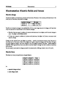
3. 1 Single high electropulse for electroporation and sensitizer delivery( A) The highest lethality of practically 100 % of cancer cells can be reached by the synergism of the pulse and light applied nearly simultaneously on cell suspension containing the sensitizer[4 ] . However , in order to see the proportionated efficacy of pulse and light stepwise actions were performed. Fig. 1 shows the results determined by trypan blue staining. Applying the pulse ( 1. 4 kV/ cm ,1. 6 ms) about 37 % of K2562 cells are blue , however , after 20 min only 16 % are not able to reseal their membranes , that means they are dead. Actinomycin2C yields less dead cells without pulse ( control ) , but more than 50 % dead cells as the result by electroporation. Finally , 85 % of dead cell by the photodynamic activity of actinomycin2C were counted after irradia2 tion of the same suspension for 20 min. This method will be not only effective for combination with elec2 troporation for cancer therapy[2 ] , but is moreover effective to deliver also macromolecular sensitizers (Mw
≤500 000) into cells[10 ] . The response of cells is somewhat weaker for daunomycin and the mentioned anthracyclines ( not shown here) than for actinomycin2C.
・264 ・
电 化 学 2004 年
The FACScan determination shows more details as trypan blue staining. For instance , a double pulse ( 1. 4 kV/ cm , 3 ms) decreases 95 % of vital cells from the be2 ginning ( besides 2. 1 % necrotic , 2. 4 % apoptotic) to 60. 5 % vital , 19. 5 % necrotic and 8. 7 % apoptotic cells , be2 sides about 11 % ruptured cells.
3. 2 A. C. bet ween electrodes in cell suspension( B) Optimal condition were found for frequency ( 16 kHz) , amplitude ( 55 mA) and time ( 20 min ) . Fig. 2 shows the percentage of lethality for U2937 cells by this field alone and in the presence of actinomycin2C and daunomycin in comparison to both cytostatic agents without any current treatment ( control ) . In the FACScan diagram ( Fig. 3) about 1 hour after Fig. 1 Step by step electroporation and irradia2 this A. C. treatment more details can be seen than with trypan blue staining indicating only the necrotic part of the cell population. According to Fig. 3 this soft method B) produces more
tion of K2562 cells. Cell death by 4 × 10 - 4 mol/ L actinomycin2C ( ■) 20 min after the pulse ( 1. 4 kV/ cm) and fol2 lowed by the additional photodynamic killing effect of further 20 min duration
apoptotic and necrotic cells than the short pulse yielding electroporation A ) , however , without ruptured cells.
um and phosphate buffer (pH 6. 5) .
Therefore the sum of all cells in the quadrants is 99. 97 %.
● dead
Additional necrosis can be produced by visible light irradiation , too.
3. 3 Inductive coupling by Helmholtz2coils around the cell suspension( C) This noninvasive method in contrast to [2 ] induces also apoptosis and necrosis not only of suspended cancer cells but also inside animal tumors[12 ,13 ] besides inhibition of
(40min after pulse) . Nutritional medi2 cells by pulse after 20min wait2
ing time for resealing the membrane without actinomycin2C
□ dead cells by actinomycin2C alone without pulse in the dark. Between ● there
■
( shortly after the pulse) and
is the resealing time of 20 min.
angiogenesis[13 ] . The following levels of application have been studied :
—C1 the synergism of field effect with hyperthermia and hyperacidity. —C2 synergism of field effect and actinomycin2C in the dark. —C3 synergism of field effect and the photodynamic activity of actinomycin2C. ( 1) C1 level In agreement with results from[11 ] the induction of apoptosis by PEMF can be determined by trypan blue staining of dead cells , however , mostly later after the treatment , e. g. 24 h.
第 3 期 Maria Radeva 等 : 电场或电磁场和光动力的协同效应对癌细胞失活和坏死的作用 ・265 ・
Fig. 2 Influence of frequency on the U2937 cell viability.
Fig. 3 Percentages of apoptotic and necrotic U2937 cells
The lethal effect by penetration of the drugs into
according to the FACScan determination :
the cells for 20min without field below left ( □) for
Control ( upper curve ) : suspension before the
actinomycin2C (1 ×10
treatment : UR2. 7 % necrotic cells ,LL 89. 86 %
mycin ( 1 ×10
- 5
- 5
M) and ( ○) for dauno2
M) as control. Lethal Effect of
vital cells ,LR 7. 17 % apoptotic cells
20 min a. c. field alone 55 mA , 16 kHz ( ◆) .
Experiment ( lower curve ) : 20 min treatment by
Synergism of 20min field and drugs ( ■) for a. c.
55 mA at 16 kHz : UR22. 4 % necrotic cells ,LL
field plus actinomycin2C ( 1 ×10
32. 71 % vital cells ,LR 44. 67 % apoptotic cells
- 5
M) and ( ●)
for field plus daunomycin (1 ×10 - 5 M) . ( The K2 562 cells show about the same sensitivity)
The results are presented in Tab. 1 at 37 ℃ and 42 ℃. The efficacy rises , if PEMF is combined with hyperthermia ( ≥42 ℃) or hyperacidity (pH was decreased from 7. 4 of nutritional medium to 6. 5) or both according to the therapy conception of M. V. Ardenne , under which conditions cancer cells react more sensitive by both changes than normal cells[15 ] , which is the reason for a quite strong difference be2 tween their vitality. Whereas the necrotic effect of B = 10 mT at 37 ℃ during 1 h is negligible but not af2 ter 3d fermentation. For B = 39 mT a doubling of E/ C can be reached during 3 h treatment under condi2 tions of hyperthermia and lowered pH2value ( hyperacidity) . FACScan diagrams correspond to Fig. 3.
电 化 学 2004 年
・266 ・
Tab. 1 Synergism of PEMF and hyperthermia and hyperacidity depending on temperature and pH B/ mT
temperature/ ℃
time/ min
pH
C/ %
E/ %
E/ C ( after 24 h)
Ef / %
39
37
180
7. 4
9. 3 ±2
14 ±2. 2
1. 5 (27 ℃)
+ 50
37
180
6. 5
12. 3 ±2. 5
23. 5 ±3. 5
1. 9 (27 ℃)
+ 90
42
180
6. 5
19. 4 ±3. 5
52. 3 ±4. 5
2. 7 (27 ℃)
+ 170
Note :percentage of lethal effect of PEMF itself on K2562 cells , measured by trypan blue after 24 h at room temperature (27 ℃) . E means with field , C without field. Nutrition medium : pH 7. 4 or with Phosphatbuffer pH 6. 5. Ef : the field effect itself. SD values from 4 countings1P < 0. 05
( 2) C2 level In this combination of actinomycin2C and PEMF both mechanisms yield apoptosis besides by necro2 sis. Typical results are shown in Tab. 2. The E/ C values at both temperatures are similar because the apoptose induction activity by actinomycin2C increases proportionally with concentration. Tab. 2 Synergism of PEMF and cytotoxic effects , percentage of lethal ( mecrotic) U2937 cells by PEMF plus actinomycin2C (1 ×10 - 4 mol/ L) B/ mT
temperatur/ ℃ time/ min
39
39
pH
C/ %
E/ %
E/ C ( after 4 h)
Ef / %
37
30
7. 4
17 ±3. 9
23. 5 ±2. 7
1. 4
+ 40
42
30
7. 4
24 ±2. 6
34. 7 ±3. 0
1. 5
+ 50
37
60
7. 4
31. 2 ±3. 1
42. 3 ±3. 0
1. 3
+ 30
42
60
7. 4
40 ±4. 0
61. 0 ±2. 2
1. 5
+ 50
Note : E means with field , C without field ; Ef : the field effect itself. K2562 shows similar E/ C values. Highest E values are about 60 % dead cells. SD(4 countings) = ±3. 6 ; P < 0. 05
( 3) C3 level Due to the similarity of chromophores of methylene blue and anthraquinone derivatives on one side
[4 ,5 ]
and actinomycin2C and the anthracycline derivatives on the other their photodynamic activities
were tested additionally in order to enhance their cytotoxic effects. Actinomycin2C combinations with PEMF take place as arrangements ( Tab. 3 and Fig. 4) : a) 2. 3 h PEMF without actinomycin2C , ( counted dead cells after 2 h and 6h) , b) 2. 3 h PEMF plus actinomycin2C including 0. 3 h light treatment ( counted after 2. 3 h and 6 h) .
第 3 期 Maria Radeva 等 : 电场或电磁场和光动力的协同效应对癌细胞失活和坏死的作用 ・267 ・ Tab. 3 Synergistic lethal effects of PEMF , actinomycin2C (1 ×10 - 4 mol/ L) . and 0. 3 h irradiation by visible light ( after 2 h field exposition) B/ mT
temperatur/ ℃
10
27 ℃
39 39
+ field time/
+ light time/ min
E/ C after ( h)
1
60
1. 2 (2h)
37 ℃
2. 3 3
20
1. 5 (3h)
37 ℃
2. 3 3
20
2 (immediately)
h
3
Note : Percentages of dead U2937 and K2562 cells are practically the same. The highest E values reache ≥85 % dead cells after storage for 24 h at 25 ℃ ( not shown here) . Control data C with actinomycin , but without field are about 8 %~ 12 % necrotic cells after 2. 3 h.
3 ) 2 h field without actinomycin2C , but 0. 3 h light afterwards. 3 3 ) 2. 3 h field with actinomycin2C , including 0. 3 h light before the end of field treatment
For comparison to Fig. 1 ( electroporation) , the Fig. 4 presents PEMF and light effects according to the scheme C3 ) . In spite of rather long PEMF treatment for sufficient apoptose induction and necrosis the final lethal effect can reach the same order of magnitude as for electroporation according to method ( A) . The morphological changes of cell mem2 brane and inner organelles can be seen on Fig. 5 for five cells after treatment , wheras the three in the corners of fig. 5 are still living.
4 Discussion With the exceptions of continuous D. C. treatment ( voltammetry ) [16 ] and capacity coupling , combinations of these Fig. 4 Step by step exposition of K2562 cells by : three basic bioelectrochemical methods —2 hours treatment with 39mT in presence of 1 ×10 - 4 mol/ L for cancer cell killing were presented on actinomy cin2C and also after 4 hours waiting without field the three levels A) , B) , C) . The results of second determination ( ●) PEMF level C1 agrees with the paper [11 ] , however , under our conditions the necro2 sis effects were found also before 24 h waiting. The results of PEMF plus cyto2
—0. 3 hours with light ( ○ , after 2 h PEMF) and 4 h later oc2 curs the second determination by trypan blue.
—0. 3 hours only light after field : the white triangle (Δ) For comparison with the electroporation efficacy see Fig. 1
・268 ・
电 化 学 2004 年
toxic agents ( level C2 ) confirm the tendency of paper [17 ] using adri2 amycin against adenocarcinoma. But the results of PEMF plus photody2 namic effects ( level C3 ) show novel possibilities for the synergistic en2 hancement of killing cancer cells. Due to so many parameters the optimal conditions must be evaluated in each laboratory. Nevertheless the principal relations between Control and Experiment ( E/ C and Ef ) will be qualitatively the same. It doesn’ t matter which other PEMF or light sources ( LASER ) , cuvettes ( glass , plastic) or filters are used. But the physiological behavior of cells after Fig. 5 Phase2contrast photo ( total magnification 1 000 x) of U2937 cells fermentation and the constant temper2 ature inside the cuvette are essential
after treatment by 10mT for 6h ( 39 ℃) . 5 Apoptotic and necrotic cells in the middle , 3 still living cells can be seen in the corners
for the reproducibility. Besides weakening of cells by apoptosis induction also channels of cell membranes become disturbed by PEMF , which facilitates penetration and yields even fu2 sion of cells and blastomers inside the zona pellucida[18 ] . According to our experiences the highest values of apop2 tosis and necrosis under PEMF conditions can be ob2 tained by B ≥39 mT , e. g. also 100mT[19 ] , and temper2 ature ≥42 ℃, pH ≤6. 5 and t ≥1h , but not below 2 mT[20 ] and that means hyperthermia and hyperacidity in2 crease stress response (formation of heat2shock proteins) and destabilize membranes additionally as well as the visible2light irradiation (photodynamics) in presence of a
Fig. 6 Frequency dependence of the field efficacy on K2562 cells in 0. 3 mol/ L manmtol solu2 tion. Ef : 9 mT during 20 min irradiation of methylene blue , (5 ×10 - 4 mol/ L) at room
dye reacting according to type I or II. Besides the appli2
temperature.
cation of 50/ 60 Hz a frequency dependence has been
without field. Field effect itself Ef = 100 %
found on proliferation determined by dehydrogenase pro2 duction [21 ] . For future research the frequency depen2
( E/ C - 1)
Control 20 min irradiation
第 3 期 Maria Radeva 等 : 电场或电磁场和光动力的协同效应对癌细胞失活和坏死的作用 ・269 ・
dence should be taken into account because the synergism according to C3 is not effective below 50Hz as can be seen on Fig. 6 using methylene blue as sensitizer (preliminary results) . Recently we confirmed that normal lymphocytes are much more stable against PEMF treatments than the cancer cells2an essential advantage of this method[21 ] ! Because PEMF acts noninvasively contrary to [2 ] and without contamination by electrode products it will be an ideal additional method for tumor therapy in the future[12 ,14 ] .
Reference s : [1 ] Jaroszeske M , Heller R , Gilbert R. Electrochemotherapy , Electrogenetherapy/ Electrically Mediated Delivering of Molecules to Cells and Transdermal Drug Delivery [M] . Totowa : Humana Press , 2000. [ 2 ] Dev S , Rabussy D , Widra G et al. Medical applications of electroporation [J ] . IEEE Trans. Plasma Sci , 2000 , 28 : 206~223. [3 ] Spikes J . Photodynamic action from paramecium to photochemotherapy [J ] . Photochem. Photobiol. B , 1997 ,55 : 142~147. [4 ] Zhou A , Liu M , Baciu C , et al. Membrane elecdtroporation increases photodynamic effects [J ] . J . Electroanalyt. Chem. , 2000 , 486 : 220~224. [ 5 ] Pang L , Baciu C , Traitcheva N , et al. Photodynamic effect in cancer cells influenced by electromagnetic fields [J ] . J . Photochem. Photobiol. B , 2001 , 64 , 21~26. [6 ] Studzinski G, Cell Growth and Apoptosis2 a Practical Approach , Ed ,[M] . Oxford : IRL2Press , 1998. [7 ] Neumann E , Sowers A , Jordan C. Electroporation and Electrofusion in Cell Biology Eds , [M] . Plenum Press , N. Y. , 1989. [8 ] Binhi V. Magetobiology , Underlying Physical Problems [M] . San Diege : Academic Press , 2002. [9 ] Gollmick F A , Berg H. ü ber den Mechanismus der photosensibilisicrten Oxydation des Guanins durch Thiopyronin [J ] . Photochem. Photobiol. , 1972 , 16 : 447~453. [ 10 ] Lambreva M , Gluck B , Berg H. Photodynamic effects of chromophor dextrans supported by electroporation[J ] . Bio2 chemistry , 2004 , in press. [11 ] Hisamitsu T , Narita K, Kasahara T , et al. Induction of apoptosis in human leukemic cells by magnetic fields [J ] . J . Physiology , 1997 , 47 : 307~310 ( In Japanese) . [ 12 ] Tofani S , Cinforino M , Barone D , et al. Increased mouse survival , tumor growth inhibition and decreased immunre2 active p53 after exposure to magnetic fields[J ] Bioelectromagnetics , 2002 ,23 :230~238. [13 ] Williams C , Markov M. Therapeutic electromagnetic field effect on angiogenesis during tumor growth : a pilot study in mice [J ] . Electro. Magnetobiology , 2001 , 20 : 323~329. [14 ] Mangiacasale R , Tritavelli A , Sciamanna I , et al. Normal and cancer2prone human cells respond differently to ex2 tremely low frequency magnetic fields[J ] . FEBSS Letters , 2001 , 487 : 397403. [15 ] Ardenne M V. Systemische Krebs2Mehrschritt2 Therapie , Hyperthermie and Hyperglykamie als Therapiebasissm. Stuttgart : Hippokratis Verlag : 1997. [ 16 ] Nordenstrom B. Survey of mechanisms in electrochemical treatment ( ECT) of cancer[J ] . Eur. J . Surgery. Suppl. , 1994 ,574 : 93~109. [17 ] Gray J , Frith Ch , Parker D. In vivo enhancement of chemotherapy with static or magnetic fields [J ] . Bioelectro2 magnetics , 2000 , 20 : 575~583.
电 化 学 2004 年
・270 ・
[18 ] Kurischko A , Berg H. Electrofusion of rat and mouse blastomeres [J ] . Bioelectrochem. Bioenerg. 1986 , 15 : 513
~519. [19 ] Seze R , Tuffet S , Moreau JM ,et al. Effects of 100mT time varying magnetic fields on the growth of tumors in mice [J ] . Bioelectromagnetics , 2000 , 21 : 107~111. [20 ] Galloni P , Marino C. Effects of 50Hz magnetic field exposure on tumor experimental models [J ] . Bioelectromag2 netics , 2000 ,21 :608~614. [ 21 ] Zhou A , Liu M , Wang X , et al. Effects of ELE inductively coupled weak magnetic field on proliferation of 6B1 cells [J ] . Electro2Magnetobiol. , 1999 , 18 : 325~331.
电场或电磁场和光动力的协同效应 对癌细胞失活和坏死的作用 Maria Radeva , Maya Lambrera , Plamena Angelova , Nelly Traitcheva , Hermann Berg (Beutenberg 大学 生物电化学研究室 耶纳 德国)
摘要 : 应用弱正弦波电磁场改变细胞膜的穿透性并引起其失活和坏死 . 研究中 , 将人类癌细胞 U2937 和 K2562 放置于强度为 10 mT 和 39 mT(50 Hz) 的正弦波电磁场内 ,并依次结合细胞毒素放线
菌素2C 以及其独特的光动力活性分别进行试验 .
关键词 : 正弦波电磁场 ;电生孔 ;放线菌素2C ;光动力学 ;协同效应 ;癌细胞 U2937 ; K2562