Seizure 20 (2011) 616–627
Contents lists available at ScienceDirect
Seizure journal homepage: www.elsevier.com/locate/yseiz
Epilepsy surgery in children with developmental tumours Marta Garcı´a-Ferna´ndez a,*, Concepcio´n Fournier-Del Castillo b, Arturo Ugalde-Canitrot c, A´ngeles Pe´rez-Jime´nez a, Juan A´lvarez-Linera d, Inmaculada De Prada-Vicente e, Jesu´s Sua´rez-Rodrı´guez f, Jordi Bernabeu-Verdu´ f, Francisco Villarejo-Ortega g a
Epilepsy Monitoring Unit, Neurophysiology Section, Hospital Infantil Universitario Nin˜o Jesu´s, Madrid, Spain Neuropsychology Section, Neuropsychiatry and Psychology Service, Hospital Infantil Universitario Nin˜o Jesu´s, Madrid, Spain Epilepsy Monitoring Unit, Neurophysiology Service, Hospital Universitario La Paz, Madrid, Spain d Neuroradiology Section, Diagnostic Imaging Service, Hospital Ruber Internacional, Madrid, Spain e Pathology Service, Hospital Infantil Universitario Nin˜o Jesu´s, Madrid, Spain f Research Methods and Diagnosis in Education, University of Valencia, Valencia, Spain g Neurosurgery Service, Hospital Infantil Universitario Nin˜o Jesu´s, Madrid, Spain b c
A R T I C L E I N F O
A B S T R A C T
Article history: Received 8 May 2010 Received in revised form 6 April 2011 Accepted 9 June 2011
We report our experience regarding evaluation, surgical treatment and outcomes in a population of 21 children with histopathologically confirmed developmental tumours [nine dysembryoplastic neuroepithelial tumours (DNET), ten gangliogliomas (GG) and two gangliocytomas (GC)] and related epilepsy, analyzing video-EEG, MRI and neuropsychological data, before and after surgery. Most children had focal epilepsy correlating well with lesion location. One patient had epileptic spasms and generalized discharges. Tumours were located in the temporal lobe in 13 patients. Mean age at surgery was 11.16 years. Postsurgical MRI showed residual tumour growth in one DNET. One child had a recurrent ganglioglioma with anaplastic transformation. At latest follow-up (mean 4.68 years) 95.2% of patients were seizure-free and no significant neuropsychological declines were observed. Evidence from our study suggests that, in this setting, surgery should be performed before criteria for refractory epilepsy are met, particularly in cases with early seizure onset, in order to optimize cognitive outcome. ß 2011 British Epilepsy Association. Published by Elsevier Ltd. All rights reserved.
Keywords: Epilepsy surgery Low-grade brain tumour Tumour-associated epilepsy Neuropsychological outcome Video-EEG Seizure outcome
1. Introduction Around twenty to thirty per cent of long-standing medically intractable epilepsies are caused by tumours of neuroepithelial tissue, especially neuronal–glial tumours.1 Gangliogliomas (GGs) and dysembryoplastic neuroepithelial tumours (DNETs) are the most common causes and constitute a major pathologic substrate of children referred for epilepsy surgery. Magnetic resonance imaging (MRI) reveals characteristic findings, although rarely specific.2 The identification of these tumours is of particular importance, with therapeutic and prognostic implications, due to their benign behaviour and tendency to mimic more aggressive tumours, such as oligoastrocytomas.3 DNETs and GGs, together with gangliocytomas, have been included in Barkovich’s classification of malformations of cortical development, as malformations due to abnormal neoplastic neuronal and glial proliferation with abnormal cell types, associated with disordered cerebral cortex.4
* Corresponding author at: Epilepsy Monitoring Unit (Neurophysiology Section), ˜ o Jesu´s, Avenida Mene´ndez Pelayo, 65, 28009 Hospital Infantil Universitario Nin Madrid, Spain. Tel.: +34 91 503 59 00; fax: +34 91 574 46 69. E-mail address:
[email protected] (M. Garcı´a-Ferna´ndez).
Epilepsy is usually the main manifestation of developmental tumours and, in most cases, the presenting feature. Its electroclinical findings are usually, but not always, concordant with the location of the lesion. Children with these tumours can be cognitively normal or present specific deficits or epilepsy-related deterioration, including psychopathological manifestations. Surgery in this setting offers very good results overall, although some controversy still exists over the best surgical strategies. In selected cases, intracranial electroencephalogram (EEG) recordings and brain mapping may aid in tailoring the resection. Seizure frequency and oncological morbidity are the most frequently assessed outcome measures in the literature. However, the consequences on the neurocognitive and behavioural functioning domains have been insufficiently analyzed to date. We report our experience regarding the evaluation and surgical treatment in a paediatric population with developmental tumours and related epilepsy. Our purpose was to review their pre-surgical and post-surgical clinical, neurophysiological, neuroimaging and neuropsychological data and to analyze and specifically delineate the findings with regard to each other. Outcomes were assessed in terms of a variety of measures, including thorough neuropsychological testing, in addition to seizure control and oncological morbidity.
1059-1311/$ – see front matter ß 2011 British Epilepsy Association. Published by Elsevier Ltd. All rights reserved. doi:10.1016/j.seizure.2011.06.003
M. Garcı´a-Ferna´ndez et al. / Seizure 20 (2011) 616–627
2. Materials and methods 2.1. Study population and data collection All children (2 cm) or small (2 >2 >2 >2 >2 >2 2 2 >2 >2 >2
FLE TLE TLE TLE PsTLE PCE FLE TLE TLE TLE PsTLE PRE PRE TLE TLE TLE TLE TLE TLE TLE FLE
3 2 1 2 2 1 1 1 1 1 2 1 1 1 2 3 2 1 1 1 1
No No No Yes No Yes No No Yes No No No No No No No No No No No No
Yes Yes Yes No Yes Yes No No No No No No No No Yes Yes No No Yes No No
Yes Yes Yes Yes Yes No No Yes No No Yes No No Yes Yes Yes Yes No Yes No Yes
No.
No. AEDs at surgery
Epilepsy duration (years)
Age at surgery (years)
Type surgery
MRI 6 m postsurgery
Seizure outcome 6–12 m post surgery
Reason/type reoperation
Follow-up (years)
Seizure outcome at latest follow-up
No. AEDs at latest follow-up
/ Recurrence + Malign/Les / / / Seizures + Residual tum/Les / / / / / Residual tumour growth/Les / Seizures + FCD/Lobs / Seizures + Residual tum/Les / / / / /
3.33 6.16
I I
7.00 7.75 7.58 4.50
1 2
2 2
3.0 3.3
4.4 16.3
Lob Les
Total resect Total resect
I I
3 4 5 6
2 2 1 1
1.8 11.0 4.3 1.0
2.0 11.9 5.6 3.6
Lob Les Les Les
Total resect Total resect Total resect Residual tum
I I I IV
7 8 9 10 11 12
1 2 1 1 2 2
1.0 1.0 0.9 0.5 7.5 3.0
16.0 9.5 2.1 10.5 19.5 11.0
Les Les Lob Les Les Les
Total resect Total resect Total resect Total resect Total resect Residual tum
I I I I I I
13 14 15 16
1 2 3 2
2.0 7.8 10.0 9.0
18.0 14.8 16.0 14.0
Les Les ExtS Les
Residual tum FCD Total resect Residual tum
I IV I III
17 18 19 20 21
2 1 3 1 4
3.0 3.0 4.0 0.6 8.0
9.0 17.0 16.0 10.6 12.0
Les Les ExtS Les Les
Total resect Total resect Total resect Residual tum Total resect
I I I I I
Pre FIQ
Post FIQ
2 1
51 97
88 100
I I I I
0 1 1 1
68 45 70 92
68 77 67 70
1.00 1.50 7.00 1.91 6.08 3.66
I I I I I I
0 2 0 1 0 2
117 82 96 94 90 85
106 91 66 84 107 88
6.33 4.41 4.66 4.16
I I I II
0 2 2 3
123 104 51 78
123 106 66 89
3.50 2.16 5.58 7.41 2.58
I I I I I
1 1 1 0 3
88 100 65 119 75
99 109 70 112 75
M: male, F: female, GG: ganglioglioma, DNET: dysembryoplastic neuroepithelial tumour, GC: gangliocytoma, FCD: focal cortical dysplasia, L: left, R: right, T: temporal, PR: perirolandic, FLE: frontal lobe epilepsy, TLE: temporal lobe epilepsy, PsTLE: pseudotemporal epilepsy, PCE: posterior cortex epilepsy, PRE: perirolandic epilepsy, AED: antiepileptic drugs, Lob: lobectomy, Les: lesionectomy, Ext: extended lesionectomy, s: subdural electrodes, *: at re-intervention, FIQ: full intellectual quotient.
abnormalities of pseudo-cystic appearance were present in 88.88% (8/9)–this highly characteristic feature was not found in GGs; focal contrast enhancement was seen in 22.22% (2/9); no oedema nor calcifications were found. With regard to histopathologically proven GGs, a wider variety of findings were seen: MRI signals on T1 sequences were isointense in 50.0% (5/10), mixed in 40.0% (4/ 10) and hypointense in 10.0% (1/10); on T2 and FLAIR sequences, MRI signals were hyperintense in 70.0% (7/10) and mixed in 30.0% (3/10); mass effect was found in 20.0% (2/10), gadolinium enhancement in 80.0% (8/10), calcifications in 20.0% (2/10) and cerebral oedema in 20.0% (2/10). Adjacent FCD was confirmed by histopathology in three cases, all of which were either type IA or IIA, according to Palmini’s classification.7 Such FCD had been previously detected by MRI in two of them. In one patient, with a DNET located in the posterior cingulum and history of status epilepticus during infancy,
preoperative MRI showed associated mesial temporal sclerosis, which was not removed. 3.3. Pre-surgical non-invasive video-EEG findings and epilepsy classification Seizures were recorded in 16 patients (76.2%), adding up to a total of 60 seizures available for analysis. Temporal lobe epilepsy (TLE) was diagnosed in 13 cases (61.9%). Seizures with typical semiological features were observed in 10 out of 11 cases with recorded seizures, including some of the following: abdominal aura, impaired consciousness, psychomotor arrest, and oral and/or manual automatisms. In most patients, ictal EEG revealed typical seizure patterns, with a well-defined regional onset. However, one patient, with epilepsy onset within the first year of life, presented with asymmetric infantile spasms and
M. Garcı´a-Ferna´ndez et al. / Seizure 20 (2011) 616–627
619
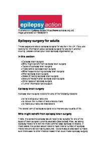
Fig. 1. Frontal DNET (patient 1). Left frontal lesion, with a well defined, slightly lobulated border, predominantly hypointense on T1 (A) and hyperintense on T2 (B), but mixed on FLAIR (C). The lesion consists on multiple small nodules (pseudocysts) with slightly variable signal, mainly on FLAIR sequence, very characteristic of DNETs. There is also a subtle triangular white matter hyperintensity, pointing to the ventricle, suggesting associated FCD.
generalized EEG abnormalities. In all cases, regional interictal epileptiform discharges were found over the temporal lobe involved by the tumour. In four patients, two independent EEG foci were found within the affected temporal lobe, usually involving a dominant focus at the lateral temporal region and a secondary focus consisting of sporadic anterior-medial temporal discharges (