What is Epilepsy Surgery
Nicholas M. Wetjen, MD, FAANS, FACS, FAAP Associate Professor of Neurosurgery and Pediatrics
[email protected] ©2015 MFMER | slide-1
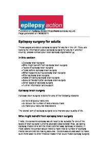
What causes epilepsy? DEGENERATIVE , 4%
INFECTION, 3%
TUMOR , 4% TRAUMA, 6%
CONGENITAL, 8% VASCULAR , 10%
UNKNOWN, 65%
Wetjen NM, et al, Resective surgery of neoplastic lesions for epilepsy. In Shorvon S, ed. The Treatment of Epilepsy, 2nd Edition, 2004 ©2015 MFMER | slide-2
Who gets epilepsy surgery? “A skilful healer does not howl incantations when a wound is crying for the knife .” Sophocles Ajax: 582.
Patients with intractable epilepsy
Epilepticus sic curabitur Sloane Manuscript, ~1190 A.D. ©2015 MFMER | slide-3
Intractable Epilepsy Statement of the Problem It is estimated that 15-35% of patients with seizure disorders are considered medically refractory. Inca ruler Capac Yupanqui's wife having an epileptic seizure (~1500). CP1208045-9 ©2015 MFMER | slide-4
The Argument for Epilepsy Surgery
Jan Sanders van Hemessen (~1500-1575): ”The stone-cutter” ©2015 MFMER | slide-5
Medically Refractory Epilepsy Brodie et al. Neurology 2002;58:S2-8
470 newly diagnosed, untreated
©2015 MFMER | slide-6
PARTIAL EPILEPSY seizure-free on medications
Partial Epilepsy: Temporal Lobe Epilepsy: Lesional Pathology: Mesial Temporal Sclerosis:
55% 35% 10% 10% ©2015 MFMER | slide-7
Pediatric Epilepsy Surgical Procedures Resective Surgical Procedures “remove epileptic brain”
Disconnective Surgery “disconnect epileptic brain from normal brain”
Stimulation Procedures “disrupt epileptic network with electrical stimulation” ©2015 MFMER | slide-8
Epilepsy Surgery: Resective • Removal of a portion of the brain from which seizures arise • Need to document: • Seizures are medically intractable and disabling • Seizures arise only from the region to be resected • The area can be resected without causing significant disability to the child • Child needs careful evaluation to see if this is an option which includes: • MRI • Video-EEG study in hospital to record typical seizures • Neuropsychology evaluation • May need other tests – SISCOM, PET, MEG, TMS, fMRI ©2015 MFMER | slide-9
Resective Surgical Procedures “remove epileptic brain” • Diagnostic surgery (Phase I) • Intracranial monitoring - subdural grids and strip electrodes • Stereoencephalography (SEEG) • Therapeutic Surgery • Image-guided resection • Electrocorticography (ECoG) guided resection
©2015 MFMER | slide-10
©2015 MFMER | slide-11
The location of seizure onset and spread is determined by the type of symptoms or behavior that occur during the seizure.
©2015 MFMER | slide-12
ECS
Increase hgEEG
SSEP
fMRI
©2015 MFMER | slide-13
Limitations of Subdural Grid Monitoring • Inadequate coverage of intrasulcal, deep brain, and interhemispheric regions • Difficulty of multilobar sampling • Inadequate functional network sampling • Large craniotomy – swelling, hematoma
• Wound complications and infection • Difficult re-operation with subdural-cortical adhesions ©2011 MFMER | ©2015 MFMER |slide-14 slide-14
SEEG - Stereoelectroencephalography
©2015 MFMER | slide-15
©2015 MFMER | slide-16
©2015 MFMER | slide-17
SEEG
©2015 MFMER | slide-18
©2015 MFMER | slide-19
©2015 MFMER | slide-20
Many electrodes that have targeted the insula during SEEG investigations have been placed using a standard orthogonal “ Talairach” approach. MRIs with electrodes in position are usually not obtained in this situation (Marc Guénot, personal communication). An alternative oblique strategy of depth electrode placement is illustrated in this figure. (Courtesy of Santiago Gil Robles and Philippe Coubes.) Upper three images: (a) occipitoinsular electrode, (b–d) frontal electrodes; lower three images: (a, b) hippocampal electrodes, (c) right inferior insular electrode; (d) right superi or insular electrode, (e) left inferior insular electrode.
©2015 MFMER | slide-21
Engel Classification • Group A • Class I: Seizure-free • Class II: Rare, nondisabling Seizures • Group B • Class III: Improvement in seizure frequency (> 80%) • Class IV: No significant improvement in seizure frequency ©2015 MFMER | slide-22
Mayo Surgical Results N=491 • Group A: 356 patients (72%)
• Group B: 135 patients (28%) • Most seizures occurred the first year following surgery • If there is mesial temporal lobe sclerosis and EEG concordance94% have an excellent outcome (Engel Class I and II) • Excellent durability • Major operative M+M 1% ©2015 MFMER | slide-23
Complications of Epilepsy Surgery 25 Centers/1,911 Patients No.
%
9