Journal of Pediatric Surgery (2005) 40, 251 – 255
www.elsevier.com/locate/jpedsurg
Results of multimodal treatment for desmoplastic small round cell tumors Dave R. Lal, Wendy T. Su, Suzanne L. Wolden, Kenneth C. Loh, Shakeel Modak, Michael P. La Quaglia* Department of Surgery, Memorial Sloan-Kettering Cancer Center, New York, NY 10021, USA Department of Pediatric Surgery, Memorial Sloan-Kettering Cancer Center, New York, NY 10021, USA
Index words: Desmoplastic small round cell tumor Chemotherapy; Resection; Radiation therapy Surgery; Survival
Abstract Purpose: Desmoplastic small round cell tumors (DSRCTs) are rare aggressive neoplasms that frequently present with large symptomatic intraabdominal masses. We examined the effects of multimodal therapy including induction chemotherapy, aggressive surgical debulking, and external beam radiotherapy on patients with DSRCT. Methods: Institutional Review Board permission was obtained. Sixty-six patients were diagnosed by histology, immunohistochemistry, and or cytogenetics as having DSRCT at our institution from July 1, 1972, to July 1, 2003. Data were collected on patient demographics, presenting symptoms, tumor location and extent, treatment regimen, and overall survival. Results: A majority of patients were male (91%), Caucasian (85%), and with a median age of 19 (7-58) years old at diagnosis. The most common presenting complaint was an intraabdominal mass (64%). In 63 patients (96%), the primary tumor was located in the abdomen or pelvis. Thirty-three (50%) had positive lymph nodes and 27 (41%) had distant parenchymal metastases at diagnosis. Overall, 3- and 5year survivals were 44% and 15%, respectively. Twenty-nine of these patients (44%) underwent induction chemotherapy (P6), surgical debulking, and radiotherapy. Three-year survival was 55% in those receiving chemotherapy, surgery, and radiotherapy vs 27% when all 3 modalities were not used ( P b .02). Gross tumor resection was highly significant in prolonging overall survival; 3-year survival was 58% in patients treated with gross tumor resection compared to no survivors past 3 years in the nonresection cohort ( P b .000 01). Ten patients (15%) have no evidence of disease with a median follow-up of 2.4 years (range, 0.4-11.2 years). Conclusions: Multimodal therapy results in improved survival in patients with DSRCT. Aggressive surgical resection of these extensive intraabdominal neoplasms correlates with improved patient outcome. D 2005 Elsevier Inc. All rights reserved.
Presented at the 35th Annual Meeting of the American Pediatric Surgical Association, Ponte Vedra, Florida, May 27-30, 2004. * Corresponding author. Department of Surgery, Memorial Sloan-Kettering Cancer Center, New York, NY 10021, USA. Tel.: +1 212 639 7002; fax: +1 212 717 3373. E-mail address:
[email protected] (M.P. La Quaglia). 0022-3468/05/4001-0045$30.00/0 D 2005 Elsevier Inc. All rights reserved. doi:10.1016/j.jpedsurg.2004.09.046
252
D.R. Lal et al.
Desmoplastic small round cell tumor (DSRCT) is an aggressive mesenchymal malignancy that usually occurs in Caucasian males in adolescence [1-3]. Although rare, increasing numbers of patients have been diagnosed with DSRCT since discovery of its specific chromosomal translocation t (11;22)(p13;q12) [4,5].This translocation is unique to DSRCT and confirms the diagnosis. Band 22q12 is the site of the EWS (Ewing’s sarcoma) gene, and 11p13 contains the WT1 (Wilms’ tumor gene) site. These genes are fused in DSRCT. The resultant chimeric protein is thought to act as a transcriptional activator that fails to suppress tumor cell growth [6,7]. Crampy abdominal pain and an associated abdominal mass is the most common presentation seen with these patients [1,8]. DSRCT has a propensity for serosal surfaces, especially in the peritoneal cavity. At diagnosis, patients commonly have numerous peritoneal implants involving the abdomen and pelvis. Primary DSRCT has also been reported in the ethmoidal sinuses, scalp, hand, paratesticular region, pleura, chest, and posterior cranial fossa [7,8]. Metastases occur most commonly to the liver, lung, bone marrow, and lymph nodes [1,3,8]. Due to the diffuse nature of the tumor, resection with negative microscopic margins is often impossible. We have therefore advocated multimodal treatment of these complex patients consisting of chemotherapy, surgical tumor debulking, and radiotherapy. In this study, we investigate the continued efficacy of aggressive surgical therapy and multimodal treatment in our previously reported series of 40 patients and add 26 new patients for analysis.
1. Materials and methods After approval was obtained from the Institutional Review Board in compliance with The Health Insurance Portability and Accountability Act, a retrospective review of all patients with DSRCT treated at Memorial Sloan-Kettering Cancer Center from July 1, 1972, to July 1, 2003 was compiled. Sixty-six patients were identified, 40 of whom had been previously reported. Diagnosis of DSRCT was established by
Fig. 1
histology, immunochemistry, and/or cytogenetics. The median length of follow-up was 2.0 years (range, 0.05-11.2 years). Tissue diagnosis was established before treatment with sufficient tissue for histology, immunochemistry, and cytogenetics. Once the diagnosis of DSRCT was established, initial treatment involved high-dose systemic polychemotherapy using alkylating agents, P6 protocol. The P6 protocol consists of 7 courses involving cyclophosphamide, doxorubicin, vincristine, ifosfamide, and etoposide [9]. Addition of irinotecan, topotecan, carboplatin, and cisplatin were added in selected patients. After 3 to 4 cycles of chemotherapy, response was assessed by computerized axial tomography scan. A majority of patients had a good response with marked diminution of the tumor. Due to the stromal composition of these tumors, a minority showed little response to chemotherapy. Resection was still performed, and tumor necrosis and decreased tumor vascularity was found upon exploration. Surgical debulking was carried out in an attempt to achieve greater than 90% resection of the gross tumor burden. The resection percentage was determined by review of operative notes and comparative imaging. Attempts were not made to achieve microscopic negative margins. Omentectomy, stripping of the parietal and visceral peritoneum, splenectomy for hilar involvement, and local resection of the diaphragm were often required. Consolidative radiotherapy to a dose of 30 Gy was delivered by external beam to the whole abdomen and pelvis. Myeloablative chemotherapy with thiotepa and carboplatin followed by autologous bone marrow or peripheral stem cell rescue was used in 16 patients. Time intervals were taken from the date of diagnosis to date of death from disease or last follow-up. Disease-specific overall survival distributions were calculated using the Kaplan-Meier method and were compared with the log-rank test.
2. Results 2.1. Patient characteristics The study included 60 male (91%) and 6 female (9%) patients. A majority of patients were Caucasian (85%); the
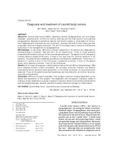
DSRCT site involvement at time of presentation (n = 66).
Results of multimodal treatment for desmoplastic small round cell tumors
253
remainder were Hispanic (9%) and African American (6%). The median age at diagnosis was 19 years (range, 7-58 years). Sixty-three patients (96%) had their primary tumor located in the abdomen or pelvis; other sites included the thoracic cavity (1) and testicle (2). Local or distant lymph node involvement was seen in 33 (50%) patients at presentation. Twenty-seven (41%) patients presented with distant parenchymal metastases and sites are displayed in Fig. 1. A majority of patients (n = 42, 64%) presented with a palpable abdominal, pelvic, testicular, or umbilical mass, and associated symptoms are displayed in Fig. 2.
2.2. Statistical analysis
Fig. 2 Presenting symptoms reported by patients at the time of diagnosis of DSRCTs.
The overall survival for all 66 patients was 44% (95% CI, 30%-58%) at 3 years and 15% (95% CI, 5%-25%) at 5 years (Fig. 3A). In 47 patients (71%), greater than 90% tumor resection was possible, and the overall survival in this group is depicted in Fig. 3B. Greater than 90% tumor resection was highly significant in prolonging overall survival when compared to patients with a lesser resection (P b .000 01). Forty-seven patients (71%) underwent treatment with the P6 protocol (Fig. 3C), and this improved survival compared to
Fig. 3 These Kaplan-Meier curves demonstrate overall survival since time of diagnosis of DSRCT. Tick marks indicate last follow-up. A, Overall survival of all patients treated. B, Overall survival comparing patients undergoing complete and incomplete reaction. Complete reaction resection is defined as greater than 90% tumor resection. C, Overall survival comparing patients treated with P6 protocol compared with other regimens. See text for definition of P6 protocol. D, Overall survival in patients treated with multimodal therapy vs those not treated. Multimodal therapy is defined as P6 protocol, greater than 90% tumor resection, and adjuvant radiotherapy.
254 other regimen ( P b.004). Treatment with multimodal therapy including P6 protocol, greater than 90% resection, and adjuvant radiotherapy was accomplished in 29 patients (44%). Multimodal therapy was also significant in increasing survival as shown in Fig. 3D ( P b .02). The presence of node positive or metastatic disease at presentation did not correlate with overall survival. Ten patients (15%) are alive and have no evidence of disease with a median follow-up of 2.4 years (range, 0.4-11.2 years).
3. Discussion Desmoplastic small round cell tumor is an aggressive malignancy with few long-term survivors. Due to the diffuse serosal spread of DSRCT, systemic chemotherapy utilizing the P6 regimen has been the cornerstone of initial treatment. Debulking surgery is then attempted with a goal of at least 90% reduction of tumor bulk. Aggressive surgical resection continues to be a major determinant in patient survival. With continued follow-up of these patients, surgical resection greater than 90%, P6 protocol and adjuvant radiotherapy all contribute to improved survival at 3 and 5 years. Our last publication of this series involved 40 patients and had a 3-year survival of 29% [1]. Although this study reports improved survival at 3 and 5 years, the overall prognosis remains grim. In rare cases, long-term survival is possible. In our series, we have 4 patients that have no evidence of disease with extended long-term follow-up (5.7, 7.8, 10.1, and 11.2 years). All 4 were treated with P6 protocol and tumor debulking, and all but one received radiotherapy. Further advances in treatment of DSRCT involve the development of new therapies that specifically target the cellular regulatory mechanism of DSRCT. The fusion protein created by the characteristic chromosomal translocation, t(11;22)(p13;q12), has been shown to induce production of endogenous platelet-derived growth factor (PDGF) [10], T cell acute lymphoblastic leukemia-associated antigen 1 protein (TALLA-1) [11], and interleukin-2/15 receptor [12]. Each of these is important in tumorigenesis. Plateletderived growth factor is a chemoattractant that is thought to enhance recruitment and proliferation of fibroblast and endothelial cells. This may explain the dense stroma and fibrosis associated with DSRCT [10]. SU101 is a PDGF pathway receptor inhibitor that is currently undergoing phase 1 trials. Adamson et al [13] have reported on a patient with progressive refractory DSRCT that responded to SU101 treatment with prolonged stabilization of disease. TALLA-1 proteins are important in cell regulation, adhesion, migration, and metastases [11]. Interleukin-2/15 receptor acts in a paracrine fashion with IL-2 and IL-15 growth factors secreted by stromal cells to enhance tumor cell proliferation [12]. Additional research is focused on immunotherapies that target DSRCT-specific antigens. Two DSRCT cell surface antigens that can be targeted by monoclonal
D.R. Lal et al. antibodies have been identified GD2, recognized by the antibody 3F8, and a novel 58 kDa glycoprotein, recognized by the antibody 8H9 [14]. Ninety-six percent of 46 DSRCT tumors examined tested positive with 8H9 and 70% with GD2 [14,15]. Current research is focused on developing targeted immunotherapy using monoclonal antibodies directed at these unique DSRCT antigens. This strategy may have the potential advantage of decreased toxicity because both of these antibodies specifically target antigen-expressing neoplastic cells and have restricted cross-reactivity with most normal tissues. Desmoplastic small round cell tumor remains a vexing disease with little long-term survival. Current treatment prolongs life and rarely achieves cure. Neoadjuvant chemotherapy, greater than 90% tumor debulking, and radiotherapy have been shown to prolong survival. Future efforts must focus on cell-specific treatments.
References [1] La Quaglia MP, Brennan MF. The clinical approach to desmoplastic small round cell tumor. Surg Oncol 2000;9:77 - 81. [2] Gerald WL, Rosai J. Desmoplastic small cell tumor with multiphenotypic differentiation. Zentralbl Pathol 1993;139:141 - 51. [3] Schwarz RE, Gerald WL, Kushner BH, et al. Desmoplastic small round cell tumors: prognostic indicators and results of surgical management. Ann Surg Oncol 1998;5:416 - 22. [4] Rodriguez E, Sreekantaiah C, Gerald W, et al. A recurring translocation, t(11;22)(p13;q11.2), characterizes intra-abdominal desmoplastic small round-cell tumors. Cancer Genet Cytogenet 1993; 69:17 - 21. [5] Sawyer JR, Tryka AF, Lewis JM. A novel reciprocal chromosome translocation t(11;22)(p13;q12) in an intraabdominal desmoplastic small round-cell tumor. Am J Surg Pathol 1992;16:411 - 6. [6] Liu J, Nau MM, Zucman-Rossi J, et al. LINE-I element insertion at the t(11;22) translocation breakpoint of a desmoplastic small round cell tumor. Genes, Chromosomes & Cancer 1997;18:232 - 9. [7] Gerald WL, Ladanyi M, de Alava E, et al. Clinical, pathologic, and molecular spectrum of tumors associated with t(11;22)(p13;q12): desmoplastic small round-cell tumor and its variants EWS-FLI1 fusion transcript structure is an independent determinant of prognosis in Ewing’s sarcoma. J Clin Oncol 1998;16:3028 - 36. [8] Lae ME, Roche PC, Jin L, et al. Desmoplastic small round cell tumor: a clinicopathologic, immunohistochemical, and molecular study of 32 tumors. Am J Surg Pathol 2002;26:823 - 35. [9] Kushner BH, LaQuaglia MP, Wollner N, et al. Desmoplastic small round-cell tumor: prolonged progression-free survival with aggressive multimodality therapy. J Clin Oncol 1996;14:1526 - 31. [10] Lee SB, Kolquist KA, Nichols K, et al. The EWS-WT1 translocation product induces PDGFA in desmoplastic small round-cell tumour. Nat Genet 1997;17:309 - 13. [11] Ito E, Honma R, Imai J, et al. A tetraspanin-family protein, T-cell acute lymphoblastic leukemia-associated antigen 1, is induced by the Ewing’s sarcoma-Wilms’ tumor 1 fusion protein of desmoplastic small roundcell tumor. Am J Pathol 2003;163:2165 - 72. [12] Wong JC, Lee SB, Bell MD, et al. Induction of the interleukin-2/15 receptor beta-chain by the EWS-WT1 translocation product. Oncogene 2002;21:2009 - 19. [13] Adamson PC, Blaney SM, Widemann BC, et al. Pediatric phase I trial and pharmacokinetic study of the platelet-derived growth factor (PDGF) receptor pathway inhibitor SU101. Cancer Chemother Pharmacol 2004;536:482 - 88.
Results of multimodal treatment for desmoplastic small round cell tumors [14] Modak S, Gerald W, Cheung NK. Disialoganglioside GD2 and a novel tumor antigen: potential targets for immunotherapy of desmoplastic small round cell tumor. Med Pediatr Oncol 2002;39:547 - 51. [15] Modak S, Kramer K, Gultekin SH, et al. Monoclonal antibody 8H9 targets a novel cell surface antigen expressed by a wide spectrum of human solid tumors. Cancer Res 2001;61:4048 - 54.
Discussion
255
perform omentectomies if tumor is involved in the omentum. The time we do remove organs is, the spleen was removed, if it involves the hilar area of the spleen. Usually, they have nodes in this region and we will take out the spleens occasionally, but we try to avoid removing solid organs. A.-X. Holterman (Chicago, IL): I have a quick question on how you handle the metastases after debulking.
S. Emil (Orange, CA): After having 2 teenagers this past year with this tumor, I learned that it is probably one of the most challenging tumors that a surgeon will have to face. I was wondering if you could expand a little bit on the debulking. I will just give you a practical example. I have a child now who has had some response to the MSK protocol but now has a tumor that is really stuck in the pelvis and is very intimately associated with the bladder and the rectum. Would you sacrifice any organs in trying to remove this tumor? I am not sure I could get 90% debulking by just chiseling away at it.
D.R. Lal (response): We enroll them back in the P6 protocol. They finish off the treatment regimen. Usually, when they go on to have metastatic disease, they succumb to the disease.
D.R. Lal (response): Typically, we try to avoid mutilating surgery. These are serosal-based tumors, and for example, that patient with tumor to anterior rectum, they commonly have tumor in this location. You can peel off the serosa with the tumor, and usually you can do that without even resecting bowel. Again, our goal is not mutilating surgery, just to get grossly negative margins if possible or debulk as much as possible without sacrificing structures. We do
D.R. Lal (response): We did not. Whole abdomen/pelvic radiation is administered after finishing adjuvant P6 therapy according to protocol. Because these are such diffuse tumors, located throughout the entire abdomen and pelvis, focused intraoperative radiotherapy is not practical. Therefore, we wait until the conclusion of their chemotherapy and then usually administer whole abdomen and pelvic radiation.
Unidentified Speaker: Just a quick question about your local tumor control. It is good to see that it does have some impact to try to get it near complete resection, but I would wonder about the way the radiation was given as part of local tumor control. Did you do any intraoperative radiation at all?