fC
al Pathologi linic st
PAT H O L O G Y
0 201 al-
athm andu
of Nepal
www.acpnepal.com
ad ,K
l ca edi Nepal M
As soc iatio
Journal of
of N
Asso
no
ep
ci a ti o
Journal of Pathology of Nepal (2011) Vol. 1, 87 - 91
i on n Building Exhibit
Ro
Original Article
Role of immunohistochemistry in the diagnosis of malignant small round cell tumors Bashyal R1, Pathak TB1, Shrestha S1, Pun CB1, Banstola S1, Neupane S1, Lee MC1 Department of Pathology, BP Koirala Memorial Cancer Hospital, Bharatpur, Nepal
1
Keywords: Immunohistochemistry; Malignant Small Round Cell Tumor; Non Hodgkin’s Lymphoma; Ewing’s Sarcoma; Rhabdomyosarcoma
ABSTRACT Background: Immunohistochemistry is a key tool for the analysis of localization of target molecules within tissues. It has a significant role in the identification of tumors lacking evidence of lineage differentiation on the basis of routine light microscopic morphology alone. Approximately 90% of tumors posing diagnostic difficulties by morphology could be accurately classified by exploiting immunohistochemistry. The aim of this study is to identify the true identity of malignant small round cell tumors by immunohistochemical analysis. Materials and Methods:This was a retrospective study done in Department of Histopathology of B.P.Koirala Memorial Cancer Hospital from January 2010 to April 2011.A total of 40 cases small round cell tumors were selected for immunostaining. The immunohistochemistry technique used is the Polymer detection-EnvisionTM System, a two step staining technique based on Horse Radish Peroxidase labeled dextran polymer technology (DAKO Company). Results: Out of 40 cases of malignant small round cell tumors, there were 21 cases (52.5%) of NonHodgkin Lymphoma , 11 cases (27.5%) of Ewing’s Sarcoma/Primitive Neuroectodermal Tumor, 1 case (2.5%) of Lymphoblastic Lymphoma , 1 case (2.5%) of Rhabdomyosarcoma, 2 cases (5%) of Low grade neuroendocrine tumor, 1 case (2.5%) of Neuroblastoma, 2 cases (5%) of Poorly differentiated Synovial Sarcoma (small cell variant), 1case (2.5%) of Malignant Melanoma (small cell variant). Conclusion: Immunohistochemistry is a valuable adjunct to routine hematoxylin and eosin staining for adequate and accurate categorization of malignant small round cell tumors.
INTRODUCTION Malignant small round cell tumors (MSRCT) are a group of highly undifferentiated neoplasm composed of monotonous population of round cells with high nuclear to cytoplasmic ratio. The histological similarity and lack of differentiating Correspondence: Dr. Reeta Bashyal, MD Department of Pathology, BP Koirala Memorial Cancer Hospital, Bharatpur, Nepal Email:
[email protected]
features on hematoxylin and eosin (HE) sections in most of them makes definite diagnosis difficult, and requires the additional support of immunohistochemical (IHC) and molecular studies. In 80-90% of cases diagnosis can be made on HE staining slides coupled with IHC. However in 1520% there is a need for molecular or electron microscopic diagnosis. The (MSRCT) that cause potential diagnostic confusion include Non Hodgkin Lymphoma (NHL), Lymphoblastic Lymphoma (LL), Ewing sarcoma/Primitive Neuroectodermal tumor (ES/PNET), Rhabdomyosarcoma
88
(RMS), Neuroblastoma, Desmoplastic small round cell tumor, Poorly differentiated Synovial sarcoma(small cell variant), Mesenchymal Chondrosarcoma, Small cell Osteosarcoma, Wilm’s tumor, Malignant Melanoma(small cell variant), and Small cell carcinoma. For treatment purposes and prognostic evaluation it is crucial to determine whether the (MSRCT) is epithelial, mesenchymal, neuroendocrine, melanocytic or hematopoietic in nature.1 The anatomic locations and microscopic details of these tumors and other aspects of their clinical presentations have a strong impact on the relative likelihood of respective diagnosis, and immunohistochemical analysis may be tailored according to such considerations.2 MATERIALS AND METHODS This was a retrospective study done in Department of Histopathology of BP Koirala Memorial Cancer Hospital from January 2010 to April 2011. Data was obtained by retrieving the filed surgical biopsy reports in the department of Pathology. The immunohistochemistry technique used was the Polymer detection-EnvisionTM System, a two step staining technique based on Horse Radish Peroxidase labeled dextran polymer technology (DAKO Company). The immunoprofiles selected for this study were Pancytokeratin, S-100, CD45, HM-B45, CD99, Vimentin, EMA, Synaptophysin and Chromogranin.The immunohistochemistry staining protocol employed in our laboratory is as follows: 1. Cut 4µ sections and spread wrinkle free on to the polyL-lysine coated glass slides. 2. Place the sections in the incubator overnight at 370c. 3. Deparaffinize the sections in 3-4 changes of xylene for 5 mins each 4. Rehydration in 3-4 changes of alcohol for 5 minutes each. 5. Wash under running tap water for 10-15 minutes. 6. Quenching of endogenous peroxidases: Treat the sections in methanol - 3% hydrogen peroxide for 20 min at room temperature. 7. Wash under running tap water for 10-15 minutes. 8. Antigen Retrieval: Arrange slides accordingly for antigen retrieval using appropriate buffer (sodium citrate/ Tris EDTA) and method for antigen retrieval (pressure cooking or microwave irradiation). 9. Bring the buffers and sections to room temp after antigen retrieval – cooling 20 minutes. 10. Rinse the slides in working Tris Buffer Saline (TBS) for 5 minutes containing 0.01-0.02% tween 20. 11. Tap and wipe off excess buffer and arrange slides in a humidity chamber. 12. Incubate the sections with the appropriate primary antibody for 30 minutes at room temperature. 13. Rinse slides with working TBS containing tween 20 for 5 minutes. 14. Incubate the sections with Dako Real TMEnvsion TM
Bashyal R et al.
15. 16.
17. 18. 19. 20. 21. 22. 23.
(Horse Radish Peroxidase Rabbit/Mouse) Polymer (enzyme conjugated secondary antibody) for 30 minutes at room temperature. Rinse the sections with working TBS containing tween 20 for 5 minutes. DAB (diaminobenzidine) substrate reaction: Mix 1 part concentrate DAB with 49 parts of substrate buffer (20µl DAB concentrate + 980 µl substrate buffer for 1 ml chromogen reagent). Incubate the sections with the above reagent for 3-5 minutes at room temp. Wash the slides under running tap water for 1-2 minutes. Counterstain: Dip the slides in hematoxylin few dips/ few seconds as per the strength of the solution. Wash the slides under running tap water for 1-2minutes. Dip the slides in working TBS without tween 20 for 2-3 minutes. Wash once again under running tap water and air dry/ or incubator for 5-10 minutes. Dehydrate and clear the sections in 3-4 changes of alcohol of 5 minutes each.
Table-1: Distribution of cases on the basis of morphology Category of malignant small round cell tumor
Total number of cases
Percentage of cases (%)
Non Hodgkin Lymphoma
21
52.5
Ewing Sarcoma/ Primitive Neuroectodermal Tumor
11
27.5
Synovial Sarcoma
2
5.0
Neuroectodermal Tumor
2
5.0
Lymphoblastic Lymphoma
1
2.5
Rhabdomyosarcoma
1
2.5
Neuroblastoma
1
2.5
Malignant Melanoma
1
2.5
Table 2: Distribution of cases on the basis of site of origin Non Hodgkin Lymphoma Lymphoblastic Lymphoma
Nasopharynx(6), stomach (3), Oropharynx(2), Neck(2), tonsil(2), Supraglottis(1), Infraclavicular(1), Inguinal(1), Colon(1), Mesentry(1), Bone(Iliac crest)(1) Maxilla(1)
Ewing Sarcoma/ Primitive Neuroectodermal Tumor
Nasopharynx(2), chest wall (2), Bone(Humerus) (2), (Clavicle )(1), Maxilla(1), Supraclavicular(1), Inguinal(1), Forearm(1)
Rhabdomyosarcoma
Soft Tissue(elbow)(1)
Neuroectodermal Tumor Neuroblastoma
Nasopharynx(1), Jejunum(1) Neck(1)
Synovial Sarcoma
Forearm(1), Knee(1)
Malignant Melanoma
Anorectum (1)
IHC in the diagnosis of malignant small round cell tumors
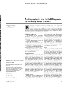
24. Clear the sections in 3-4 changes of xylene of 5 min each. 25. Mount the sections in DPX and view under the light microscope. The immunostaining was considered positive only if the signal localization is correct for the antibody used. RESULTS Out of 40 cases of MSRCT, the most common tumor was NHL (21 cases, 52.5%) followed by Ewing’s Sarcoma/ Primitive Neuroectodermal Tumor (11 cases, 27.5%). Varieties of MSRCT and its location are given in Table 1 and Table 2. Each tumor was adequately categorized using the appropriate antibody panel. The recommended immunohistochemical panel for small round cell tumors is summarized in Table-3. Immunohistochemical staining was interpreted in the context of morphology in the appropriate cells concerned by comparing with the corresponding HE section. DISCUSSION Immunohistochemistry is playing an increasing role in modern surgical pathology.3,4 The objective of performing immunohistochemistry (IHC) is to recognize cell constituents (antigens) and, consequently, to identify and classify specific cells within a cell population whose morphology is heterogeneous or apparently homogenous. The visualization of the antigen-antibody complex is made possible through the addition of either a fluorochrome conjugate or an enzyme to the antibody, which is then viewed under microscopy.5 The use of extensive panels of antibodies in all malignant undifferentiated neoplasm’s allows accurate histological diagnosis in more than 89% cases.6 As shown in our results in Table 2, NHL in organs other than the lymph nodes is the most common MSRCT. IHC was performed in all cases of suspected NHL. The tumor cells show cytoplasmic and membranous immunoreactivity to CD45 (Fig.1A and 1B). Cytokeratin (CK) was negative which excluded poorly differentiated and undifferentiated carcinomas. CD45 is a surface antigen expressed by virtually all hematolymphoid proliferations, and monoclonal antibodies for this marker are reliably specific.7, 8 EWS/PNET is the second most common MSRCT. The tumor cells show cytoplasmic immunoreactivity to vimentin and membranous immunoreactivity to CD99 (Fig 2A and 2B). CD45 was negative which ruled out lymphoma. Immunohistochemically, strong membrane staining for CD99 is consistently seen in almost all cases of EWS/ PNET, although it is not very specific because it is shown in several other soft tissue sarcomas and lymphoblastic lymphomas.9-12 There was one case of LL which showed membrane positivity for CD99 but negative staining for CD45. This excluded the diagnosis of NHL but EWS/
89
PNET was considered in the differentials. LL is especially a diagnostic challenge because of its overlap with EWS both morphologically and immunohistochemically.13 LL may be negative for CD45, but positive for CD99. In our study, LL showed negative staining to vimentin hence the possibility of EWS/PNET was eliminated. We found one case of rhabdomyosarcoma which showed cytoplasmic immunoreactivity for vimentin, desmin and CD99, hence subcategorized as alveolar rhabdomyosarcoma. Immunohistochemically, rhabdomyosarcoma express skeletal muscle markers, specifically, desmin, myoD1, myogenin and CD99(the alveolar type).14,15 It’s due to the unavailability of the following markers in our laboratory, There were 2 cases of atypical carcinoid tumor and one case of neuroblastoma which showed cytoplasmic positivity for NSE, synaptophysin and chromogranin. Absence of positive staining for CD99 and CD45 was helpful in differentiating it from EWS/PNET and lymphoma. The markers CD56, synaptophysin, and chromogranin A are restricted to neuroendocrine neoplasms; nevertheless, it should be reemphasized that they are seen in only 30-50% of cases.16 Neuroblastoma shows immunoreactivity with NSE, synaptophysin, chromogranin-A, CD56, and NB84. Tumor cells usually do not react with vimentin, CD99, actin or desmin.17 We reported 2 cases of small cell variant of synovial sarcoma which was difficult to distinguish from EWS/PNET. However, small cell SS can be differentiated from EWS/PNET by positive staining for EMA(95-100%) of cases, CK(50%); it may also express CD99.18 These markers EMA and Ck are conversely negative in most EWS/PNET.1922 In our study the tumor cells labeled for EMA(cytoplasmic), CD99, and vimentin, but CK was negative (Fig 3B-D). There was one case of malignant melanoma with small cell morphology and absence of melanin pigment. The tumor cells showed cytoplasmic staining for HMB45, S100 and vimentin which confirmed the diagnosis of melanoma (Fig 4A-C). The S100 is a sensitive, albeit not a specific, melanoma marker, decorating more than 95% of MM’s of primary and metastatic sites. The diagnosis of MM requires confirmation with a melanocyte-specific marker, including HMB-45, Melan-A.1 Although wilm’s tumor, desmoplastic small round cell tumor, mesenchymal chondrosarcoma, small cell osteosarcoma and small cell carcinoma fall in the category of SMRCT, as we did not encounter these tumors during our study. CONCLUSION The SMRCTs are a heterogeneous group of malignant neoplasms. IHC represents a tool that can provide a clear distinction among the various tumor types. Its purpose is to categorize patients in order to ensure appropriate and specific treatment, as well as to identify tumors at higher risk of recurrence and fatal outcomes. Because IHC can provide such important information, it must be performed at a high standard so that the results are meaningful and reproducible. In recent years, a better understanding of molecular genetic
90
Bashyal R et al.
1A
1B
2A
2B
3A
3B
3C
3D
4B
4C
4A
Figure 1A: Non Hodgkin Lymphoma composed of diffusely arranged neoplastic cells (HE stain, X20) Figure 1B: Tumor cells showing cytoplasmic and membranous immunoreactivity to CD45 (CD45 stain, X20) Figure 2A: Microphotograph showing tumor cell nests comprising of small round cells (HE stain, X10) Figure 2B: Tumor cells showing membranous CD99 positivity confirming the diagnosis of Ewing’s sarcoma/PNET (CD99 stain, X10) Figure 3A: Microphotograph showing hyperchromatic diffuse small round tumor cells (HE stain, X40) Figure 3B: Tumor cells of synovial sarcoma (small cell variant) showing cytoplasmic vimentin positivity (Vimentin stain, X20) Figure 3C: Tumor cells of synovial sarcoma showing membranous CD99 positivity (CD99 stain, X20) Figure 3D: Tumor cells of synovial sarcoma showing cytoplasmic EMA positivity (EMA stain, X20) Figure 4A: Tumor cell nest with absence of melanin pigment (HE stain,X40) Figure 4B: Tumor cells of malignant melanoma showing cytoplasmic immunoreactivity to S100 (S100 stain, X20) Figure 4C: Tumor cells of malignant melanoma showing cytoplasmic immunoreactivity to HMB 45 (HMB 45 stain, X20)
IHC in the diagnosis of malignant small round cell tumors
studies of these tumors allows molecular testing as a further valuable tool for definitive diagnosis in questionable cases. AKNOWLEDGEMENT The authors wish to express their thanks to Mr. M Sapkota of the pathology department of BP Koirala Memorial Cancer Hospital for the technical assistance and support in the preparation of the IHC slides. REFERENCES 1.
Bahrami A, Truong L. True Identity by Immunohistochemistry. Arch Pathol Lab Med 2008;132:326-48
2.
Wick MR. Immunohistochemical approaches to the diagnosis of undifferentiated malignant tumors. Ann Diagn Pathol 2008;12:72-84.
3.
Chan JK. Advances in immunohistochemistry: Impact on surgical pathology practice. Semin Diagn Pathol 2000;17:170–7.
4.
Jagirdar J. Immunohistochemistry: then and now. Arch Pathol Lab Med 2008;132: 323–5.
5.
Capelozzi VL. Role of immunohistochemistry in the diagnosis of lung cancer. J bras pneumol 2009;35:4.
6.
Ahmed Z, Azad NS, Bhurgari Y et al. Significance of immunohistochemistry in accurate characterization of malignant tumors. J Ayub Med Coll Abbottabad 2006; 18:38-43.
7.
Kurtin PJ, Pinkus GS. Leukocyte common antigen-a diagnostic discriminant between hematopoietic and nonhematopoietic neoplasms in paraffin sections using monoclonal antibodies: correlation with immunologic studies and ultrastructural localization. Hum Pathol 1985;16:353-65.
8.
9.
Wick MR. Monoclonal antibodies to leukocyte common antigen, In: Monoclonal antibodies in diagnostic immunohistochemistry. Wick MR, Siegal GP, editors: New York: Marcel Dekker;1988.pp 285-307. Ambros IM, Ambros PF, Strehl S et al. MIC2 is a specific marker for Ewing’s sarcoma and peripheral primitive neuroectodermal tumors. Evidence for a common histogenesis of Ewing’s sarcoma and peripheral primitive neuroectodermal tumors from MIC2 expression and specific chromosome aberration. Cancer 1991;67:1886-93.
91 10. Granter SR, Renshaw AA, Fletcher CD et al. CD99 reactivity in mesenchymal chondrosarcoma. Hum Pathol 1996;27:1273-6. 11. Lae MW, Roche PC, Jin L et al. Desmoplastic small round cell tumor: a clinicopathologic, immunohistochemical, and molecular study of 32 tumors. Am J Surg Pathol 2002; 6:823-35. 12. Ozdemirli M, Fanburg-Smith, JC, Hartmann DP et al. Differentiating lymphoblastic lymphoma and Ewing’s sarcoma: lymphocyte markers and gene rearrangement. Mod Pathol 2001;14:1175-82. 13. Ozdemirli M, Fanburg-Smith JC, Hartmann DP et al. Precursor B-lymphoblastic lymphoma presenting as a solitary bone tumor and mimicking Ewing’s sarcoma: a report of four cases and review of the literature. Am J Surg Pathol 1998;22:795-804. 14. Wang NP, Marx J, McNutt MA et al. Expression of myogenic regulatory proteins (myogenin and MyoD1) in small blue round cell tumors of childhood. Am J Pathol 1995;147:1799-1810. 15. Cessna MH, Zhou H, Perkins SL et al. Are myogenin and myoD1 expression specific for rhabdomysarcoma? A study of 150 cases, with emphasis on spindle cell mimics. Am J Surg Pathol 2001;25:1150-7. 16. Guinee Jr, Fishback NF, Koss MN et al. The spectrum of immunohistochemical staining of small cell lung carcinoma in specimens from transbronchial and open-lung biopsies. Am J Clin Pathol 1994;102:406-14. 17. Shaoying Li, Siegal GP. Small Cell Tumors of Bone. Adv Anat Pathol 2010;17:1-11. 18. Swanson PE, Wick MR. Soft tissue tumors. In: Colvin R, Bhan A, McCluskey R, (eds). Diagnostic immunopathology. 2nd ed. Lippincott-Raven: Philadelphia; 1995. pp599-632. 19. Weiss SW, Goldblum JR. Enzinger and Weiss’s Soft Tissue Tumors. 4th ed. St Louis, Mo: Mosby; 2001.pp235-49. 20. Garin-Chesa P, Fellinger EJ, Huvos AG et al. Immnuohistochemical analysis of neural cell adhesion molecules: differential expression in small round cell tumors of childhood and adolescence. Am J Pathol 1991;139:275-86. 21. Miettinen M, Cupo W. Neural cell adhesion molecule distribution in soft tissue tumors. Hum Pathol 1993;24:62-6. 22. Mechtersheimer G, Staudter M, Moller P. Expression of the natural killer cell associated antigens CD 56 and Cd57 in human neural and striated muscle cells and in their tumors. Cancer 1991;51:1300-7.