The University of Pennsylvania Orthopaedic Journal 13: 10–17, 2000 © 2000 The University of Pennsylvania Orthopaedic Journal
Rehabilitation of the Shoulder Following Rotator Cuff Surgery BRIAN G. LEGGIN, M.S., P.T., O.C.S.
AND
Introduction
MARTIN J. KELLEY, M.S., P.T., O.C.S.
therapist once in the hospital, then at subsequent postoperative visits with the surgeon. These visits are usually at seven to 10 days, four to six weeks, three months, and six months postoperatively. More frequent visits to a therapist may be necessary if the patient is not progressing as expected. The rehabilitation program we implement consists of a core set of stretching and strengthening exercises. The imitation of these exercises depends on the type of procedure performed and the size of the rotator cuff tear (Tables 1–3). It is useful to break down the rehabilitation process into phases. The goals of each phase are as follows: phase I consists of patient education, controlling pain and inflammation, restoring normal ROM, and decreasing atrophy; phase II consists of optimizing ROM and restoring strength and endurance; phase III consists of optimizing strength and neuromuscular control, improving endurance, and preparing for return to work or sport; and phase IV consists of returning to work or sport and promoting the concept of prevention and modification. We will discuss rehabilitation of rotator cuff surgery in reference to these phases and the nuances of rehabilitation based on the size of the rotator cuff tear.
Effective rehabilitation of the shoulder following rotator cuff surgery is considered essential for the patient to return to an acceptable level of function. Controversy exists regarding how much rehabilitation is required and whether it should be performed under a therapist’s supervision or directed completely by a physician [1,2]. Our philosophy is that rehabilitation is most effective when a team approach is utilized with excellent communication among team members. The team comprises the surgeon, the therapist, and the patient. The surgeon communicates to the patient and therapist the specifics of the procedure and necessary precautions. It is extremely useful for the therapist to receive a copy of the operative report prior to seeing the patient. At the very least, prescriptions for therapy should include diagnosis, surgical intervention, range of motion (ROM) limitations, and precautions. The therapist can then educate the patient on precautions, proper performance of exercise, and use of rest and modalities for pain relief. The therapist must also communicate to the surgeon the patient’s progress and any complications involved in the rehabilitation process. The patient’s responsibility is to diligently perform the exercises while at home and follow through on the precautions that have been outlined. In addition, the patient must provide feedback to both the surgeon and therapist regarding progress toward his/her goals. When the patient does not progress as expected, the therapist and surgeon must discuss and implement program modifications in order to achieve the desired outcome. We have found that this team approach is most successful when the therapist regularly spends time with the surgeon during office hours and observes surgery. Rehabilitation following rotator cuff repair may vary based on surgical technique, cuff tear size, tissue quality, amount of tension at repair site, patient age, patient goals, functional demands of the patient, and systemic disease processes. The prognosis following repair has been correlated to the rotator cuff tear size [3,4], presurgery atrophy [5], and presurgery ROM restrictions [5]. The amount of interaction the patient has with the surgeon and therapist is dictated by individual patient need. Typically, the patient is seen by the
Phase The therapist must take into consideration many factors of a patient’s history prior to initiating the rehabilitation program. These include surgical procedures performed including acromioplasty and/or cuff repair, the size of the tear and tendon involvement, quality of the tissue and ease of tendon mobilization, surgical technique, presurgery treatment, and the patient’s goals. Patients who have had an acromioplasty with an intact cuff and/or small and medium-sized cuff repairs are immobilized in a sling for comfort. If patients are comfortable, they can go without the sling as early as the first postoperative day. Patients who have had large or massive rotator cuff repairs are immobilized in an abduction pillow or brace for the first three to six weeks postoperatively. They are allowed to come out of the brace for exercise, bathing, and dressing, but must keep the arm passively supported at 45 degrees in the plane of the scapula. We recommend that a patient use a slightly deflated beach ball for support during bathing. The rationale for bracing in this position stems from the work by Hersche and Gerber [6] in which they measured the passive tension generated in the supraspinatus musculotendinous unit at the time of repair in
From the University of Pennsylvania Health System, Penn Therapy and Fitness, Philadelphia, PA. Address correspondence to Brian G. Leggin, M.S., P.T., O.C.S., University of Pennsylvania Health System, Penn Therapy and Fitness, 3624 Market Street, Philadelphia, PA 19104. E-mail:
[email protected]
10
REHABILITATION patients undergoing repair of long-standing rotator cuff rupture. They found that shoulder adduction increases the amount of passive tension of the muscle. Positioning the arm at 45 degrees of elevation in the plane of the scapula allows healing while reducing the risk of damage to the rotator cuff repair site. Patients should also be instructed to avoid leaning on their elbows, sleeping on the involved side, making sudden movements, heavy lifting and carrying, reaching behind their backs, and pushing or pulling. They may use their affected arm for waist-level activities in the plane of the body and basic activities of daily living such as feeding. Patients are also instructed in the importance of only performing those home exercises outlined by the thera-
OF THE
SHOULDER
11
pist or surgeon and benefits of using modalities such as the use of ice for pain relief. All patients, regardless of type of surgery, are instructed in pendulum exercises, elbow active ROM, and hand squeezes on the first postoperative day. Those who have had an acromioplasty with an intact rotator cuff or those who have had a small rotator cuff repair may also be instructed in supine passive forward elevation using the opposite hand for assistance. At the first postoperative visit (typically seven to 10 days after surgery) the therapist reviews the precautions and monitors the patient’s performance of the exercises. Patients are instructed in phase I stretching exercises that include supine passive forward elevation and su-
Table 1. Acromioplasty guidelines: intact rotator cuff Phase I: 0–4 weeks Goals 1. Patient education 2. Permit healing 3. Control pain and inflammation 4. Initiate ROM exercises Treatment Immediately postoperative or postoperative day 1 1. Immobilized in sling (use sling for comfort) 2. Pendulums 3. Hand squeezes 4. Elbow active ROM Supine passive ROM forward elevation (in appropriate patient) 7–10 days postoperatively 1. Pendulums 2. Supine passive ROM forward elevation and external rotation 3. Heat and ice 4. Active scapular exercises (shoulder shrugs, scapular retraction)
Phase II: 4–6 weeks postoperatively Goals 1. Improve to full ROM 2. Improve neuromuscular control and strength Treatment 1. Continue all stretches 2. Add phase II stretches if not yet performing (extension, internal rotation, and cross body adduction) 3. Progress to phase II strengthening exercises when at green for all phase I strengthening (abduction, forward elevation, and external rotation at 45° in POS with arm supported) 4. Advanced scapular strengthening 5. Manual resistance for rotator cuff, deltoid, and PNF 6. Bodyblade below 45°
2–4 weeks postoperatively 1. Continue all stretches 2. Passive ROM internal rotation, cross body abduction, and extension 3. Phase I strengthening (internal rotation, external rotation, extension) Phase III: 6–12 weeks postoperatively Goals 1. Full painfree ROM 2. Optimize neuromuscular control 3. Improve endurance 4. Initiate return to functional activities Treatment 1. Continue all stretches and strengthening (progress rotator cuff exercises into POS abduction) 2. Appropriate variable resistance and/or free weight resistance 3. Strengthening above 90° 4. Plyometrics*/Bodyblade 5. Work/sport-specific exercise* *Applies to athlete or laborer.
Phase IV: 12–16 weeks postoperatively Goals 1. Return to sport,* occupation,* or desired activities 2. Promote concept of prevention Treatment 1. Work hardening* 2. Sport-specific training*
12
LEGGIN
pine passive external rotation. For the latter, a stick may be used for assistance (Fig. 1). Passive forward elevation is performed with the arm in the plane of scapula and with the elbow bent at 90 degrees in order to reduce the lever arm and to allow neutral tension of the soft tissues of the shoulder. Passive external rotation is performed with the arm positioned at 45 degrees in the plane of the scapula because external rotation with the arm adducted has been shown to increase tension across the repaired tissues [7,8]. The patient is instructed to perform 10–20 repetitions with at least a 10-second hold, four to six times per day at home. Emphasis is placed on the patient achieving a tolerable, submaximal stretch several times per day rather than aggressive short bouts of stretching. Patients may be referred to supervised therapy if the surgeon or therapist believes that they
AND
KELLEY do not have the ability to adequately perform the exercises at home or if they have excessive stiffness or pain. Patients who have had an acromioplasty or small rotator cuff repair will be instructed in phase II stretching (Fig. 2) exercises at two to four weeks postoperatively. Phase II stretching consists of passive extension with a stick, passive functional internal rotation with the opposite hand or a towel, and cross body adduction with the opposite hand. These patients will be instructed in phase I strengthening exercises (Fig. 3) at four weeks postoperatively if manual resistance of the rotator cuff is painfree. However, if minimal manual resistance causes pain, phase I strengthening will be delayed until at least six weeks postoperatively. Phase I strengthening exercises consist of internal rotation, external rotation, and extension of the adducted arm against
Table 2. Rehabilitation guidelines for small/medium rotator cuff tears following surgical repair Phase I: 0–6 weeks Goals 1. Patient education 2. Permit healing 3. Control pain and inflammation 4. Initiate ROM exercises Treatment Immediate postoperative or postoperative day 1 1. Immobilized in sling (use sling for comfort) 2. Pendulum 3. Hand squeezes 4. Elbow active ROM Supine passive ROM forward elevation (in appropriate patient)
Phase II: 6–8 weeks postoperatively Goals 1. Improve to full ROM 2. Improve neuromuscular control and strength 3. Emphasize normal scapulohumeral rhythm Treatment 1. Continue all stretches 2. Add phase II stretches (extension, internal rotation, and cross body adduction) 3. Phase I strengthening (external rotation, internal rotation, extension) 4. Resisted scapular strengthening (with arms below shoulder height)
7–10 days postoperatively 1. Pendulums 2. Supine passive ROM forward elevation and external rotation 3. Heat and ice 4. Active scapular exercises (shoulder shrugs and scapular retraction) Phase III: 8–12 weeks postoperatively Goals 1. Full painfree ROM 2. Optimize neuromuscular control 3. Improve endurance 4. Initiate return to functional activities Treatment 1. Resisted scapular strengthening 2. Manual resistance for rotator cuffs and deltoid 3. Bodyblade below 45° 4. Progress to phase II strengthening (abduction, forward elevation, and external rotation at 45° in POS with arm supported) when at green for all phase I exercises 4. Appropriate variable resistance and/or free weight resistance 5. Strengthening above 90° 6. Bodyblade *Applies to athlete or laborer.
Phase IV: 16 weeks–6 months postoperatively Goals 1. Return to sport, occupation, or desired activities* 2. Promote concept of prevention Treatment 1. Work on sport-specific exercises* 2. Work hardening* 3. Gradual return to sport or desired activities*
REHABILITATION elastic resistance. A pillow or towel roll may be used under the arm to help alleviate passive tension across the rotator cuff during exercise. In order to standardize the resistance provided by the elastic band, the patient is instructed to begin in the starting position with no tension on the band and step away until there is tension on the band. We always start with the yellow Thera-Band威 for 10 repetitions. If the patient can perform the first set of 10 repetitions without difficulty, a second set of 10 is performed. When patients can perform both sets of 10 without difficulty, they perform a third set. Once all three sets of 10 repetitions are performed with ease, they progress to the next color (usually red) and repeat the sequence. During phase I, all rotator cuff patients may be instructed in active scapular exercises such as shoulder shrugs and scapular retraction. These exercises to maintain or improve scapulothoracic mobility and strength.
OF THE
SHOULDER
13
Phase II Phase II of the rehabilitation process typically begins six weeks after surgery. Patients are asked to continue performing phase I stretching exercises. Those who have had medium, large, or massive rotator cuff repairs progress to phase II stretching exercises and phase I strengthening exercises. Caution must be employed with patients who have had massive rotator cuff repairs. Restrictions in internal rotation are expected due to the nature of the repair; therefore, overstretching to regain this ROM should be avoided! In addition, pain with phase I strengthening exercises may necessitate shorter arcs of motion. Patients who do not progress as expected may be referred for supervised therapy. Glenohumeral mobilizations and gentle, manual stretching can be performed to help advance passive ROM. The elastic resistance strengthening exercises may be augmented with manual resistance performed with
Table 3. Rehabilitation guidelines for large/massive rotator cuff tears following surgical repair Phase I: 0–6 weeks postoperatively Goals 1. Patient education 2. Permit healing 3. Control pain and inflammation 4. Initiate ROM exercises Immediate postoperative or postoperative day 1 1. Patients may be immobilized in sling or abduction brace If sling, use for comfort If abduction brace, immobilized for 3–6 weeks 2. Pendulums 3. Hand squeezes 4. Elbow active ROM
Phase II: 6–12 weeks postoperatively Goals 1. Improve to full ROM 2. Improve neuromuscular control and strength Treatment 1. Continue all stretches 2. Add phase II stretches (internal rotation, cross body adduction, and extension) 3. Rotator cuff isometrics (submaximal) 4. Phase I strengthening (external rotation, internal rotation, extension) 5. Resisted scapular strengthening (with arms below shoulder height)
7–10 days postoperatively 1. Pendulums 2. Supine passive ROM forward elevation and external rotation above level of brace 3. Heat and ice 4. Active scapular exercises (shoulder shrugs and scapular retraction) Phase III: 12–16 weeks postoperatively Goals 1. Full painfree ROM 2. Optimize neuromuscular control 3. Improve endurance 4. Initiate return to functional activities Treatment 1. Continue all stretches and strengthening 2. Progress to phase II strengthening when at green for all phase I exercises (abduction, forward elevation, external rotation at 45° in POS with arm supported) 3. Manual resistance for rotator cuff and deltoid 4. Bodyblade in nonprovocative positions *Applies to athlete or laborer.
Phase IV: 16 weeks–6 months postoperatively Goals 1. Return to work, sport, or desired activities (in appropriate patient) 2. Promote concept of prevention Treatment 1. Work hardening* 2. Gradual return to work or desired activity 3. Progress Bodyblade into elevated positions 4. Work/sport-specific exercises
14
LEGGIN
multiangle isometrics beginning at 45 degrees in the plane of the scapula. Four directions of submaximal isometric resistance are performed by the patient in sequence: abduction/external rotation, adduction/internal rotation, elevation, and extension. This technique allows the therapist to assess the patient’s strength and pain level with resistance and continues the process of neuromuscular training of the glenohumeral and scapular muscles. If pain is encountered, the position and/or resistance level is modified or the exercise is deferred until another session. Depending on the patient’s response to resistive exercise and time from surgery, the position of resistance is progressed toward 90 degrees in the
AND
KELLEY plane of the scapula. This progression continues to more provocative positions as tolerated. Rehabilitation of the patient who has undergone latissimus dorsi transfer for treatment of massive rotator cuff tear presents a unique challenge. We have discovered that although the insertion of the latissimus dorsi has been altered such that it is now in the position of an external rotator, it is still activated by adduction and extension of the arm. The muscle must be reeducated to work as an external rotator. To do so, the patient’s arm is placed at 45 degrees in the plane of the scapula and supported with a foam wedge or pillow on a table. The patient adducts the arm by pressing in to the wedge or pillow. Once this is done, the latissimus fires and the arm is placed in 20 degrees of external rotation. The patient is asked to hold this position for three to five seconds. This is done for 10 repetitions at 20 degrees, 30 degrees, 45 degrees, and 60 degrees. Once patients have mastered the isometric hold at each of these positions, they are asked to go through the motion actively while simultaneously adducting the arm. Elastic resistance can later be added to help increase strength. We are currently studying at what point, if ever, the latissimus dorsi can actively externally rotate the arm without simultaneous adduction. One factor affecting outcome in this population is that the patient must have an intact and well-functioning subscapularis in order to achieve adequate forward elevation [9,10]. The addition of scapular strengthening is also part of this phase of rehabilitation. Elastic resistance below shoulder height is utilized to enhance scapulohumeral rhythm. Emphasis is on slow, controlled movements to integrate the scapular and glenohumeral muscles. A device that we have found useful in rehabilitating patients with shoulder pathology is the Bodyblade (Hymanson Inc., Playa Del Ray, CA). This device enhances strength, dynamic control, proprioception, and endurance training. Small to large oscillations of a fiberglass rod are performed in multiple positions and at various time intervals. Oscillating the blade requires short excursion, high-speed cocontraction muscle activity of the rotator cuff-deltoid-biceps complex in addition to the scapular muscles. In selected patients, this device can be used at 8 to 10 weeks postoperatively in nonprovocative positions. Typically, patients begin in a slightly abducted position and are asked to oscillate the blade primarily through elbow flexion and extension. This requires proximal stabilization of the glenohumeral and scapulothoracic joints while the distal elbow is moved. The Bodyblade exercise progresses to internal/ external rotation at 30 degrees with the elbow flexed and to various positions below shoulder height. Careful attention must be paid to increasing pain or soreness. Phase III
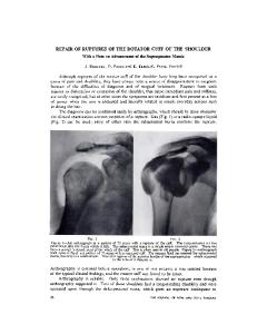
Fig. 1. Phase I stretching exercises. (A) Passive elevation. (B) Active-assisted elevation. (C) Passive external rotation in 45 degrees of elevation in plane of scapula.
This phase begins six weeks postoperatively for patients with an intact cuff, eight weeks for patients with mediumsized cuff repairs, and 12 weeks for patients with large or massive cuff repairs. Patients should have near full passive ROM unless they have had massive rotator cuff repair re-
REHABILITATION
OF THE
SHOULDER
15
Fig. 2. Phase II stretching exercises. (A) Extension. (B) Internal rotation. (C) Cross body adduction.
quiring significant lateral mobilization of the tendon. This latter group of patients may have limitations in external rotation and functional internal rotation. In these cases, we encourage patients to continue slow progression of their passive ROM exercises. It is important to assess whether the patient can achieve painfree, end range of active motion. Commonly, abnormal scapular elevation (shrugging) is present and requires scapulohumeral rhythm to be normalized. Many patients, especially those with large or massive rotator cuff repairs, may not be able to achieve greater than 90 degrees of active forward elevation. The therapist must evaluate whether this limitation is due to weakness, stiffness, or lack of neuromuscular control. If near full passive ROM is present and there is good rotator cuff strength with the arm at the side,
lack of neuromuscular control is the suspected culprit. It may have been six to 12 months since the rotator cuff worked in functional positions. Therefore, it is reasonable to suspect that it has “forgotten” how to work in those elevated or end range positions. In addition, the deltoid has not been used in this manner and most likely lacks the necessary strength to abduct or flex the arm against gravity. Patients who are unable to actively elevate the arm against gravity should be trained in the supine position. The therapist should apply light resistance to external rotation while the patient is asked to elevate the arm with the elbow flexed. This is repeated in the supine position until patients can elevate the arm independent of external rotation resistance. Gravity is gradually introduced by placing the patient in the semi-reclined position. The position is progressively
Fig. 3. Phase I strengthening exercises. (A) External rotation. (B) Extension. (C) Internal rotation. A towel roll or bolster can be used if the patient is experiencing pain.
16
LEGGIN
inclined when the patient can elevate the arm without external rotation resistance. This process is repeated until the patient can elevate the arm in the erect position. This exercise is enhanced by having the patient hold a ball (preferably a medicine ball) with both hands. The patient holds the ball with the palms of the hands to promote proximal contraction of the shoulder muscles. Another way to add resistance to this exercise is to tie an elastic band around the patient’s foot. With both the medicine ball and elastic resistance, begin in the supine position and gradually introduce the resistance of gravity. In this phase, all patients progress to phase II strengthening exercises (Fig. 4). These exercises include abduction to 45 degrees, forward elevation, and external rotation supported at 45 degrees in the plane of the scapula. These exercises are designed to begin training the rotator cuff and deltoid for functional demands. As always, pain is respected and the intensity of the exercises must be monitored. The patient may also now be advanced to isolated triceps and biceps exercises with the arm at the side; row-type pulls at chest level and latissimus pull downs (modified to the front in the plane of the scapula) are also added. Phase IV This phase typically begins at 16 weeks and continues to six months. At this time, most lower demand patients gradu-
AND
KELLEY ally progress their home exercise program. Patients are encouraged to approach overhead activities with caution and to, whenever possible, use ladders or stools to raise their hand closer to the task so that the elbow can remain below shoulder level. They are again instructed in the biomechanics of lifting in an effort to reduce the risk of rotator cuff overload. In the athlete, plyometric training using weighted balls can be used to enhance neuromuscular control, strength, and proprioception by reproducing the physiologic stretchshortening cycle of muscle in multiple shoulder positions. By catching and/or throwing a weighted ball (2 to 10 pounds), the adductors/internal rotators are eccentrically loaded, and thus stretched, which is followed by a concentric shortening phase. These exercises appear to enhance muscle performance by neuromuscular control. Initially, concentric activity may be utilized by an activity simulating a chest pass. A lighter weight ball can be used to simulate the throwing motion. Also, catching the ball from a vertical drop while standing or side lying is a technique to eccentrically load the posterior cuff and scapular decelerators. The patient who must return to work is gradually progressed to work-simulated activities. Emphasis is placed on simulating work activities in a safe and effective manner. The patient is educated on proper lifting mechanics, ergonomic modifications, and common sense. Summary Proper rehabilitation of the shoulder following rotator cuff surgery is essential to the recovery of patients who have had rotator cuff surgery. Successful rehabilitation is dependent on effective communication and interaction among the surgeon, therapist, and patient. Each of these team members has a defined role in the rehabilitation process and must fulfill their responsibilities in order for the desired outcome to be achieved. This article presented principles and rationale for rehabilitation of patients after rotator cuff surgery. Whether the patient will be followed in supervised therapy or seen in the office at specific intervals, extensive patient education is essential for a successful rehabilitation. Constant reevaluation by the surgeon and therapist is important to make necessary program modifications if the patient is not achieving preset goals. The common goals of the rehabilitation process include reduction of pain and inflammation, facilitation of collagen healing, improvement in ROM and strength, and optimization of proprioception and endurance. This is achieved by gradually increasing the program from nonprovocative to provocative positions. It is the responsibility of the therapist and surgeon to identify when to implement the appropriate modalities or exercises in order to improve the impairment and thereby increase the patient’s function. References
Fig. 4. Phase II strengthening exercises. (A) Abduction. (B) Forward elevation.
1. Andersen N, et al: Self-training versus physiotherapist-supervised rehabilitation of the shoulder in patients treated with arthoscopic sub-
REHABILITATION
2.
3.
4.
5.
acromial decompression: A clinical randomized study. J Shoulder Elbow Surg 8:99–101, 1999. Kelley M and Leggin B: Shoulder rehabilitation. In: Iannotti WG (ed). Disorders of the Shoulder: Diagnosis and Management. Philadelphia: Lippincott Williams & Wilkins, pp. 979–1019, 1999. Gazielly D, et al: Functional and anatomical results after surgical treatment of ruptures of the rotator cuff. 1. Preoperative functional and anatomical evaluation of ruptures of the rotator cuff. Rev Chir Orthop 81:8–16, 1995. Habernek H, et al: A new approach to the subacromial space: Technique and 2-year results in 28 rotator-cuff repair cases. Acta Orthop Scand 64:92–94, 1993. Gshwend N, Bloch H, Bischof A: Long-term results of surgical management of rotator cuff rupture. Orthopade 20:255–261, 1991.
OF THE
SHOULDER
17
6. Hersche O and Gerber C: Passive tension in the supraspinatus musculotendinous unit after long-standing rupture of its tendon: A preliminary report. J Shoulder Elbow Surg 7:393–396, 1998. 7. Harryman DT II et al: The role of the rotator interval capsule in passive motion and stability of the shoulder. J Bone Joint Surg 74A: 53–66, 1992. 8. Zuckerman JD, et al: The effect of arm position and capsular release on rotator cuff repair: A biomechanical study. J Bone Joint Surg 73B: 402–405, 1991. 9. Gerber C, et al: Latissimus dorsi transfer for the treatment of massive tears of the rotator cuff. Clin Orthop 232:51–61, 1988. 10. Gerber C: Latissimus dorsi transfer for the treatment of irreparable tears of the rotator cuff. Clin Orthop Rel Res 275:152–160, 1993.