Musculoskelet Surg (2009) 93:S55–S63 DOI 10.1007/s12306-009-0003-9
Post-operative rehabilitation after surgical repair of the rotator cuff Marco Conti · Raffaele Garofalo · Giacomo Delle Rose · Giuseppe Massazza · Enzo Vinci · Mario Randelli · Alessandro Castagna
Published online: 16 March 2009 © Springer-Verlag 2009
Abstract Today advances in techniques and materials for rotator cuff surgery allow the repair of a large variety of types or extensions of cuff lesions in patients from a wide range of age groups who have different kinds of jobs and participate in different kinds of sports, and who have widely different expectations in terms of recovery of functions and pain relief. A large number of factors must
M. Conti (쾷) Via L. Settala 82, Milan, Italy Office: Via Locatelli 6, Milan, Italy e-mail:
[email protected] M. Conti · G. Delle Rose · M. Randelli · A. Castagna Shoulder Unit IRCCS Istituto Clinico Humanitas Rozzano (MI), Italy R. Garofalo Orthopaedic and Traumatologic Unit Regional Hospital F. Miulli Centre of Excellence Acquaviva delle fonti (BA), Italy G. Massazza Orthopaedic and Traumatologic Unit and Occupational Medicine Medicine Faculty, University of Turin Turin, Italy E. Vinci Cliniche Humanitas Gavazzeni Bergamo, Italy
be taken into account before implementing a rehabilitation protocol after rotator cuff surgery. These mainly include the technique (materials and procedure) used by the surgeon. Moreover, tissue quality, retraction, fatty infiltration and time from rupture are important biological factors while the patient’s work or sport or daily activities after surgery and expectations of recovery must also be assessed. A rehabilitation protocol should also take into account the timing of biological healing of bone to tendon or tendon to tendon interface, depending on the type of rupture and repair. This timing should direct the therapist’s choice of correct passive or assisted exercise and mobilisation manoeuvres and the teaching of correct active mobilisation movements the patient has to do. Following accepted knowledge about the time of biological tissue healing, surgical technique and focused rehabilitation exercise, a conceptual protocol in four phases could be applied, tailoring the protocol for each patient. It starts with sling rest with passive small self-assisted arm motion in phase one, to prevent post-op stiffness. In phase two passive mobilisation by the patient dry or in water, integrated with scapular mobilisation and stabiliser reinforcement, are done. Phase three consists of progressive active arm mobilisation dry or in water integrated with proprioceptive exercise and “core” stabilisation. In phase four full strength recovery integrated with the recovery of work or sports movements will complete the protocol. Because of the multi-factorial aspects of the problem, the best results can be obtained through a full transfer of information from the surgeon to the therapist to optimise timing and sizing of the individual rehabilitation protocol for each patient. Keywords Rehabilitation · Rotator cuff · Shoulder · Rotator cuff surgery · Functional recovery · Passive motion
S56
Introduction It is well known that rotator cuff tendon lesions are frequently found in the framework of shoulder pathologies [1], especially among patients aged over 45. However, especially in sports and overhead working activities, younger people are increasingly subjecting their scapular-humeral joints to significant stress and can therefore suffer from this type of lesion. The presence of a rotator cuff tendon lesion does not necessarily suggest the need for surgical operations [2–5]. There is in fact increasing agreement that surgery is indicated after failure of preventive rehabilitation treatment carried out for a period of at least 3–4 months, or in the cases in which significant and progressive rotator cuff tendon insufficiency occurs [6]. Today, the surgical repair of a rotator cuff tendon tear (RCT) can be carried out using a variety of methods, open, mini-open and arthroscopic techniques [7], and the post-op results are generally good and substantially comparable [8–11]. At present, one of the great topics of discussion is not therefore the surgical technique to use but rather post-surgery recurrence. When minor re-ruptures occur, patients may still show improvement of the clinical symptoms compared with the pre-operative period, while patients with massive re-ruptures do not benefit in any way from the surgical operation [5–12]. The clinical results therefore seem to be linked to the level of healing of the repaired tendon [13], a factor which increasingly appears to be the linchpin for therapeutic success. Many factors can influence healing of the repaired tendon [14, 15]: we can in fact distinguish between surgical factors associated with recognition of the type of lesion (forms and number of tendons involved), the size of the lesion, the technique as such, the mobilisation of the tendon tissue it was possible to obtain intraoperatively, the tendon quality, the degree of muscular hypo-atrophy, as well as adequate subacromial decompression, correct preparation of the humeral tuberosity, and the anchoring and suturing method. Then a series of factors related to the patient such as age, lifestyle and the presence of other shoulder complaints or systemic illnesses [7] are by no means secondary either. In this light, the post-surgery rehabilitative treatment assumes great importance as it must be able to protect the repair in the early stages, to prevent post-op stiffness and then restore the function of the scapular-humeral joint [15, 16]. Many rehabilitation protocols have been proposed, often based only on empirical experiences, without fully taking into account biological aspects relative to the healing steps of the repaired tendon [17–19]. Post-surgical rehabilitation of the suture of rotator cuff tendons, furthermore, can vary from patient to patient, bearing in mind the surgical technique, the patient’s expectations and functional demands, the number of tendons repaired
Musculoskelet Surg (2009) 93:S55–S63
and therefore the grade of the lesion, the quality of the tissue and any associated surgical actions [16].
Tendon healing One of the most important aspects the rehabilitation protocol must take into account is biological time for the tendon to heal. This process involves tendon healing at the bone footprint if the tendon has been reinserted into its anatomical place using a technique with anchors and sutures or alternatively tendon with tendon healing if a suture technique with latero-lateral stitches has been carried out. The reparative phenomena follow a cascading series of mechanisms in an orderly manner one after another in healthy individuals [20] (Table 1). The first stage is the inflammatory one: during the first week it is characterised by inflammatory cells, leukocytes, lymphocytes and monocytes, which release histamine and bradykinin, which increase vascular permeability and therefore allow the plates to reach the level of the repair site. The fibrin along with the fibronectin form a fragile scar which reduces the haemorrhaging process without any real adhesion between tendon and bone. The inflammatory stage lasts for a period which varies between 1and 2 weeks and which is generally transformed into the proliferative stage. During the proliferative stage, the inflammatory tissue is gradually replaced by fibroblasts, myofibroblasts and endothelial cells, which organise themselves with the Table 1 Stages of the tendon healing process Stage (duration)
Evolution of the process
Inflammatory (0–14th day)
Leucocytes, lymphocytes, monocytes Release of histamine and bradykinin which increase vascular permeability Increase of platelets in situ Initial scar thanks to fibrin and fibronectin
Proliferative (2nd–3rd/4th week)
Inflammatory tissue replaced by fibroblasts, myofibroblasts and endothelial cells Formation of granulation tissue Tighter tendon-bone adhesion Production of collagen III (immature) by fibroblasts (after 15 post-op days)
Maturation and remodelling (3rd–4th week – 12th/26th week)
Maturation of the scar tissue Collagen III replaced by mature collagen I Formation of dense connective tissue Integration of the tendon in the bone
Musculoskelet Surg (2009) 93:S55–S63
new extracellular matrix to form a granulation tissue which guarantees tighter adhesion between the tendon and the bone surface. In this stage, the fibroblasts produce type III collagens, which are therefore immature, and glycosaminoglycans; then there is significant neoangiogenesis. This stage lasts about 10 days and commences after the first 15 days after surgical repair. This stage is followed by maturing and remodelling, which therefore begins around the third week and is characterised by maturing of the scar tissue: the immature type III collagen is replaced by type I with the formation of dense connective tissue. By now the fibroblastic cells will have replaced the inflammatory cells. The process continues during the following weeks until the tendon is completely integrated with the bone surface. Tendon healing studies have mainly been carried out on animal models and therefore the remodelling and maturing stages have been seen to vary in duration depending on the animal model used. Some authors have described periods of 12–16 weeks for the tendon to recover its tensile strength [20], while other authors who have studied sheep models have reported times of as much as 26 weeks (4 months) [21]. These studies obviously reveal a set of limits linked to the model studied while, furthermore, human tendons that are ruptured reveal a series of degenerative alterations which can negatively influence and therefore prolong the healing time. The recognition of the biological timing is very important and must be properly recognised by the doctor so as to modulate the rehabilitative timing.
Immobilisation and sling
S57
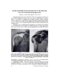
a
b Fig. 1a,b Sling positioning in the neutral or rest position
Bearing in mind the biological healing stages of the repaired cuff tendon tissue, it is clear that precocious and aggressive mobilisation or violent muscular contraction can exceed the mechanical strength of the repair and damage it, even though precocious mobilisation could reduce the risks of articular stiffness. The purpose of a reasoned rehabilitative process after a rotator cuff repair is therefore to obtain cuff tendon healing by recovering mobility and shoulder function gradually. From this standpoint, an adequate immobilisation of the limb is very important during the initial post-operative stage in order to guarantee effective tendon healing. A study on rats has in fact highlighted that the cuff tendons that were immobilised after surgical suturing revealed excellent orientation of the collagen fibres and enhanced organisation of the extracellular matrix compared with rats that were left free after the repair [22]. Adequate immobilisation must however take into account the vascular and biomechanical characteristics of the rotator cuff [23]. The use of an abduction sling during the early weeks seems capable of reducing tension at suture level and improving vascularisation of the scar. We do in fact
know that the hypovascular zone of a healthy supraspinatus tendon is about 1.5 cm from the greater tuberosity of the humerus and the position of the head of the humerus influences tendon vascularisation significantly [24]. Assessing tendon microcirculation in relation to the head position, Rathbun and Macnab showed that there is a reduction of the haematic flow to the tendon when the arm is in a total adduction position [25]. Based on these observations, it seems prudent to recommend post-op immobilisation in a sling with the arm abducted at least to 30° and an external rotation of 0° (a neutral or rest position) for the first 4–6 post-operative weeks in order to improve microcirculation and reduce the stress on the operated tendon, especially in the case of a repair carried out on an inveterate tendon lesion [26, 27] (Fig. 1a,b).
Continuous passive mobilisation There are not many studies in the literature that help us to understand if this therapeutic aid (obtained today with
S58
Musculoskelet Surg (2009) 93:S55–S63
movement only, while it remains to be seen whether it can have a positive effect on faster recovery of working activities or of common everyday activities. It is also not yet known if the use of CPM can influence healing of the repaired tendons to any extent. Certainly, however, this method can be used in patients who have a cuff lesion associated with adhesive capsulitis in the pre-operative period and who can therefore benefit from a recovery, or at least from a non-loss, of the range of movement already gained during the surgical operation.
Post-operative functional rehabilitation
Fig. 2 Example of an automatic programmable CPM system
electronically controlled automatic programmable mobilisers, see Fig. 2) can be of benefit in the rehabilitation of patients operated by suturing the rotator cuff. As a general rule this continuous passive mobilisation (CPM) can be begun in the immediate post-op period on condition that stress in the repair zone is kept low. Hatakeyama et al. have shown that the safety position after this surgery is 30° of elevation on the scapular plane with an external rotation range between 0 and 60° [28]. In a double-blind randomised study of patients treated with repair of the cuff and subacromial decompression, Raab et al. [29] showed that three months after the surgical operation there were no differences in the various scores between patients treated with physical therapy and CPM and those treated with physical therapy only; however the range of movement and pain level were better in patients in the first group. Recently, Michael et al. [30] seemed to confirm these data and also showed how the recovery of the range of movement is faster in patients treated with CPM in the post-operative period. In another randomised prospective study on 31 patients operated for repair of the rotator cuff, Lastayo et al. [31] compared 2 groups, one treated with CPM in the first 4 weeks while the other was subjected in the same period of time to a physical therapy programme with passive recovery of mobility. A follow-up carried out after 22 months found that there were no statistically significant differences in the scores of the two groups or in pain and isometric muscular strength. Our figures, currently being published [32], relating to a randomised prospective study on 100 patients seem to indicate that the precocious use of CPM for at least two hours daily overall, for one month after the operation, can permit better recovery of the passive ROM in both abduction and external rotation and in forward flexion with significant data already at two and a half months. It therefore seems, from the analysis of the literature, that in the medium and long term, CPM succeeds in substantially influencing the recovery of the range of
From what we have said so far, it can be deduced that the rehabilitative management after RCT repair must take into account multiple factors, which the surgeon and rehabilitation therapist must share. This information is made up of the processes and biological timing of tendon healing, the size of the tendon lesion treated, the quality of the treated tendon, the type of repair made (type of cuff suture made, if partial or total, if a monoor pluri-tendon suture, and if the repair is of the tendon–tendon or tendon–bone type; knowledge of the type of implant used – reabsorbable or not reabsorbable – is also important), any associated surgical actions (acromion plastic surgery, resection of the distal clavicle or, in younger patients, repair of an associated lesion of the SLAP type, tenotomy or tenodesis of the long end of the biceps and possibly knowledge of the type of tenodesis technique – whether static or dynamic – to the soft parts of the cuff), the physiological age and expectations of the patient, and the range of pre-op movement of the operated shoulder. In particular, account must be taken of the fact that the prognosis after repair was significantly related to the degree of fatty degeneration of the tendon and of muscular atrophy, the size of the lesion treated and the extent of reduction of the range of movement in the pre-op period [12, 13, 33–35]. In this sense, communications and coordination between the surgeon, physiatrist and physiotherapist are fundamental in order to obtain an optimum result for the patients themselves. The current general consensus in the literature [14, 15, 19, 36–39] is to subdivide the post-operative rehabilitative treatment into four stages, each one with different aims (Table 2). The aim of the first stage is to prevent articular blockage because of post-surgical adherences by means of exercises of the passive type which help to minimise loading at the repair site. The aim of the second stage is the progressive recovery of the passive range of movement without scapular compensation by means of exercises of the assist-
Musculoskelet Surg (2009) 93:S55–S63 Table 2 Objectives of the various rehabilitation stages Stage
Objectives
1
Prevention of joint stiffness due to postoperative adherences Progressive recovery of the range of passive movement without scapular compensation Recovery of strength and of physiological scapulohumeral rhythm Complete the recovery of the power and normal actions for both work and sports
2 3 4
ed/active type which begin gradually to apply work loads on the repaired tendons. The aim of the third stage is to recover strength and physiological scapular-humeral rhythm by means of toning exercises focussing on the recovery of power and the strength of the rotator cuff tendons. The aim of the fourth stage is the best recovery of the strength and normal actions for both work and sports. These stages naturally interweave and overlap without any break and it is possible, in the same stages, to find a series of variables linked to all the conditions regarding the patient and type of lesions treated and to the type of surgical technique used. The first rehabilitation stage runs from the immediate post-op period until the 4th–6th week. During this stage the patient wears the abduction sling (up to 6 weeks for complete lesions; up to 4 weeks for partial and incomplete lesions) and it is only removed 3–4 times per day to carry out passive abduction, front flexion and external rotation mobilisation exercises. During this stage the loads on the repair made must be minimal and, in fact, this stage is characterised biologically by a slight coagulation of fibrin with type III collagen; therefore exercises with active muscular contraction on the operated limb must be avoided at this stage. The recovery of the passive movement must be carried out inside a safety range and the patient must work without pain and with the avoidance of maximum stretching. In the event of a subscapular repair, external passive rotation must be limited to 0° and no more. Therefore patients can make active movements of the wrist, hand and elbow. The active flexion–extension of the latter must be modulated and limited in this stage if tenodesis of the long end of the biceps has been carried out, especially if of the dynamic type at the rotator cuff. Pendulum exercises are useful at this stage, to be carried out with extreme relaxation of the musculature and with the trunk tilted 30° forward. Furthermore, preference must be given to active and proprioceptive work of the scapular/thoracic joint. Once the suture stitches have been removed the passive mobilisation and slight stretching exercises can also be carried out in a pool (2–3 times per week, for 15–20 min per session) [26]. Ice is a useful anti-inflammatory aid for use in this stage, especially in the first 10–15 days and after the
S59
physiotherapy sessions [39]. CPM can be useful in this stage, particularly with patients who have had a reduction of the physiological range of movement or capsulitis. The second stage runs from the 4th–6th week until the 12th (3rd month). This is because from the 6th week after the operation, the extent of healing of the tendon to the bone and of the tendon to tendon begins to be sufficient to allow the introduction of active movements at a minimum load. At this stage, the mobilisation exercises can also be carried out by a therapist and it is possible to begin greater and greater stretching, with decoaptation of the humeral head to prevent a subacromial iatrogenous conflict in order to begin to recover the range of movement towards the greatest angles. It is possible at this stage to begin to use aids such as pulleys and sticks. In this case too it is necessary to continue to maintain certain stratagems. For example, if the top fibres of the subscapular muscle have been sutured, the recovery of external rotation should preferably be obtained by means of an abduction of the limb to 45º (and with the elbow raised 4–6 cm from the couch to reduce stress on the sutures to a minimum if the execution is carried out in a supine position). At 6–8 weeks it is very useful to begin the active mobilisation exercises in water; this should be deemed to be an active mobilisation exercise assisted in a situation of reduced force of gravity and, as a consequence, with low loading at the level of the operated tendon. The patient can be allowed to swim breaststroke and when front passive flexion reaches around 130º some modified backstroke can be added, without submerging the limb but ending the movement at surface level. At this stage, therefore, it is possible to start active movements without forcing and therefore the use of the arm is permitted in everyday activities. The proprioceptive exercises on the scapular-thoracic joint are intensified as is active toning of the active scapular fixator muscles. Particularly important at this stage is the use of neuromuscular biofeedback systems which, though simple, help the therapist to get the patient to “relearn” the ability of voluntary and coordinated control of the fundamental muscle groups for scapulohumeral stabilisation and which, in general, have been dysfunctioning for some time because of profound alteration of the motor patterns induced by the cuff lesion and by the compensation mechanisms implemented instinctively so as to permit the spatial positioning of the hand for as long as possible on the basis of living needs (see Fig. 3). The third stage begins around the third month (10th–12th week) and is the muscular toning stage with progressive functional recovery. This stage lasts until the 12th week and beyond. The start of this stage obviously depends on various factors. As we have already said, one of the most important factors is the type of lesion repaired. This is because the more serious the tendon lesion repaired, the further this stage is put back.
S60
Fig. 3 Simple neuro-muscular sound/visual biofeedback system
Fig. 4 Exercise in a closed kinetic chain
Furthermore, the start of this rehabilitation stage is secondary to recovery of a satisfactory range of active movement of the operated limb, especially in terms of front flexion and external rotation. This is because repeated attempts to tone a shoulder which is still stiff can give rise to pain, subacromial conflict and excess stress on the repair itself. Patients who are unable to actively raise the arm against gravity at this stage should begin to carry out reinforcement exercises without resistance in a supine position. In this position, gravity has virtually been eliminated and the patient begins to raise the limb over 90º and reinforce the deltoid. This exercise can be carried out at the beginning with the elbow flexed and then gradually increasing the lever arm by extending the elbow. This
Musculoskelet Surg (2009) 93:S55–S63
exercise can be carried out progressively with a small weight in the hand or using an elastic resistance system [40]. At this rehabilitation stage it is important to respect the pain while the intensity of the exercises must be properly monitored. At the beginning it is possible to carry out isometric contractions which permit the application of controlled force through the repaired tendon. If the supraspinatus has been repaired, toning is carried out first in an attempt to reinforce the pair of front (subscapular) and rear (subspinatus) forces by means of exercises with the limb abducted to 30°–45° and 60º, so as to limit the possibility of a subacromial conflict that can entail pain as well as mechanical stress on the repair made. Isometric reinforcement is followed by a reinforcement stage with elastic bands and it is initially necessary to concentrate on the execution of many repetitions with low resistances. Remembering that muscular toning is dependent on the articular angle, it is necessary to seek different angular positions of the humerus at which to carry out different exercises so that, with each exercise, you can select in turn the subspinatus, the teres minor, the subscapular, the deltoid, first front and then rear, the mid and inferior parts of the trapezius and the rhomboideus or costoscapularis muscles. The exercises must be modulated to spare the repaired tendon as much as possible at the beginning. Takeda et al. [41] have shown that with the arm abducted on the scapular plane the supraspinatus is isolated and that this would therefore be the ideal position for the reinforcement of this tendon. However it is necessary to be very careful with the humeral rotation in this position. Some NMR studies have shown that in the abduction and internal rotation position the subacromial space is reduced in a dynamic manner and gives rise to stress on the repair [42], and this is why abduction positions of less than 90º are recommended if internal or external rotations are associated with them [26]. In this rehabilitation stage it is also necessary to continue to improve the range of movement with exercises for stretching the capsulo-ligamentous structures, in particular on the antero-inferior and postero-inferior capsule. The proprioceptive work of the scapular stabilisers must be intensified without forgetting “core” stabilisation (the muscular system of the abdominal, oblique, dorsal and gluteus muscles), fundamental for correct positioning of the scapula. A crucial role in progressive functional recovery is played by proprioceptive exercises in a closed kinetic chain first below and then above the breast (Fig. 4). The fourth and last rehabilitation stage begins around the 16th week and continues until the 6th month. This stage is a progression of the third stage, and its end point is different depending on the type of patient [43]. This is because at this stage a patient with a low functional demand will continue to improve in a progressive manner in a programme of exercises prevalently at home and in
Rotator cuff suture operation
Rotator cuff suture operation
Possible CPM for weeks 5/6 (no pain range) with progressive PROM increase Passive/assisted mobilis. in water Proprioceptive in closed kinetic chain below the breast Use of the limb for everyday activities below the breast No loads No fast gestures
CPM (no pain range) with progressive PROM increase
Scapular fixator muscle activation NB: biceps long head spared if tenotomy/tenodesis
Cont. passive / assisted mobilisation for PROM recovery (Max 45º ER if subscapular suture)
Stage 2 4th/6th–12th week
Passive auto-mobilisation Passive pendular exercises (body tilted forward 30º) Active hand-wrist mobilisation Assisted elbow mobilisation in case of biceps long head tenodesis Max 0º ER if subscapular suture
Sling (Abd. 30° and ER 0°)
Stage 1 0–4th/5th week
Breaststroke, plus modified backstroke from weeks 14/16 (see text) Use of the limb for everyday activities also above the breast No loads No fast gestures Musc. isometric reinforcement Humeral ADD + ER + IR (respecting the sutured muscles more) Active reinforcement with elastic bands of the scapular fixator muscles From weeks 14/16, isotonic reinforcement with elastic bands NB: do not stress the supraspinatus (and biceps long head if tenotomy or tenodesis Proprioceptive in closed kinetic chain above the breast “Core” stabilisation: active reinforcement of the gluteus, low abdominal, oblique and dorsal muscles muscular electrostimulation
Stage 3 12th–16/18th week
Table 3 Logical plan of the stages and contents of the rehabilitation process for post-operative rehabilitation of the rotator cuff
Functional recovery – specific sports training
Complete return to everyday activities – specific sports training
> 20th week: simulation of sports gestures in the gym Recovery of work movements (ergotherapy)
Proprioceptive in open kinetic chain, simulating sports gestures
Use of the limb for everyday activities also above the breast
Stage 4 16th/18th– 36th week
Musculoskelet Surg (2009) 93:S55–S63 S61
S62
Musculoskelet Surg (2009) 93:S55–S63
References
Fig. 5 Exercise in an open kinetic chain
an increasingly complete recovery of the normal activities of everyday life and the resumption, with great caution, of overhead activities. With regard to young patients and athletes, these begin exercises first in an open kinetic chain (Fig. 5) and then go on to specific sports recovery exercises first for the gesture and then with force applied to the specific sports gesture itself, while workers begin to carry out activities which simulate the working activity in a specific and progressive manner. It may be the therapist’s job to teach patients appropriate stratagems for limiting stress on the repaired tendon to a minimum during the sports or work activity. See Table 3 for a logical-temporal diagram of the above illustrated rehabilitation process.
Conclusions Evident from the analysis set out above is the consensus about how post-operative rehabilitation constitutes a progressive, integrated and personalised process (rather than a “protocol”) in which a fundamental role is played by the passage of information between the surgeon and the physiotherapist, as well as the sharing of knowledge regarding the characteristics of each individual operation both in terms of surgical technique and in terms of the biological and anatomical characteristics of the repaired tissues. Only through the integration of this information with data regarding the patient’s lifestyle and expectations will it be possible to establish a rehabilitation programme which, though personalised as regards the executive procedures, cannot ignore the times and processes of biological tissue healing in order to achieve the best possible result both in terms of functional recovery and of management of the symptoms. Conflict of interest The authors declare that they have no conflict of interest related to the publication of this manuscript.
1. Sher JS, Uribe JW, Posada A et al (1995) Abnormal findings on magnetic resonance images of asymptomatic shoulders. J Bone Joint Surg Am 77:10–15 2. Worland RL, Lee D, Orozco CG et al (2003) Correlation of age, acromial morphology and rotator cuff tear pathology diagnosed by ultrasound in asymptomatic patients. J South Orthop Assoc 12:23–26 3. Hawkins RH, Dunlop R (1995) Non operative treatment of rotator cuff tears. Clin Orthop Relat Res 321:178–188 4. Ainsworth R (2006) Physiotherapy rehabilitation in patients with massive irreparable cuff tears. Musculoskeletal Care 4:140–151 5. Rockwood CA Jr, Williams GR Jr, Burkhead WZ Jr (1995) Debridement of degenerative irreparable lesions of the rotator cuff. J Bone Joint Surg Am 77:857–866 6. Arroyo J, Flatow EL (1999) Management of rotator cuff disease: intact and repairable cuff. In: Iannotti J, Williams G (eds) Disorders of the shoulder: diagnosis and management. Lippincott Williams and Wilkins, Philadelphia, pp 31–56 7. Accousti KJ, Flatow EL (2007) Technical pearls on how to maximize healing of the rotator cuff. Instr Course Lect 56:3–12 8. Ide J, Maeda S, Takagi K (2005) A comparison of arthroscopic and open rotator cuff repair. Arthroscopy 21:1090–1098 9. Liem D, Bartl C, Lichtemberger S et al (2007) Clinical outcome and tendon integrity of arthroscopic versus mini-open supraspinatus tendon repair: a magnetic resonance imaging controlled matched pair analysis. Arthroscopy 23:514–521 10. Sauerbrey AM, Getz CL, Piancastelli M et al (2006) Arthroscopic versus mini open rotator cuff repair: a comparison of clinical outcome. Arthroscopy 21:1415–1420 11. Bess E, Steuber KU, Waibl B (2005) Open versus arthroscopic rotator cuff repair: a comparative view of 96 cases. Arthroscopy 21:597–604 12. Castagna A, Conti M, Markopoulos N et al (2008) Arthroscopic repair of rotator cuff tear with a modified Mason-Allen stitch: mid-term clinical and ultrasound outcomes. Knee Surg Sports Traumatol Arthrosc 16:467–503 13. Boileau P, Brassart N, Watkinson DJ et al (2001) Arthroscopic repair of full-thickness tears of supraspinatus: does the tendon really heal? J Bone Joint Surg Am 87-A:1229–1240 14. Sonnabend DH, Watson EM (2002) Structural factors affecting the outcome of rotator cuff repair. J Shoulder Elbow Surg 11:212–218 15. Jackins S (2004) Postoperative shoulder rehabilitation. Phys Med Rehabil Clin N Am 15:643–682 16. Bruzga B, Sleer K (1999) Challenges of rehabilitation after shoulder surgery. Clin Sports Med 18:769–793 17. Noyes FR, DeMaio M, Mangine RE (1991) Evaluation-based protocols: a new approach to rehabilitation. Orthopedics 14:1383–1385 18. Glasoe WM, Fischer CJ, Murty D (2004) Treatment for an acute large rotator cuff repair. Physiotherapy 90:217–220 19. Delbrouck C, Dauty M, Huguet D, Dubois C (2002) Rehabilitation after shoulder rotator cuff surgery: in-patient or day-hospitalization (about 76 cases). Ann Readapt Med Phys 46:207–213 20. Carpenter JE, Thomopoulos S, Flanagan CL et al (1998) Rotator cuff defect healing: a biomechanical and histologic analysis in an animal model. J Shoulder Elbow Surg 7:599–605 21. Lewis CW, Sclegel TF, Hawkins RJ et al (2001) The effect of immobilization on rotator cuff healing using modified MasonAllen stitches: a biomechanical study in sheep. Biomed Sci Instrum 37:263–268
Musculoskelet Surg (2009) 93:S55–S63 22. Thomopoulos S, Williams GR, Soslowsky LJ (2003) Tendon to bone healing: differences in biomechanical, structural, and compositional properties due to a range of activity levels. J Biomech Eng 125:106–113 23. Rathbun JB (1970) The microvascular pattern of the rotator cuff. J Bone Joint Surg Br 52-B:540 24. Determe D, Rongieres M, Kany J et al (1996) Anatomic study of the tendinous rotator cuff of the shoulder. Surg Radiol Anat 18:195–200 25. Rathbun JB, Macnab I (1970) The microvascular pattern of the rotator cuff. J Bone Joint Surg Br 52:540–553 26. Millett PJ, Wilcox RB, O’Holleran JD, Warner JJP (2006) Rehabilitation of the rotator cuff: an evaluation-based approach. J Am Acad Orthop Surg 14:599–609 27. Hersche O, Gerber C (1998) Passive tension in the supraspinatus musculotendinous unit after long-standing rupture of its tendon: a preliminary report. J Shoulder Elbow Surg 7:393–396 28. Hatakeyama Y, Itoi E, Pradhan RL et al (2001) Effect of arm elevation and rotation on the strain in the repaired rotator cuff tendon: a cadaveric study. Am J Sports Med 29:788–794 29. Raab MG, Rzeszutko D, O’Connor W, Greatting MD (1998) Early results of continuous passive motion after rotator cuff repair: a prospective, randomized, blinded, controlled study. J Bone Joint Surg Am 80:1002–1011 30. Michael JW, Konig DP, Imhoff AB et al (2005) [Efficacy of a postoperative treatment after rotator cuff repair with a continuous passive motion device (CPM)]. Z Orthop Ihre Grenzgeb 143:438–445 31. Lastayo PC, Wright T, Jaffe R, Hertzel J (1998) Continuous passive motion after repair of the rotator cuff. A prospective outcome study. J Bone Joint Surg Am 80:1002–1011 32. Conti M, Garofalo R, Castagna A, Borroni M (2008) Effects of one month of CPM after arthroscopic rotator cuff repair.
S63
33. 34.
35.
36.
37.
38. 39. 40.
41.
42.
43.
Preliminary results of prospective randomized study. SECEC meeting (submitted for publication) Iannotti J (1994) Full-thickness rotator cuff tears: factors affecting surgical outcome. J Am Acad Orthop Surg 2:87–95 Thomazeau H, Boukobza E, Morcet N et al (1997) Prediction of rotator cuff repair results by magnetic resonance imaging. Clin Orthop Relat Res 344:275–283 Schaefer O, Winterer J, Lohrmann C et al (2002) Magnetic resonance imaging for supraspinatus muscle atrophy after cuff repair. Clin Orthop Relat Res 403:93–99 Rubin BD, Kibler WB (2002) Fundamental principles of shoulder rehabilitation: conservative to postoperative management. Arthroscopy 18:29–39 Kibler WB, McMullen J, Uhl T (2001) Shoulder rehabilitation: strategies, guidelines and practice. Orthop Clin N Am 32:527–538 Kibler WB (2003) Rehabilitation of rotator cuff tendinopathy Clin Sports Med 22:837–847 Browning DG, Desai MM (2004) Rotator cuff injuries and treatment. Prim Care Clin Office Pract 31:807–829 Speer KP, Warren RF, Horowitz L (1996) The efficacy of cryotherapy in the postoperative shoulder. J Shoulder Elbow Surg 5:62–68 Takeda Y, Kashiwagushi S, Endo K et al (2002) The most effective exercise for strengthening the supraspinatus muscle. Evaluation by resonance imaging. Am J Sports Med 30:374–381 Graichen H, Bonel H, Sammberger T et al (1999) Subacromial space width changes during abduction and rotation. A 3-D MR imaging study. Surg Radiol Anat 21:59–64 Leggin BG, Kelley MJ (2007) Disease-specific methods of rehabilitation. In: Iannotti J, Williams G (eds) Disorders of the shoulder: diagnosis and management, Vol. 2. Lippincott Williams and Wilkins, Philadelphia, PA, pp 1265–1275