American Journal of Botany 84(9): 1042–1046. 1997.
QUANTITATIVE
DETERMINATION OF CALCIUM OXALATE
AND OXALATE IN DEVELOPING SEEDS OF SOYBEAN
(LEGUMINOSAE) H. ILARSLAN,1,5 R. G. PALMER,2 J. IMSANDE,3
AND
H. T. HORNER4,6
1Department of Agronomy, Iowa State University, Ames, Iowa 50011-1010; USDA ARS FCR and Departments of Agronomy and of Zoology/Genetics, Iowa State University, Ames, Iowa 50011-1010; 3Department of Agronomy, Iowa State University, Ames, Iowa 50011-1010; and 4Department of Botany and Bessey Microscopy Facility, Iowa State University, Ames, Iowa 50011-1020. 2
Developing soybean seeds accumulate very large amounts of both soluble oxalate and insoluble crystalline calcium ( Ca) oxalate. Use of two methods of detection for the determination of total, soluble, and insoluble oxalate revealed that at 116 d postfertilization, the seeds were 24% dry mass of oxalate, and three-fourths of this oxalate (18%) was bound Ca oxalate. During later seed development, the dry mass of oxalate decreased. Crystals were isolated from the seeds, and X-ray diffraction and polarizing microscopy identified them as Ca oxalate monohydrate. These crystals were a mixture of kinked and straight prismatics. Even though certain plant tissues are known to contain significant amounts of oxalate and Ca oxalate during certain periods of growth, the accumulation of oxalate during soybean seed development was surprising and raises interesting questions regarding its function. Key words:
calcium; crystals; development; legume; Leguminosae; oxalate; quantification; soybean.
Calcium (Ca) oxalate crystals are prevalent in fungi (Arnott, 1995) and many higher plants (Horner and Wagner, 1995), including legumes (Arnott and Webb, 1983; Zindler-Frank, 1987). The shapes, locations, and hydration forms of these crystals are specific for a species, and developmentally determined by the type of cell, tissue, and organ in which they occur. Their formation and presence constitute a portion of a larger field of study called biomineralization that encompasses processes by which organisms are capable of converting ions into solid minerals (Simkiss and Wilbur, 1989). Ca oxalate is a product of environmentally derived Ca ion and metabolically formed oxalate ion. In contrast to the Ca oxalate stones that form in the urinary tracts of animals and humans, oxalate does not constitute a pathological condition in plants. Therefore, the occurrence and location of crystals within cells, or in or on their cell walls, do not seem to adversely affect normal development and function. In fact, their presence may be beneficial in one or more phases of a plant’s life cycle. Manuscript received 27 August 1996; revision accepted 21 January 1997. The authors thank B. L. Wagner for his technical assistance (Bessey Microscopy Facility); A. M. Cody for performing the X-ray diffraction analysis (Department of Geologic and Atmospheric Sciences); Dr. M. H. Spalding for use of spectrophotometer (Department of Botany); and H. J. Arnott, A. M. Cody, C. Leopold, and N. R. Lersten for reading the manuscript and making helpful suggestions. This is a joint contribution of the USDA ARS FCR Unit and Journal Paper number J-16999 of the Iowa Agriculture and Home Economics Experiment Station, Ames, IA 50011; Project 3352. The mention of a trademark or proprietary product does not constitute a guarantee or warranty of the product by the United States Department of Agriculture or Iowa State University and does not imply its approval to the exclusion of other products that may be suitable. 5 Current address: Ankara University, Science Faculty, Department of Biology, 06100 Tandogan, Ankara, Turkey. 6 Author for correspondence.
Some gymnosperms and angiosperms contain significant amounts of Ca oxalate and other Ca salts (Horner and Wagner, 1995). Only a few higher plant families show little or no oxalate accumulation (Zindler-Frank, 1976). The shapes of Ca oxalate crystals vary considerably and are typically described as raphides (needles), druses (spherical aggregates), crystal sand, styloids, and prisms (Arnott, 1982; Cody and Horner, 1984; Horner and Wagner, 1995). These crystals consist of one of two hydration forms, monohydrate or dihydrate (Frey-Wyssling, 1981), and they can be identified as such by X-ray diffraction, infrared spectroscopy, differential interference contrast microscopy, and polarizing microscopy. Ca oxalate and free or solution oxalate can be quantitatively identified in the urine of animals and humans by various methods, including an enzymatic and colorimetric procedure developed by the SIGMA Chemical Company. This latter procedure has been used to qualitatively identify the presence of oxalate in white rot fungus (Connolly and Jellison, 1995). The SIGMA method involves degradation of oxalate by oxalate oxidase and combining the degradation product, hydrogen peroxide, with two substrates that form a colored complex, measurable by colorimetry. Using this procedure, the amount of oxalate can be determined in milligrams and by percentage. In this communication, we present methods for preparing plant tissues (seeds of soybean) that can be used with the Urinalysis Diagnostics Kit by SIGMA to quantitatively determine oxalate concentration. To our knowledge, this is the first time these procedures have been used together to follow developmental changes in the concentration of oxalate and Ca oxalate in higher plants. Their combined use could facilitate studies of organic acid dynamics in other plant systems.
1042
August 1997]
ILARSLAN
ET AL.—DETERMINATION OF OXALATE IN SOYBEAN
MATERIALS AND METHODS Normal (N) soybean seeds [Glycine max (L.) Merr. cv. Harosoy, Legiuminosae] were studied at intervals from 13 d to 160 d postfertilization. Flower buds at different developmental stages were collected from plants induced to flower in a growth chamber with a temperature photoperiod regime of 18 h light (298C) during the initial 4 wk, 16 h during the 5th wk, and 14 h until maturity. Nighttime temperature was always 268C. Seed clearing method—This method was modified from ZindlerFrank (1974) to retain the crystals. Seeds were dissected out of floral buds and placed in 95% ethyl alcohol (EtOH) overnight. Older seeds were hand sectioned longitudinally to provide thinner segments for viewing. To dissolve and remove nonwall organic materials around the crystals, dissected seeds were treated with 2.5% Clorox (sodium hypochlorite 5 NaOCl) for 8 h (small seeds), and for 1–2 d (older seeds). After these treatments, cleared seeds/segments were dehydrated through a graded EtOH series, then EtOH : xylene (1:1), pure xylene, and mounted in Permount on slides and coverslipped. The time for each step varied, depending upon the seed/segment size. Crystals in whole seed/segment clearings were viewed by using a Leitz Wetzlar Orthoplan microscope with planapochromatic lenses and fitted with polarizing filters. Kodak Ektachrome 64T and Techpan films were used to record the images. Determination of oxalate concentration—Several seeds were collected on each designated day, and their wet masses measured in milligrams; after drying at 608C for 1–2 d, their dry masses were measured. Additional seeds were collected from 116 d and 160 d and served as replicates for these 2 d. Some of the seeds from each collection day were analyzed for total oxalate (soluble and insoluble). The remaining seeds were cleared (see previous section) and dried at 608C. These seeds were analyzed for insoluble oxalate (Ca oxalate). After drying, both the cleared and uncleared samples were individually blended in 2 mL of distilled water. Two millilitres of sample diluent (EDTA 5 ethylenediamine-tetraacetic acid) were then added to the 2 mL of blended sample and mixed, and the pH was adjusted to between 5 and 7. Oxalate concentration in each sample was determined per SIGMA (Urinalysis Diagnostics Kit; Procedure Number 591, SIGMA) protocol: oxalate reagents were warmed to 378C; tubes were labeled for blank, control, standard, and sample; 1 mL oxalate reagent A [DMAB (3-dimethylamino) benzoic acid 1 MBTH (3-methyl-2-benzothiazolinone hydrazone), pH 5 3.1] was added to each tube; 50 mL of sample were added to each sample tube; 50 mL deionized water were added to the blank and control tubes; 50 mL of oxalate standard were added to the standard tube; and 0.1 mL of oxalate reagent B (oxalate oxidase and peroxidase) was added to all tubes and immediately mixed by gentle inversion. All tubes were incubated at 378C for 5 min; then absorbances (A; the concentration of oxalate in sample is determined by comparing absorbance of sample with that of oxalate standard) of blank, control, standard, and sample were determined at 590 nm in a Perkin-Elmer Lambda UV/VIS spectrophotometer. Measurements were taken two to three times to check repetitiveness of instrument. Corrected absorbances (A) were determined by subtracting blank absorbance from absorbance readings of standard, control, and sample. Calculations to determine oxalate concentration in milligrams and percentage dry mass were determined per the SIGMA Urinalysis Diagnostics Kit. Isolation of crystals and X-ray diffraction—Seeds were processed according to Horner and Zindler-Frank (1982), except an enzyme solution containing 5% cellulase and 1% pectolyase was used to aid in release of crystals from surrounding cell and crystal walls. A JEOL JSM-35 scanning electron microscope (SEM) operated at 15kV and 60 mA beam current was used to visualize isolated crystals. Images were recorded on Polaroid 665 Film. Isolated crystals were attached to a glass
1043
slide with vaseline and X-ray diffraction analysis was done according to the procedure used by Cody and Horner (1983) and Horner et al. (1985). The diffraction pattern was compared with ASTM (American Society for Testing and Materials) data for the hydration forms of Ca oxalate.
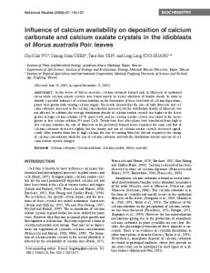
RESULTS Normal soybean seeds produce significant numbers of Ca oxalate crystals in their integument tissues beginning 3 d after fertilization and in cotyledons through 60 d (Ilarslan, Palmer, and Horner, 1997). During this time seeds greatly increase in size (Ilarslan et al., unpublished data). Clearings show that crystal numbers increase from 13 d through 116 d (Figs. 1–4), and then decrease through 160 d (data not shown). All crystals have prismatic shapes, they are twins, and typically one and, sometimes, two form per cell. The crystals often appear as pairs in clearings (typically in two adjacent cells). The long axis of each crystal is usually oriented perpendicular to the long axis of the enlarging seed (Fig. 2). The twinned crystals are kinked (Fig. 5) and they are straight (Fig. 6); both have a winged appearance with ridges running the full length of each crystal on each pair of opposing faces. The kinked crystals are identified by rotation between crossed polarizers (Figs. 7–11), where each half extinguishes at a different rotation angle. The straight crystals do not display one-half of the crystal extinguishing at different angles from the other one-half of the crystal when viewed between crossed polarizers (Figs. 7–12); at any given angle these crystals are either all bright or totally extinguished. X-ray diffraction analysis of the crystals indicates that the crystals are Ca oxalate monohydrate (whewellite; diffraction data not shown). Only peaks for whewellite were observed. Total oxalate (soluble and insoluble) concentration (in percentage) per dry mass (milligrams) of seeds, and insoluble oxalate (i.e., Ca oxalate) concentration (in percentage) per cleared seed, were calculated (Fig. 13) by using the SIGMA Urinalysis Diagnostics Kit. The difference between total oxalate and insoluble oxalate represents soluble oxalate (Fig. 13). Measurements of replicates for 116 d and 160 d measured the same as the initial measurements for these 2 d. The analyses show that as the seed size increased the concentration of total oxalate increased through 116 d, and afterwards decreased (Fig. 13). At 116 d, total oxalate reached 24 % of the dry mass of the uncleared seeds, of which threefourths were insoluble Ca oxalate. The measurements of insoluble Ca oxalate support the visual data for the presence of crystals in cleared seeds and represent a significant buildup and change in Ca oxalate/oxalate concentration during early seed development in soybean. DISCUSSION The occurrence of significant amounts of both soluble and insoluble oxalate during the life cycles of many plants has been a puzzle because of the lack of any information identifying specific functions. Oxalate in either form does not seem to be detrimental to normal plant development, a fact that suggests some selective, functional significance or adaptation. Higher plants typically form their crystals within living
1044
AMERICAN JOURNAL
OF
BOTANY
[Vol. 84
Figs. 1–12. Clearings of soybean seeds and images of isolated crystals. Figs. 1–4. Clearings of seeds showing increase in crystals during development. Crystals appear between crossed polarizers. 1. 13 d seed. 2. 15 d seed. 3. 18 d seed. 4. 116 d portion of seed cotyledon. Figs. 5, 6. Isolated prismatic crystals observed with SEM. 5. Kinked crystal showing twin plane (arrows). Note wing extending width of crystal. 6. Straight crystal. Note wing extending from crystal. Figs. 7–12. Two isolated prismatic crystals observed between crossed polarizers at different angles of rotation to demonstrate that one crystal is kinked and other is straight. Polarizer directions are 6 458 from vertical. 7. 00 rotation. 8. 100 rotation. 9. 300 rotation. 10. 550 rotation. 11. 700 rotation. 12. 1860 rotation. Bars 5 300 mm in Figs. 1–3; 1 mm in Fig. 4; 5 mm on Figs. 5, 6; and 25 mm on Figs. 7–12.
cells in the vacuoles, or to a lesser degree in seed storage protein bodies, or within or on the outer surface of cell walls. In addition, the way in which a plant deposits Ca oxalate under normal growth conditions is cell, tissue, organ, and species specific. The shape and hydration form of each crystal are also specific, and all three aspects seem to be genetically controlled (Kausch and Horner, 1982). Ca is derived from the environment, and oxalic acid, a two-carbon dicarboxylic acid, is metabolically produced from several different biochemical pathways. Two immediate precursors are glyoxylate and L-ascorbic acid; the former is converted to oxalic acid by glyoxalate oxidase and the latter by a several-step conversion. Glyoxylate, however, is formed in large quantities in a variety of tissues from different metabolic pathways (Gietl, 1992). In glyoxysomes, glyoxylate and succinate are produced from isocitrate by isocitrate lyase (Breidenbach,
Kahn, and Beevers, 1968). Subsequently, glyoxylate and acetylCoA are condensed by malate synthase to yield malate and CoA. Isocitrate lyase and malic synthase are key enyzmes in the glyoxylate cycle and are very active during fatty acid catabolism. In chloroplasts, the relative abundance of O2 and CO2 determines whether RUBISCO produces two molecules of phosphoglycerate, or one molecule each of glycolate and phosphoglycerate, from ribulose-1,5-bisphosphate. Glycolate is phosphorylated in the chloroplast and then exported to the peroxisome where it is oxidized to glyoxylate, some of which is subsequently transaminated to yield glycine. These latter reactions are components of the photorespiratory nitrogen cycle (Lea, Robinson, and Stewart, 1990). Under adverse environments, however, both glycolate and glycolatephosphate can be oxidized in the chloroplast producing glyoxylate (Goyal and Tolbert, 1996).
August 1997]
ILARSLAN
ET AL.—DETERMINATION OF OXALATE IN SOYBEAN
1045
Fig. 13. Graph showing percentage of total, insoluble, and soluble oxalate per milligram of dried and cleared soybean seeds from 13 d to 160 d postfertilization. Total oxalate was determined from dried seeds, and insoluble (Ca oxalate) was determined from cleared seeds. Difference identifies soluble oxalate.
Some N2-fixing legumes export newly reduced nitrogen from the nodules to differentiating tissues as ureides. During soybean pod fill (seed development), much of the exported ureides are directed here. Ureide catabolism produces one molecule of glyoxylate and generally two molecules of urea (Schubert and Boland, 1990). Contrary to the known roles of glyoxylate in the glyoxylate cycle and the photorespiratory nitrogen cycle, glyoxylate produced from ureides has no known function. Thus, nodulated soybeans possess three major metabolic pathways for the formation and accumulation of glyoxylate, and possibly oxalic acid. Another pathway of oxalic acid formation, not involving glyoxylate, is the metabloic transformation of L-ascorbic acid (Franceschi and Loewus, 1995; Saito, 1996). Cleavage of the six-carbon ascorbic acid molecule between carbons 2 and 3 can produce oxalic acid and L-threonic acid. This conversion does not involve glycolate, glyoxylate, or glycolate oxidase. Ascorbic acid synthesis takes place in plastids. There is little information at the present time indicating which of the pathways just described for oxalic acid formation is operational in the developing soybean seeds. However, it is clear that significant amounts of oxalic acid are produced, and combined with Ca to form crystals (Ilarslan, Palmer, and Horner, 1997). Furthermore, the reduction in the number of crystals and subsequently the oxalate later in seed development indicate there is a mechanism for eliminating and/or utilizing oxalate. One possibility is oxalate oxidase, which is abundant in certain tissues (Lane, 1994). In the presence of O2, oxalate is converted to 2CO2 1 H2O2. Legume tissue active in nitrogen assimilation can condense CO2 with phosphoen-
olpyruvate-forming oxaloacetate, which stimulates both the TCA cycle and ammonia assimilation (Schuller and Werner, 1993). Thus, the observed transient accumulation of oxalate in seed tissues could be associated with ureide degradation and subsequent amino acid synthesis, which is required for seed storage protein synthesis. It has been proposed that oxalate degradation is developmentally regulated and has important roles in plant development, including cell wall biochemistry and tissue remodeling (Lane, 1994), and salt stress and homeostasis (Hurkman and Tanaka, 1996). Also, the Ca and H2O2 released as a result of oxalate degradation possibly could play a role in plant signal transduction (Dixon, Harrison, and Lamb, 1994). One additional possibility is that during seed development there is a change in tissue pH toward the acid side, which would allow for the dissolution of the crystals and release of the Ca and oxalate ions. There is no evidence for this suggestion, and such a physical/ chemical change could adversely affect other surrounding tissues that are undergoing rapid development. In normal, developing soybean seeds we have determined that the integument tissues and the embryo cotyledons contain relatively large amounts of kinked and straight Ca oxalate monohydrate prismatic crystals, along with soluble oxalate. These integument tissues are eventually crushed as the embryo cotyledons enlarge and fill most of the mature seed (Ilarslan, Palmer, and Horner, 1997). Crystals have been found in or on the surfaces of other seeds, and suggestions have been presented as to their possible functions, including protection against invading microorganisms (Webb and Arnott, 1982, 1983; Dixon, Harrison, and Lamb, 1994). In the soybean seed, we suggest that the large accumulation of Ca oxalate and
1046
AMERICAN JOURNAL
oxalate is associated with Ca storage and seed storage protein synthesis. In this communication, the initial purposes were (1) to record the presence of seed crystals, via clearings, and where the crystals occurred; and (2) to introduce a method for determining changes in the percentage of total, soluble, and insoluble oxalate (crystals) during seed development. There are a number of methods that have been used by other investigators to test for the presence of oxalate, primarily in animals and humans (Hodgkinson, 1977), including the oxalate oxidase assay method by SIGMA. A review of the literature indicates that this latter technique is very sensitive, but has not been applied together with higher plant materials and clearings. Therefore, these techniques, when used together, represent a potentially useful method for plant biologists. In addition, the transient nature of the oxalates demonstrated by these techniques provides insights as to the possible function(s) of the crystals and oxalate in the soybean seeds during the important process of pod filling. LITERATURE CITED ARNOTT, H. J. 1995. Calcium oxalate in fungi. In S. R. Khan [ed.], Calcium oxalate in biological systems, ch. 5, 73–111. CRC Press, Boca Raton, FL. . 1982. Three systems of biomineralization in plants with comments on the associated organic matrix. In G. H. Nancollas [ed.], Biological mineralization and demineralization, 199–218. Dahlem Konferenzen, Berlin. , AND M. A. WEBB. 1983. Twin crystals of calcium oxalate in the seed coat of the kidney bean. Protoplasma 114:23–34. BREIDENBACH, R. W., A. KAHN, AND H. BEEVERS. 1968. Characterization of glyoxysomes from castor bean endosperm. Plant Physiology 43:705–713. CODY, A. M., AND H. T. HORNER. 1983. Twin raphides in the Vitaceae and Araceae and a model for their growth. Botanical Gazette 144: 318–330. , AND . 1984. Crystallographic analysis of crystal images in scanning electron micrographs and their application to phytocrystalline studies. Scanning Electron Microscopy 1984/III: 1451– 1460. CONNOLLY, J. H., AND J. JELLISON. 1995. Calcium translocation, calcium oxalate accumulation, and hyphal sheath morphology in the whiterot fungus Resinicium bicolor. Canadian Journal of Botany 73: 927–936. DIXON, R. A., M. J. HARRISON, AND C. J. LAMB. 1994. Early events in the activation of plant defense responses. Annual Review of Phytopathology 32:479–501. FRANCESCHI, V. R., AND F. A. LOEWUS. 1995. Oxalate biosynthesis and function in plants and fungi. In S. R. Khan [ed.], Calcium oxalate in biological systems, ch. 6, 113–130. CRC Press, Boca Raton, FL. FREY-WYSSLING, A. 1981. Crystallography of the two hydrates of crystalline calcium oxalate in plants. American Journal of Botany 68: 130–141.
OF
BOTANY
[Vol. 84
GIETL, C. 1992. Malate dehydrogenase isozymes: cellular locations and role in the flow of metabolites between the cytoplasm and cell organelles. Biochimica et Biophysica Acta 1100:217–234. GOYAL, A., AND N. E. TOLBERT. 1996. Association of glycolate oxidation with photosynthetic electron transport in plant and algal chloroplasts. Proceedings of the National Academy of Sciences, USA 93:3319–3324. HODGKINSON, A. 1977. Oxalic acid in biology and medicine, 1–256. Academic Press, London. HORNER, H. T., AND B. L. WAGNER. 1995. Calcium oxalate formation in higher plants. In S. R. Khan [ed.], Calcium oxalate in biological systems, ch. 4, 53–72. CRC Press, Boca Raton, FL , AND E. ZINDLER-FRANK. 1982. Calcium oxalate crystals and crystal cells in the leaves of Rhynchosia caribaea DC. (Leguminosae: Papilionoideae). Protoplasma 111:10–18. , L. H. TIFFANY, A. M. CODY AND G. KNAPHUS. 1985. Development of fungal calcium oxalate crystals associated with the basidiocarps of Geastrum minus (Lycoperdales). Scanning Electron Microscopy 1985/II:789–801. HURKMAN, W. J., AND C. K. TANAKA. 1996. Effect of salt stress on germin gene-expression in barley roots. Plant Physiology 110:971– 977. ILARSLAN, H., R. G. PALMER, AND H. T. HORNER. 1997. Calcium oxalate crystals in developing soybean seeds. Scanning Microscopy. KAUSCH, A. P., AND H. T. HORNER. 1982. A comparison of calcium oxalate crystals isolated from callus cultures and their explant sources. Scanning Electron Microscopy 1982/I:199–211. LANE, B. G. 1994. Oxalate, germin, and the extracellular matrix of higher plants. FASAB Journal 8:294–301. LEA, P. J., S. A. ROBINSON, AND G. R. STEWART. 1990. The enzymology and metabloism of glutamine, glutamate, and asparagine. In P. K. Stumpf and E. E. Conn [eds.], The biochemistry of plants, vol. 16, 121–159. Academic Press, San Diego, CA. SAITO, K. 1996. Formation of L-ascorbic acid and oxalic acid from D-glucosone in Lemna minor. Phytochemistry 41:145–149. SCHUBERT, K. R., AND M. J. BOLAND. 1990. The ureides. In P. K. Stumpf amd E. E. Conn [eds.], The biochemistry of plants, vol. 16, 197–282. Academic Press, San Diego, CA. SCHULLER, K. A., AND D. WERNER. 1993. Phosphorylation of soybean (Glycine max L.) nodule phosphoenolpyruvate carboxylase in vitro decreases sensitivity to inhibition by L-malate. Plant Physiology 101:1267–1273. SIMKISS, K., AND K. M. WILBUR. 1989. Biomineralization: cell biology and mineral deposition. Academic Press, New York, NY. WEBB, M. A., AND H. J. ARNOTT. 1982. A survey of calcium oxalate crystals and other mineral inclusions in seeds. Scanning Electron Microscopy 1982/III:1109–1131. , AND . 1983. Inside plant crystals: a study of the noncrystalline core in druses of Vitis vinifera endosperm. Scanning Electron Microscopy 1983/IV:1759–1770. ZINDLER-FRANK, E. 1974. Die Differenzierung von Kristallidioblasten im Dunkeln und bei Hemmung der Glykolsa¨ureoxidase. Zeitschrift fu¨r Pflanzenphysiologie 73: 313–325. . 1976. Oxalate biosynthesis in relation to photosynthetic pathway and plant productivity—a survey. Zeitschrift fu¨r Pflanzenphysiologie 80:1–13. . 1987. Calcium oxalate crystals in legumes. In C. Stirton [ed.], Advances in legume systematics, part 3, 279–316. Royal Botanic Gardens, Kew.