Adhesion at calcium oxalate crystal surfaces and the effect of urinary constituents Xiaoxia Sheng*, Taesung Jung*, Jeffrey A. Wesson†‡, and Michael D. Ward*‡ *Department of Chemical Engineering and Materials Science, University of Minnesota, 421 Washington Avenue SE, Minneapolis, MN 55455; and †Nephrology Division, Department of Veteran Affairs Medical Center and the Medical College of Wisconsin, 5000 West National Avenue, Milwaukee, WI 53295
Kidney stones, aggregates of microcrystals, most commonly contain calcium oxalate monohydrate (COM) as the primary constituent. The aggregation of COM microcrystals and their attachment to epithelial cells are thought to involve adhesion at COM crystal surfaces, mediated by anionic molecules or urinary macromolecules. Identification of the most important functional group– crystal face adhesive combinations is crucial to understanding the stability of COM aggregates and the strength of their attachments to epithelial cell surfaces under flow in the renal tubules of the kidney. Here, we describe direct measurements of adhesion forces, by atomic force microscopy, between various functional groups and select faces of COM crystals immersed in aqueous media. Tip-immobilized carboxylate and amidinium groups displayed the largest adhesion forces, and the adhesive strength of the COM crystal faces decreased in the order (100) > (121 ) > (010), demonstrating that adhesion is sensitive to the structure and composition of crystal faces. The influence of citrate and certain urinary proteins on adhesion was examined, and it was curious that osteopontin, a suspected regulator of stone formation, increased the adhesion force between a carboxylate tip and the (100) crystal face. This behavior was unique among the various combinations of additives and COM crystal faces examined here. Collectively, the force measurements demonstrate that adhesion of functional groups and binding of soluble additives, including urinary macromolecules, to COM crystal surfaces are highly specific in nature, suggesting a path toward a better understanding of kidney stone disease and the eventual design of therapeutic agents. atomic-force microscopy 兩 kidney stone 兩 protein
idney stone disease, which occurs in ⬇10% of the U.S. population, causes substantial suffering and occasional renal failure, yet the disease mechanism is poorly understood. Kidney stones are aggregates, most commonly containing microcrystals of calcium oxalate monohydrate (COM) as the primary inorganic constituent. Stones also contain small amounts of embedded proteins, which are thought to play an adhesive role in these aggregates, and they often are found attached to the tip of renal papilla, presumably through adhesive contacts. Although crystallogenesis is essential to stone formation, calcium oxalate crystal growth rates are sluggish, to the extent that during typical urine transit times, it is unlikely that single crystals will grow large enough to become lodged in the terminal collecting duct of the kidney based on size alone (1, 2). Therefore, crystal aggregation and attachment to interfaces at the papillary tip must play a significant role in crystal retention and stone formation. Some in vitro studies have suggested that anionic molecules (e.g., citrate) and urinary proteins with substantial anionic functionalities (e.g., aspartate and glutamate) may serve as adhesives that promote COM aggregation and attachment to epithelial cells (3–6). Anionic molecules such as phospholipids, which are embedded in epithelial cell membranes, also are thought to promote the attachment of COM to renal tubules (7). It also has been suggested that COM mineralization may occur
K
www.pnas.org兾cgi兾doi兾10.1073兾pnas.0406835101
on Randall’s plaques, which are subepithelial hydroxyapatite deposits (8). However, certain urinary molecules are thought by others to suppress crystal aggregation and cell attachment, presumably because of adsorption on COM crystal faces (9–14). Unfortunately, little is known of the actual strength of adhesion between calcium oxalate surfaces and functional groups on small molecules or on side chains of urinary proteins. Here, we describe measurements of the adhesion forces between functional groups immobilized on the tip of an atomicforce microscope (AFM) and prominent COM crystal faces. In this configuration, an AFM tip modified with certain functional groups can be viewed as a mimic of urinary protein segments or crystal-recognition sites embedded in epithelial cell membranes. Carboxylate groups on an AFM tip also can be viewed as mimics for oxalate ions on a calcium oxalate surface. The strength of the adhesion force depends on the specific functional group–crystal face combination, which can only be interpreted in terms of specific binding contribution at the crystal surface. The addition of soluble additives known to regulate stone formation (citrate and various urinary proteins) indicates face-specific binding of these additives that may be responsible for regulating the growth, aggregation, and adhesion properties of COM crystals. Methods COM Crystal Synthesis. The COM crystals used for adhesion-force
measurements were grown in vitro by one of three methods (refs. 15 and 16; A. Millan, personal communication), each intended to maximize the area of a particular crystal face so that AFM imaging was feasible. Method 1 relied on rapid mixing at room temperature of highly concentrated aqueous solutions of CaCl2 and sodium oxalate at pH ⬇7.5 and high ionic strength {[calcium oxalate] ⫽ 0.38 mM, [NaCl] ⫽ 150 mM, [4-(2-hydroxyethyl)-1piperazineethanesulfonic acid] ⫽ 10 mM} at room temperature, which generated COM crystals as elongated hexagonal plates (⬇2 m thick and 10–100 m long) with large (100) faces. Method 2 involved slow evaporation of an aqueous solution saturated with calcium oxalate at pH ⬇1.5, which produced crystals with a xiphoid (knife-like) habit (20–50 m long and 10–20 m wide) and a large (121 ) face. Method 3 involved mixing highly supersaturated solutions of 15 mM CaCl2 and 15 mM sodium oxalate at pH ⬇7 at elevated temperatures (75 ⫾ 5°C) and rapid stirring, which produced crystals with large (010) faces (up to 30 m long and 15 m wide). Osteopontin (OPN), extracted and purified from bovine milk, was donated by Esben Sorenson (University of Aarhus, Aarhus, Denmark). Human serum albumin, transferrin, and chondroitin sulfate A were obtained from Sigma–Aldrich and used as received. This paper was submitted directly (Track II) to the PNAS office. Abbreviations: COM, calcium oxalate monohydrate; AFM, atomic-force microscope; OPN, osteopontin. ‡To
whom correspondence should be addressed. E-mail:
[email protected] or jwesson@ mcw.edu.
© 2004 by The National Academy of Sciences of the USA
PNAS 兩 January 11, 2005 兩 vol. 102 兩 no. 2 兩 267–272
CHEMISTRY
Edited by Alexandra Navrotsky, University of California, Davis, CA, and approved October 28, 2004 (received for review September 15, 2004)
In Situ AFM Imaging. Characterization of the COM crystal surfaces
and adhesion-force measurements were executed with a Digital Instruments (Santa Barbara, CA) Nanoscope IIIa Multimode system. All measurements were performed in aqueous solutions saturated with calcium oxalate at pH ⬇7, which ensured that the COM crystal surfaces were stable. COM crystals were harvested by filtration of the crystallization medium through a polycarbonate membrane with 10-m pores. The crystals then were transferred to the AFM specimen disk by gently pressing the side of the membrane containing the crystals onto a partially cured UV-curable polymer film that had been coated previously onto the specimen disk. The polymer then was cured completely to affix the crystals permanently, and the crystals were washed with saturated calcium oxalate solution and then dried with nitrogen. Topographical and lattice images of the crystal surfaces were acquired with commercial Si3N4 cantilever tips. Adhesion-force measurements were performed by using gold-coated Si3N4 tips modified with various thiol molecules (total tip radius of curvature, ⬇75 nm, including gold thickness of ⬇50 nm), which were prepared by immersing the gold-coated tips in ethanol solutions containing 3 mM thiol reagent for at least 12 h. The spring constant of the AFM cantilevers, which is needed to convert the cantilever deflection to an adhesion force, was 0.074 ⫾ 0.003 N/m, as measured by a reported method (17). The working solution, which was prepared immediately before the measurements and stored in a reservoir connected to the liquid cell by a Teflon tube, was refreshed at 10-min intervals to ensure uniform solution conditions. A typical adhesion force measurement with a given modified tip involved acquisition of 1,000 individual force-distance curves recorded at 20 different locations on the crystal face. The force-distance curves were acquired at a rate of 2 Hz and with a loading force of 1–2 nN. The force-distance curves did not change perceptibly for acquisition rates in the range of 0.2–2 Hz nor did they change with loading forces up to 6 nN. The effects of these parameters outside these ranges were not examined. Individual adhesion forces were determined from the retraction portion of individual curves by using customized software that automatically calculated the change in deflection after detachment of the tip from the crystal surface. These individual adhesion forces were tabulated into histograms, and the mean values and standard deviations were determined from the normal distribution curves. These were not appreciably different from the arithmetic mean values and standard deviations. This procedure accounts for the likely possibility that the number of molecules adhering to the surface varies for individual force curves while effectively averaging contributions from crystal-surface defects and nonuniformities in the tip. The effects of citrate and proteins were evaluated by measuring the adhesion force 45 and 120 min, respectively, after their introduction to the AFM cell. The AFM cell was refreshed with additive-free calcium oxalate solution between measurements. Results and Discussion Characterization of COM Crystals. Scanning electron microscopy
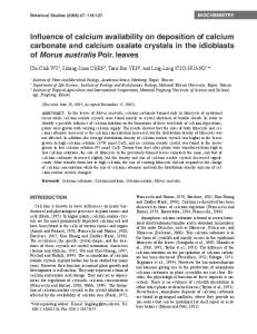
revealed the crystal habit and dominant faces of the COM crystals grown by the three methods described above (Fig. 1). The Miller indices of the upper crystal face and all adjoining faces were assigned with the aid of the crystal modeling program SHAPE (Shape Software, Kingsport, TN), which generates models of the external habit of a particular crystal structure from the perspective of the viewer based on the single crystal structure, in this case the crystal structure of COM (P21/c, a ⫽ 6.290 Å, b ⫽ 14.5803 Å, c ⫽ 10.116 Å,  ⫽ 109.46°) (18). We note that this space-group setting, reported by Tazzoli and Domeneghetti (18), differs from the setting used by Deganello and Piro (19), which has been used by others. We use the Tazzoli and Domeneghetti setting because it allows assignment of more prominent faces to 268 兩 www.pnas.org兾cgi兾doi兾10.1073兾pnas.0406835101
Fig. 1. COM crystal surfaces. (Top) Scanning electron microscopy images of COM crystals viewed perpendicular to the (100), (121 ), and (010) faces. (Middle) AFM lattice images, displayed as raw (upper half) and Fourier-filtered (lower half) data. (Bottom) Topographical images of the three crystal faces. The images were acquired in aqueous solutions (pH ⬇7) saturated with calcium oxalate (0.11 mM)
lower index values [e.g., the (1 01) in the Deganello and Piro setting is equivalent to (100) in the Tazzoli and Domeneghetti setting]. The Miller index assignments were corroborated by topographic and lattice images of the prominent faces collected with contact-mode AFM imaging in aqueous solutions saturated with calcium oxalate (0.11 mM). The COM (121 ) and COM (010) exhibited large terraces separated by well defined steps. COM (121 ) terraces were separated by 4.3-Å-high steps, essentially identical to d12-1 in the bulk crystal (4.39 Å). Lattice images of the (121 ) face revealed an oblique unit cell with lattice constants of 17.6 and 25.1 Å along [2 10] and [012] and an acute angle of 56°. These values are in reasonable agreement with the anticipated values of 19.9 Å, 24.9 Å, and 58.2°, although the discrepancy between the measured and anticipated values for the first lattice constant suggests some minor surface reconstruction (20). The step heights on the COM (010) were 14.2 Å, similar to the bulk value b ⫽ 14.58 Å. The lattice image of the (010) face revealed in-plane lattice constants of a ⫽ 6.4 Å, c ⫽ 10.5 Å, and  ⫽ 106°, which compared favorably with the anticipated values of a ⫽ 6.29 Å, c ⫽ 10.12 Å, and  ⫽ 109.5°. COM (100) has 200-nm-wide terraces bounded by irregular step edges. The 6.3-Å step height is similar to the anticipated value for COM (100) layers (d100 ⫽ 5.93 Å). Because of the roughness of the (100) face, two-dimensional periodicity was difficult to discern in its real-space lattice images. However, Fourier transforms of the AFM data, however, revealed one-dimensional features oriented along the [021] direction, coinciding with one-dimensional chains of calcium and oxalate ions. These AFM lattice images indicate that the three COM crystal faces have a high degree of crystalline order in aqueous media, which argues that these surfaces can be viewed as an extension of the bulk structure. Adhesion-Force Measurements. AFM imaging has proven to be a
powerful tool for real-time visualization of crystal growth on specific faces of organic and inorganic crystals (21–24), including COM (15, 25). AFM imaging also has been used to obtain images Sheng et al.
of kidney stones (26). Surprisingly, the force measurement capability of AFM imaging, which has been used extensively for quantitative measurement of adhesion forces between various molecular and biomolecular surfaces (27, 28), has been rather limited with respect to single crystal surfaces (29–31). To our knowledge, the adhesion properties of different faces of a crystalline material have never been examined. To identify the most adhesive functional group–crystal face combinations, various AFM tips, each decorated with a monolayer of a single type of organosulfur molecule, were brought into contact with a particular COM crystal face (Fig. 2). The adhesion force associated with an individual force curve was measured from the change in the cantilever deflection when the AFM tip detached from the crystal surface during retraction. The mean adhesion force then was calculated from 1,000 individual force curves recorded at 20 different locations on a particular crystal face. Functional group contributions to adhesion were examined by varying the substituents at the ends of the organosulfur molecules, some resembling amino acid side chains in urinary proteins. The effect of the steric environment and rigidity of the probe molecules was investigated by comparison of adhesion forces measured with arenethiols and alkanethiols. Within a given class (i.e., arenethiols or alkanethiol), the adhesion forces for tip molecules terminated with ionic groups were larger than those with uncharged functional groups irrespective of the crystal face (Fig. 3). For example, the mean adhesion force for the Au:S(CH2)10COO⫺ tip and COM (100) (4.28 ⫾ 0.62 nN) was approximately five times the value determined for Au:S(CH2)10CH2OH and Au:S(CH2)10CH3 on this face (0.83 ⫾ 0.33 and 0.66 ⫾ 0.17 nN, respectively). The adhesion forces measured for the amidinium tip Au:S(CH2)2NHC(NH⫹ 2 )NH2, a cationic probe that can be viewed as a mimic of an arginine group, were comparable with those measured for Au:S(CH2)10COO⫺. The adhesion forces for the alkane-carboxylate and amidinium tips clearly depended on the crystal face, decreasing in the order (100) ⬎ (121 ) ⬎ (010). In contrast, the adhesion force for the Au:S(C6H4)COO⫺ probe was essentially identical for all three crystal faces. The mean adhesion force for each tip did not change during the time the measurements were performed, indicating that the modified tips were robust. Replicate measurements with different tips and COM crystals demonstrated that the adhesion-force profile for the functional groups on each face and the order of adhesion of each tip among the three faces, as depicted in Fig. 3, were highly reproducible. The largest number of replicate measurements was performed with the Au:S(CH2)10COO⫺ and Au:S(CH2)2NHC(NH⫹ 2 )NH2 tips, because the differences in adhesion to the three crystal faces were most apparent for these tips. For example, data compiled from measurements with 40 Au:S(CH2)10COO⫺ tips (1,000 force curves per tip) confirmed that the order of the adhesion forces [(100) ⬎ (121 ) ⬎ (010)] was statistically meaningful. Collectively, these observations indicate that the functional group of the probe molecule, the composition and structure of Sheng et al.
Fig. 3. Mean adhesion forces measured in aqueous solutions saturated with calcium oxalate (0.11 mM) for various modified AFM tips with COM crystal faces [(100) (⽧), (121 ) (䊐), and (010) (Œ)]. Although the tip–surface interaction involves multiple contacts, only a single thiol is depicted on each tip for clarity. Each data set for a particular crystal face was collected on the same COM crystal, and each data point represents the mean value for at least 1,000 force curves acquired with a single tip. The error bars represent one standard deviation, as calculated from analysis of each force histogram. The conditions used here ensure that the anionic carboxylate and cationic amidinium forms predominate over their uncharged counterparts. The mean adhesion force for each tip did not change appreciably during these measurements, indicating that the tips were robust. The mean adhesion force for a bare gold tip was ⬍1 nN, with a very narrow histogram width.
the crystal face, and the steric environment and conformational rigidity of the probe molecule all affect adhesion. Fig. 3 illustrates that the Au:S(CH2)10COO⫺ tip is particularly effective for adhesion, reflecting its ability to bind to Ca2⫹ lattice ions exposed on the COM crystal faces. The adhesion forces measured for the Au:S(CH2)10CH2OH and Au:S(CH2)10CH3 tips were very small; the latter was comparable with unmodified gold-coated tips. Therefore, the adhesion forces for these tips, which have the same number of methylene groups in the alkane tether as Au:S(CH2)10COO⫺, can be viewed as a measure of the nonspecific binding forces. Consequently, the relatively large forces observed for the carboxylate tip reveals the contributions from specific interactions. Notably, the Ca2⫹ surface density decreases in the order (100) ⫽ 0.0542 Ca2⫹/Å2 ⬎ (121 ) ⫽ 0.0429 Ca2⫹/Å2 ⬎ (010) ⫽ 0.0333 Ca2⫹/Å2, the same order observed for the adhesion forces for the Au:S(CH2)10COO⫺ tip. This result supports specific binding between the carboxylate group and Ca2⫹ ions on the crystal surfaces. Because the oxalate ion surface densities are identical to the Ca2⫹ surface densities, specific binding of the Au:S(CH2)2NHC(NH⫹ 2 )NH2 tip is also implicated. Indeed, the typical NOH䡠䡠䡠HON distance in the amidinium group (2.25–2.35 Å) and O䡠䡠䡠O distance of the oxalate carboxyl group (2.21–2.34 Å) are ideally matched for NOH䡠䡠䡠O hydrogen bonding as a heterodimer (Scheme 1). The COM lattice contains two types of oxalate ions, denoted as types I and II, which can be distinguished by their orientations on each of the three crystal faces (Fig. 4). The long axes of both types I and II ions lay in the plane of the (100) surface. The long axes of the type I ions are perpendicular to the (010) plane, but the type II ions are parallel. Both types are canted at the (121 ) surface. It is reasonable to suggest that specific binding at the crystal surface will be optimized if the probe molecule can dock in an oxalate vacancy and bind to a Ca2⫹ ion in a manner that mimics the orientation of the missing oxalate ion (Scheme 1). Unlike the aliphatic Au:S(CH2)10COO⫺ carboxylate probe, the adhesion forces for the Au:S(C6H4)COO⫺ tip with the three COM faces are essentially identical. The conformational flexibility of the Au:S(CH2)10COO⫺ tip should make this probe less PNAS 兩 January 11, 2005 兩 vol. 102 兩 no. 2 兩 269
CHEMISTRY
Fig. 2. Schematic representation of the contact between an idealized hemispherical gold-coated AFM tip, modified with a monolayer of organosulfur molecules of the same kind, and the COM crystal surface (not drawn to scale).
Scheme 1.
sensitive to the geometric constraints associated with docking, allowing this probe to access most or all of the Ca2⫹ sites exposed at the surface on all three crystal faces. Under these conditions, it is to be expected that the adhesion force for the Au:S(CH2)10COO⫺ tip would be governed by the number of the Ca2⫹ sites available at the surface independent of geometric factors, which is consistent with the trend observed for the adhesion forces of this tip on the three faces. In contrast, the steric encumbrance and the conformational rigidity of the Au:S(C6H4)COO⫺ probe would be expected to frustrate docking in the oxalate vacancies. Consequently, the adhesion force on the (100) surface, in which all the oxalate ions are parallel to the face, would be reduced compared with the flexible aliphatic probe, as reflected by the adhesion forces. Type I sites on the (010) surface, however, are oriented along the direction of tip approach, creating a favorable orientation for docking that can compensate for the lower surface concentration of Ca2⫹ sites. The surface oxalates on COM (121 ) are canted with respect to the surface and have a surface concentration intermediate between COM (100) and COM (010). Therefore, these features may conspire to produce nearly identical adhesion forces on all three faces for this tip even though the total Ca2⫹ surface concentrations are different. The obser vation that the
Fig. 4. Lattice ion orientations. (Upper) Surface packing of the three COM crystal faces. (Lower) Crystal models of COM illustrating the approach of the modified AFM tip toward the three faces. The (100) and (010) planes are shown together (Left) to emphasize the different orientations of types I and II oxalate ions on these faces. The types I and II oxalate ions are all shown at the bottom in their respective orientations, for clarity. 270 兩 www.pnas.org兾cgi兾doi兾10.1073兾pnas.0406835101
Fig. 5. Mean adhesion forces measured for a Au:S(CH2)10COO⫺ tip in contact with different COM crystal surfaces in 0.115 mM calcium oxalate solution, pH ⬇7.0, in the presence of citrate at various concentrations. The error in the adhesion forces typically ranges from 0.2 to 0.5 nN. The adhesion forces for each face were measured at the specified concentrations with the same tip.
Au:S(CH2)10CH2OH and Au:S(C6H4)OH tips exhibited comparable adhesion forces despite their different conformational flexibility and sterics argues that only nonspecific interactions are operative here, which further supports the importance of local geometric effects during specific binding by the carboxylate and amidinium tips. We note that only the type I oxalate ions on the (010) surface are ideally oriented for linear NOH䡠䡠䡠O hydrogen bonds with the Au:S(CH2)2NHC(NH⫹ 2 )NH2 tip. Hydrogen bonds, however, can be insensitive to orientation effects such that the orthogonal orientation required for binding to oxalates on COM (100) remains strong. Under this condition, the adhesion forces would be expected to scale with the Ca2⫹ surface concentration, as observed. Adhesion forces can be affected by surface energy terms associated with the creation of new surfaces during an unbinding event (32). The constant solution conditions used here and the similar surface energies of the tips [except for the nonpolar Au:S(CH2)10CH3], however, are expected to minimize the contributions of these effects. Influence of Citrate on Adhesion. Citrate, a polyanionic molecule that is abundant in urinary solution, is recognized as an inhibitor of COM growth and aggregation at concentrations similar to those found in urine (1–2 mM) (13). Citrate also has been reported to block adhesion of COM crystals to renal epithelial cells. To probe the influence of citrate at the microscopic level, citrate was added to the AFM cell during measurement of the adhesion-force measurements between the Au:S(CH2)10COO⫺ probe and the COM (100), (121 ), and (010) faces in saturated calcium oxalate solution (Fig. 5). The adhesion force decreased monotonically with increasing citrate concentration for all three faces. The effect of citrate clearly was most significant for the COM (100) face. It is notable that the presence of citrate has been reported to enlarge COM (100) faces during COM crystallization, consistent with strong adsorption of citrate to COM (100) (33). Citrate also was reported to reduce the length-to-width ratio of COM crystals, consistent with preferred adsorption of citrate on the (121 ) and (12 1 ) faces compared with (010). These observations are supported by the adhesion-force profile, which reflects citrate adsorption decreasing in the order (100) ⬎ (121 ) ⬎ (010). The forces for the three faces converge as the citrate concentrations approach 0.5 mM, most likely because of Sheng et al.
Face-Specific Binding of Urinary Proteins. Urinary macromolecules such as OPN, human serum albumin, chondroitin sulfate, and transferrin are thought to play an important role in regulating stone formation, although the exact nature of their effects on stone formation remains largely unresolved. This uncertainty is exemplified by various studies involving OPN, which exists in vivo at levels that are known to inhibit COM crystallization (⬇4 g/ml) (34). This behavior must reflect adsorption of OPN on COM crystal surfaces, because this concentration is not sufficient for any significant reduction in calcium concentration through complexation with pendant carboxylate groups. OPN also has been demonstrated to interfere with COM attachment to renal epithelial cells in culture (35), and experiments using an OPN knockout mouse model support inhibition of stone formation by OPN (36). On the other hand, OPN, similar to other putative macromolecular inhibitors of stone formation, is often found in COM-rich kidney stones, in which it may perform as an adhesive between microcrystals. Some in vitro data support a role for OPN as a promoter of stone formation, possibly mediating attachment of calcium oxalate crystals to renal tubule cells (6). Nonetheless, the evidence suggests that urinary macromolecules regulate stone formation by mediating aggregation and attachment to renal tubule surfaces, adhesion processes that are affected by specific adsorption of these proteins on calcium oxalate crystal surfaces. To probe the effect of urinary macromolecules on adhesion, OPN, human serum albumin, transferrin, and chondroitin sulfate were added individually to the AFM cell during adhesionforce measurements with the Au:S(CH2)10COO⫺ tip and the three COM crystal faces (Fig. 6). Among the proteins examined, OPN was unique in that the adhesion force for COM (100) increased over a period of 2.5 h after addition of OPN (5 g/ml), unlike any other protein–crystal face combination examined here. Conversely, the adhesion forces on the COM (121 ) and COM (010) surfaces decreased during this period. This behavior clearly illustrates that OPN binds differently to the three crystal faces. Furthermore, it suggests a unique binding configuration of OPN to COM (100) that permits a sufficient number of unbound aspartate or glutamate residues to complex Ca2⫹ from solution, thereby creating an adhesive bridge to the carboxylate groups on the Au:S(CH2)10COO⫺ tip (see Fig. 6 Upper Inset). It is interesting to note that OPN is unique among the proteins examined here, because it contains a continuous DDEDDDD sequence (37) that may reinforce Ca2⫹-mediated binding to the AFM tip. Because we have not yet determined whether the Au:S(CH2)10COO⫺ tip simulates an oxalate-decorated COM surface, a cell surface decorated with anionic receptors, or anionic macromolecules in solution, it is difficult to draw firm conclusions at this time regarding the relationship of this result to the effect of OPN in the aforementioned stone models. Regardless of the actual mechanism, the face-specific behavior of OPN observed here indicates that its role in stone regulation may be complex. The data in Fig. 6 further support the role of specific binding by the remaining three urinary macromolecules. The adhesion forces in the presence of human serum albumin were reduced to comparable levels on all three faces. The forces were reduced by transferrin on (100) and (121 ) as well, but its effect on (010) was Sheng et al.
Fig. 6. Effects of urinary macromolecules on adhesion. (Upper) The adhesion forces measured for Au:S(CH2)10COO⫺ tips and COM crystal surfaces in 0.15 mM calcium oxalate solution (pH ⬇7.0) after addition of OPN (5 g/ml). (Lower) Comparison of the effect of several urinary proteins on the adhesion force. Because of the irreversible adsorption of the protein, each measurement in the presence of protein was performed with a new tip and new COM crystal. The adhesion force reported for each protein– crystal face combination was determined after measurement in protein-free solution. The values then were scaled to the average of all the protein-free measurements (white bar).
negligible. Conversely, the effect of chondroitin sulfate on adhesion was negligible for (100) but significant for (121 ) and (010). Combined with the observations for OPN, these results clearly demonstrate that the regulation of adhesion depends on the macromolecule–crystal face combination. The force measurements revealed that the adsorption of citrate on the crystal faces was reversible, because the adhesion forces returned to the original values after 30 min of exposure to citrate-free solutions (saturated with calcium oxalate). In contrast, the adsorption of the urinary proteins examined here was much more persistent, because the adhesion forces remained unchanged after prolonged exposure to protein-free solutions. It is notable that we observed that adsorption of poly(aspartate) and poly(glutamate) are reversible, indicating that urinary proteins behave much differently than synthetic macromolecules. The observations described here demonstrate the usefulness of AFM imaging for probing binding of specific functional groups to single crystal surfaces, and they clearly indicate that adhesion forces measured by AFM imaging are sensitive to the structure and composition of crystal faces. The adhesion forces are consistent with growing evidence that carboxylate-rich synthetic macromolecules and native urinary proteins play important roles in regulating aggregation of COM and its attachment to renal epithelial cells. The observation that the adhesion forces measured for the carboxylate and amidinium tips depend on the identity of the crystal face substantiates the role of specific binding. It is interesting to note that dumbbell-shaped aggregates of COM crystals, in which the (100) faces are bonded together as ‘‘fans’’ such that they are effectively shielded from binding to renal tubule cells (38), have been observed in asymptomatic PNAS 兩 January 11, 2005 兩 vol. 102 兩 no. 2 兩 271
CHEMISTRY
saturation of the Ca2⫹-binding sites on all three faces. Overall, the suppression of the adhesion force by citrate corroborates its role in the regulation of COM growth, aggregation, and attachment to cells through adsorption on crystal faces. Interestingly, parallel experiments revealed that propionate, a simple monocarboxylic acid, was not as effective toward suppressing adhesion, which suggests a unique adsorption mechanism for citrate, possibly involving binding of more than one of its three carboxylate groups to lattice sites on the crystal surface.
renal epithelial cells. We anticipate that additional studies of this kind eventually will identify the most relevant macromolecule– crystal surface combinations related to kidney stone formation, which can aid the development of therapeutic agents designed to reduce (or eliminate) the most adhesive COM faces or to suppress the binding of adhesive proteins.
crystalluria. However, similar structures are not common in actual stone samples, suggesting that the formation of the dumbbell morphology may protect against stone disease. The observation here that COM (100) is the most adhesive face may explain the formation of this morphology in the presence of carboxylate-rich urinary molecules. The observed effects of urinary species, including citrate and native proteins, on the adhesion force further supports specific binding of urinary molecules to the COM crystal surfaces. It is reasonable to suggest that these binding characteristics are related to the role of these urinary species in aggregation of COM microcrystals and their attachment, either as single crystals or aggregates, to
This work was supported by the Medical College of Wisconsin, the Department of Veterans Affairs, the McKnight Foundation, and the Materials Research Science and Engineering Center program of the National Science Foundation (Grant DMR-0212302). X.S. was supported by doctoral dissertation fellowship from the University of Minnesota Graduate School.
Finlayson, B. & Reid, F. (1978) Invest. Urol. 15, 442–448. Kok, D. J. & Khan, S. R. (1994) Kidney Int. 46, 847–854. Govindaraj, A. & Selvam, R. (2001) Urol. Res. 29, 194–198. Grases, F., Gil, J. J. & Conte, A. (1989) Colloids Surf. 36, 29–38. Christmas, K. G., Gower, L. B., Khan, S. R. & El-Shall, H. (2002) J. Colloid Interface Sci. 256, 168–174. Yamate, T., Kohri, K., Umekawa, T., Iguchi, M. & Kurita, T. (1998) J. Urol. 160, 1506–1512. Bigelow, M. W., Wiessner, J. H., Kleinman, J. G. & Mandel, N. S. (1997) Am. J. Physiol. 272, F55–F62. Evan, A. P., Lingeman, J. E., Coe, F. L., Parks, J. H., Bledsoe, S. B., Shao, Y., Sommer, A. J., Paterson, R. F., Kuo, R. L. & Grynpas, M. (2003) J. Clin. Invest. 111, 607–616. Lieske, J. C., Huang, R. & Toback, F. G. (2000) Am. J. Physiol. 278, F130–F137. Hess, B., Nakagawa, Y. & Coe, F. L. (1989) Am. J. Physiol. 257, F99–F106. Grover, P. K. & Ryall, R. L. (1999) Eur. J. Biochem. 263, 50–56. Bowyer, R. C., Brockis, J. G. & McCulloch, R. K. (1979) Clin. Chim. Acta 95, 23–28. Ryall, R. L., Harnett, R. M. & Marshall, V. R. (1981) Clin. Chim. Acta 112, 349–356. Springman, K. E., Drach, G. W., Gottung, B. & Randolph, A. D. (1986) J. Urol. 135, 69–71. Guo, S., Ward, M. D. & Wesson, J. A. (2002) Langmuir 18, 4284–4291. Cody, A. M. & Horner, H. T. (1984) Scan. Electron Microsc. 3, 1451–1460. Cleveland, J. P., Manne, S., Bocek D. & Hansma, P. K. (1993) Rev. Sci. Instrum. 64, 403–405. Tazzoli, V. & Domeneghetti, C. (1980) Am Mineral 65, 327–334. Deganello, S. & Piro, O. (1981) Neues Jahrb. Mineral. Monatsh. 2, 81–88. Jung, T., Sheng, X., Choi, C., Kim, W., Wesson, J. A. & Ward, M. D. (2004) Langmuir 20, 8587–8596. Hillier, A. C. & Ward, M. D. (1994) Science 263, 1261–1264.
22. Yip, C. M., Brader, M. L., DeFelippis, M. R. & Ward, M. D. (1998) Biophys. J. 74, 2199–2209. 23. Ward, M.D. (2001) Chem. Rev. (Washington, D.C.) 101, 1697–1725. 24. Teng, H. H., Dove, P. M., Orme, C. A. & De Yoreo, J. J. (1998) Science 282, 724–727. 25. Qiu, S. R., Wierzbick, A., Orme, C. A., Cody, A. M., Hoyer, J. R., Nancollas, G. H., Zepeda, S. & De Yoreo, J. J. (2004) Proc. Natl. Acad. Sci. USA 101, 1811–1815. 26. Dorian, H., Rez, P. & Drach, G. W. (1996) J. Urol. 156, 1833–1837. 27. Noy, A., Vezenov, D. V. & Lieber, C. M. (1997) Annu. Rev. Mater. Sci. 27, 381–342. 28. Takano, H., Kenseth, J. R., Wong, S., O’Brien, J. C. & Porter, M. D. (1999) Chem. Rev. (Washington, D.C.) 99, 2845–2890. 29. Abendan, R. S. & Swift, J. A. (2002) Langmuir 18, 4847–4853. 30. Danesh, A., Davies, M. C., Hinder, S. J., Roberts, C. J., Tendler, S. J., Williams, P. M. & Wilkins M. J. (2000) Anal. Chem. 72, 3419–3422. 31. Sheng, X., Wesson, J. A. & Ward, M. D. (2003). J. Am. Chem. Soc. 125, 2854–2855. 32. Skulason, H. & Frisbie, C. D. (2002) Anal. Chem. 74, 3096–3104. 33. Wierzbicki, A., Sikes, C. S., Sallis, J. D., Madura, J. D., Stevens, E. D. & Martin, K. L. (1995) Calcif. Tissue Int. 56, 297–304. 34. Shiraga, H., Min, W., VanDusen, W. J., Clayman, M. D., Miner, D., Terrall, C. H., Sherbotie, J. R., Foreman, J. W., Przysiecki, C., Neilson, E. G., et al. (1992) Proc. Natl. Acad. Sci. USA 89, 426–430. 35. Kumar, V., Farell, G. & Lieske, J. C. (2003) J. Urol. 170, 221–225. 36. Wesson, J. A., Johnson, R. J. Mazzali, M., Beshensky, A. M., Stietz, S, Giachelli, C., Liaw, L., Alpers, C. E., Couser, W. G., Kleinman, J. G., et al. (2003) J. Am. Soc. Nephrol. 14, 139–147. 37. Kiefer, M. C., Bauer, D. M. & Barr, P. J. (1989) Nucleic Acids Res. 12, 3306. 38. Shirane, Y., Kurokawa, Y., Sumiyoshi, Y. & Kagawa, S. (1995) Scanning Microsc. 9, 1081–1088.
1. 2. 3. 4. 5. 6. 7. 8.
9. 10. 11. 12. 13. 14. 15. 16. 17. 18. 19. 20. 21.
272 兩 www.pnas.org兾cgi兾doi兾10.1073兾pnas.0406835101
Sheng et al.