REVIEW ARTICLES William C. Oliver, Jr, MD Paul G. Barash, MD Section Editors
Pulmonary Hypertension and Right Ventricular Dysfunction: Physiology and Perioperative Management Johann Strumpher, MBChB, FRCPC, and Eric Jacobsohn, MBChB, MHPE, FRCPC
P
ULMONARY HYPERTENSION (PH) is a life-threatening disease with a complex pathophysiology that if undiagnosed culminates in progressive increases in the pulmonary artery pressure (PAP) and pulmonary vascular resistance (PVR), which inevitably lead to right ventricular (RV) failure and death.1-5 Unfortunately, significant delays in diagnosis and treatment occur because of nonspecific disease symptomatology, relative low incidence (2-5 persons/million/y in the United States), and general unfamiliarity with the disease. The perioperative management of these patients is complex and requires a thorough understanding of the pathophysiology of PH and RV function. A greater understanding of this condition has grown beyond considering it as a single disease entity; instead, it is acknowledged that its main pathophysiologic components of vasoconstriction, thrombus in situ and vascular remodeling, are shared by various disease states. This review discusses the historic perspective, epidemiology, classification, pathophysiology, diagnosis, and therapy of PH. To conclude, the mechanism and management of acute perioperative RV decompensation also are discussed. The normal pulmonary vascular bed is a high-flow, lowresistance circulation because of the pulmonary precapillary arterioles having a thinner media and fewer smooth muscle cells, making them highly compliant, distensible, and recruitable so as to accommodate an increase in blood flow. With a surface area of 200 m2, the pulmonary arterial endothelial cells are exposed to the entire cardiac output (CO). They sense local shear stress and initiate nitric oxide (NO) production and vasodilatation to accommodate rapid changes in blood flow.6 Under normal circumstances, increases in CO distend open pulmonary arteries and recruit previously closed vessels so that PVR actually decreases with an increase in CO. The normal systolic, diastolic, and mean PAP is 25 mmHg, 10 mmHg, and 15 mmHg, respectively; the normal range for PVR is 0.9 to 1.4 Wood units or 90 to 120 dynes · s · cm⫺5. The PVR is the quotient represented by PVR ⫽ (⌬P)/flow, where ⌬P represents the mean PAP (mPAP) minus the left atrial pressure (LAP). This gradient commonly is referred to as the transpulmonary gradient (TPG). If the TPG is elevated, there is an increase in the PVR. On the contrary, if the TPG is not elevated, the increase in PAP is caused by an elevated LAP (implicating left-heart pathology). Flow is the blood flow through the pulmonary circulation or CO. Thus, PVR ⫽ (mPAP ⫺ LAP) ⫼ CO. Transposed, this yields an expression
for mPAP as follows: mPAP ⫽ LAP ⫹ (CO ⫻ PVR). Therefore, only 3 factors can be responsible for an increase in mPAP: (1) an increase in LAP (left ventricular [LV] systolic/diastolic dysfunction and mitral valve stenosis/ regurgitation), (2) an increase in CO (congenital heart disease [CHD] with left-toright shunt, fluid overload, and hyperdynamic states), and (3) an increase in PVR (pulmonary parenchymal/airway disease, hypoxia, interstitial lung disease, thromboembolic disease, and idiopathic pulmonary artery hypertension). Because of pulmonary vascular remodeling, any of these etiologies eventually could lead to an increased PVR. A case in point would be patients with mitral valve stenosis who have an increased mPAP solely because of an increased LAP (without increased PVR, ie, “reversible” PH); after valve replacement, these patients usually have uncomplicated perioperative courses. In comparison, patients with mitral stenosis and increased mPAP because of increased LAP as well as increased PVR (secondary to pulmonary vascular remodeling, ie, “fixed” PH) may present with severe RV failure after mitral valve replacement and may have difficulty in weaning from cardiopulmonary bypass (CPB). Acute on chronic increases in PVR during the perioperative period can be caused by hypoxia, hypercarbia, acidosis, hypothermia (shivering), increased sympathetic tone (pain, anxiety), and exogenous or endogenous pulmonary vasoconstrictors such as catecholamines, serotonin, thromboxane, and endothelin. Early recognition and reversal of these causes of acute deterioration could be lifesaving. Unless otherwise specified, the term “PH” collectively describes pulmonary “arterial” hypertension (elevated PAP, normal pulmonary capillary wedge pressure) and pulmonary “venous” hypertension (elevated PAP, elevated pulmonary capillary wedge pressure).
From the Department of Anesthesia and Perioperative Medicine, University of Manitoba, Winnipeg, Manitoba, Canada. Address reprint requests to Johann Strumpher, MBChB, FRCPC, Department of Anesthesia and Perioperative Medicine, University of Manitoba, St Boniface Hospital, 409 Tache Avenue, Winnipeg, Manitoba R2H 2A6, Canada. E-mail:
[email protected] © 2011 Elsevier Inc. All rights reserved. 1053-0770/2504-0017$36.00/0 doi:10.1053/j.jvca.2011.02.022 Key words: pulmonary hypertension, right ventricular dysfunction, perioperative management
Journal of Cardiothoracic and Vascular Anesthesia, Vol 25, No 4 (August), 2011: pp 687-704
687
688
STRUMPHER AND JACOBSOHN
HISTORIC PERSPECTIVE
In 1891, Ernest von Romberg described “pulmonary vascular sclerosis” based on the histologic observations made in patients who presumably died from PH.3 In 1901, Abel Ayerza coined the term “cardiacos negros” to describe a syndrome of PH causing RV failure. His colleagues in Argentina designated the syndrome “Ayerza disease” and suggested syphilis as an etiology. Between 1901 and 1925, case reports supported the view that pulmonary vascular sclerosis described by Romberg was caused by “syphilitic pulmonary vasculitis.” In the 1940s, Oscar Brenner, a pathologist from Birmingham, England, proposed the small muscular arteries and arterioles as the seat of PH, disputing the widespread belief in syphilis as the cause of PH. He, however, failed to recognize the important role of pulmonary vasoconstriction in the pathogenesis of PH and also failed to appreciate the causal relationship between pulmonary vascular lesions and RV failure.4 With the advent of right-heart catheterization (RHC) by Cournand and Richards during this same period, PH was discovered to be the consequence of a large variety of diseases that either raised pressure beyond the pulmonary capillaries (backpressure), increased blood flow through the lungs, induced vasoconstriction, or caused obstruction of the pulmonary vessels by embolism or fibrosis. They were aware of an earlier publication by Werner Forssman in 1929 when he published a picture of a catheter in his heart after he catheterized his own right heart via an antecubital vein, an “experiment” that damaged his career because this procedure was thought to be grossly unsafe. However, Forssman, together with Cournand and Richards, won the Nobel Prize in physiology in 1956, being the first to perform RHCs and for leading the revolution in invasive cardiopulmonary medicine. In 1950, Paul Wood, a British cardiologist, made an important contribution in explaining the pathophysiology of PH in congenital and acquired heart disease, introducing the concept of “reactive” PH, suggesting vasoconstriction as an etiology. In 1951, Dresdale et al5 showed that pulmonary vasoconstriction was involved in the pathogenesis of PH and coined the term “primary” PH. They showed a dramatic decrease in PAP by the administration of intravenous tolazoline, a pulmonary as well as systemic vasodilator. Subsequently, Paul Wood substituted tolazoline for acetylcholine, which is eliminated exclusively within the pulmonary circulation, and showed a pulmonaryspecific hemodynamic improvement in patients with primary PH. Wagenvoort and Wagenvoort7 described the histologic changes seen in 156 patients with primary PH as extensive vascular injury and remodeling, introducing the term “plexogenic pulmonary arteriopathy,” which was thought to be the pathognomonic hallmark of the disease. Histologic findings are no longer the cornerstone of the clinical classification of PAH because few of the pathologic patterns are truly disease-specific and biopsies are now performed rarely. The current approach to the classification of PH is based on the hemodynamic definition coupled with etiologic and clinical characteristics.8 DEFINITIONS
PH is a hemodynamic and pathophysiologic state that can be found in multiple clinical conditions. The European Society of Cardiology and the European Respiratory Society define PH as
follows9: precapillary PH is defined as a persistent increase in mPAP ⱖ25 mmHg at rest as assessed by RHC in the setting of a normal PCWP of ⱕ15 mmHg, a PVR of ⬎3 Wood units, and normal or reduced CO. Postcapillary PH is defined as a persistent increase in mPAP ⱖ25 mmHg at rest as assessed by RHC in the setting of an increased PAWP ⱖ15 mmHg, PVR ⬎3 Wood units, and normal or a reduced CO.10 A recent re-evaluation of available data has shown that the normal mPAP at rest is 14 ⫾ 3 mmHg, with an upper limit of normal of ⫾20 mmHg.11 The significance of mPAP between 21 and 24 mmHg is unclear. The definition of PAH with exercise as an mPAP ⱖ30 mmHg as assessed by RHC is not supported by published data. According to the American College of Cardiology/American Heart Association 2009 Expert Consensus Document on PH, the diagnosis of PH requires confirmation by RHC, with the hemodynamic definition of a resting mPAP ⬎25 mmHg, a PCWP/LAP ⱕ15 mmHg, and a PVR ⬎3 Wood units.12 CLASSIFICATION, INCIDENCE, AND DEMOGRAPHICS
The classification of PH has undergone a series of changes since it was first proposed in 1973.13 The initial classification designated only 2 categories: primary PAH or secondary PAH depending on the presence or absence of identifiable causes or risk factors. Twenty-five years later (1998), the 2nd World Symposium on PAH developed the “Evian classification” that had 5 categories of PAH that shared common pathologic, clinical, and therapeutic options.14 This classification system allowed clinical trials to occur among patients with shared underlying pathogenesis. In 2003, the 3rd World Symposium was held in Venice, Italy. It resulted in the Venice Classification that notably omitted the term primary PH in favor of “idiopathic” PAH (IPAH);8,15 it introduced the terms “familial” PAH when there was a family history of PAH and “associated” PAH if there was another cause such as a connective tissue disease present. The most recent classification was established in Dana Point, CA, in 2008 (Table 1). The term “familial” PAH was abandoned in exchange for “heritable” PAH because 11% to 40% of idiopathic cases have the bone morphogenetic protein type-2 gene mutation (BMPR2), which is found in about 70% of “familial” cases; thus, the distinction between idiopathic and familial cases was artificial. In addition, in 30% of families with PAH, no BMPR2 mutation has been identified. Heritable forms of PAH include IPAH with germline mutations and familial cases with or without germline mutations. The 5 main categories are: (1) pulmonary arterial hypertension, (2) pulmonary (venous) hypertension because of left-heart disease, (3) PH associated with lung diseases and/or hypoxemia, (4) PH because of chronic thrombotic and/or embolic disease (chronic thromboembolic PH [CTEPH]), and (5) PH with unclear multifactorial mechanisms. Since the National Institutes of Health conducted the first registry for “primary” PH in the 1980s, results from the National Registry of PH in France in 2006 described the minimum prevalence, incidence, clinical, and hemodynamic characteristics from a registry of 674 patients.16 Subsequently, the Surveillance of North American PH and Surveillance of PH in America17 registries focused on evaluating associated environmental factors, particularly anorexigens, with PH. The
PULMONARY HYPERTENSION AND RV DYSFUNCTION
Table 1. The Dana Point Classification of PH 1. Pulmonary arterial hypertension (PAH) 1.1. Idiopathic PAH 1.2. Heritable 1.2.1. BMPR2 1.2.2. ALK1, endoglin (with or without hereditary hemorrhagic telangiectasia) 1.2.3. Unknown 1.3. Drug- and toxin-induced 1.4. Associated with 1.4.1. Connective tissue diseases 1.4.2. HIV infection 1.4.3. Portal hypertension 1.4.4. Congenital heart diseases 1.4.5. Schistosomiasis 1.4.6. Chronic hemolytic anemia 1.5. Persistent PH of the newborn 1.6. Pulmonary veno-occlusive disease (PVOD) and/or pulmonary capillary hemangiomatosis (PCH) 2. PH caused by left-heart disease 2.1. Systolic dysfunction 2.2. Diastolic dysfunction 2.3. Valvular disease 3. PH caused by lung diseases and/or hypoxia 3.1. Chronic obstructive pulmonary disease 3.2. Interstitial lung disease 3.3. Other pulmonary diseases with mixed restrictive and obstructive patterns 3.4. Sleep-disordered breathing 3.5. Alveolar hypoventilation disorders 3.6. Chronic exposure to high altitude 3.7. Developmental abnormalities 4. CTEPH 5. PH with unclear multifactorial mechanisms 5.1. Hematologic disorders: myeloproliferative disorders, splenectomy 5.2. Systemic disorders: sarcoidosis, pulmonary Langerhans cell histiocytosis, lymphangioleiomyomatosis, neurofibromatosis, vasculitis 5.3. Metabolic disorders: glycogen storage disease, Gaucher disease, thyroid disorders 5.4. Others: tumoral obstruction, fibrosing mediastinitis, chronic renal failure on dialysis Abbreviations: ALKI, activin receptor-like kinase type 1; PH, pulmonary hypertension; CTEPH, chronic thromboembolic PH. Reprinted with permission from Simmonneau et al.8
Registry to Evaluate Early and Long-term PH Disease Management (REVEAL Registry) was developed to delineate the demographics, clinical course, hemodynamic characteristics, and disease management of patients with World Health Organization group-1 PH.18 RHC criteria required for inclusion were an mPAP ⬎25 mmHg at rest or ⬎30 mmHg with exercise, PCWP or LV end-diastolic pressure ⱕ18 mmHg, or PVR ⱖ240 dynes · s · cm⫺5. Roughly half of the patients had IPAH, and the other half had (associated) APAH. The most common associated conditions were connective tissue disease and CHD. One striking finding was the female preponderance: 4.1:1 female-to-male ratio among patients with IPAH, 3.8:1 ratio in APAH, and 5.4:1 ratio among black women compared with black men. This should be compared with the findings of the
689
National Institutes of Health registry for primary PH in the 1980s that reported a female-to-male ratio of only 1.7:1 in the total cohort and a 4.3:1 ratio among blacks. The reason for this marked increase in incidence among women is unclear, but the role of hormonal replacement therapy (estrogen) among women in the United States has been considered because estrogen may promote cellular proliferation. Lymphangioleiomyomatosis, a disease that affects only women and promotes proliferation of pulmonary artery smooth muscle cells (PASMCs), also could play a role. The registry further shows that most patients with PAH are between 45 and 54 years old, tend to be overweight (body mass index ⬎29 kg/m2), and there is an increased prevalence of recreational drug use, particularly cocaine and amphetamines. The median delay of 1 year between symptom onset and RHC likely is caused by the nonspecific symptomatology of the disease. Cardiopulmonary hemodynamics were similar between IPAH and APAH, and patients with CHD had higher PAPs but better CO and exercise tolerance. This finding supports the relative prognostic importance of RV function and CO as compared with PAP alone. Systemic hypertension was the most commonly reported comorbid condition (40.2%), perhaps reflecting either a generalized vasculopathy or common substrate affecting both the pulmonary and systemic circulations, such as sleep apnea or diastolic dysfunction. Thyroid disease was seen relatively frequently and may signal an underlying autoimmune process. PATHOLOGY AND PATHOGENESIS
PH is a syndrome resulting from a pathologic increase in PVR, which leads to restricted flow through the pulmonary arterial circulation and, ultimately, RV failure. The loss of vascular luminal cross-section because of vascular remodeling (excessive cell proliferation compared with reduced rates of apoptosis) is the main cause for the increased PVR. Excessive vasoconstriction may be a significant contributing factor in about 20% of patients.19 Histologic examination reveals that PAH is a pan-vasculopathy that predominantly affects small “resistance” pulmonary arteries (distal to lobar arteries and proximal to capillaries) and involves intimal hyperplasia, medial hypertrophy, adventitial proliferation, thrombus in situ, varying degrees of inflammation, and plexiform arteriopathy.20 Thus, the PAH “phenotype” is characterized by endothelial dysfunction that causes excessive proliferation/impaired apoptosis of the PASMCs (rather than vasoconstriction) and a thickened, disordered adventitia in which there is excessive activation of adventitial metalloproteases.2 PAH is inherited in 6% to 10% of cases. Segregation studies have shown an autosomal-dominant pattern of inheritance with reduced (10%-20%) penetrance. Inheritance of the appropriate genetic mutation shows genetic anticipation; in each successive generation in which the disease develops, it occurs at a younger age and with greater severity than the preceding generation. Mutations in 3 genes in the transforming growth factor- superfamily receptor pathway, namely, BMPR-2, activin receptor–like kinase-type 1(ALK-1), and endoglin, have been implicated in the pathogenesis of heritable PAH.21 Mutations of BMPR-2 gene and dysfunctional BMPR signaling have been found in approximately 80% of cases of heritable PAH and 11% to 40% of cases of “sporadic” PAH. It recently has been
690
suggested that this subgroup of patients generally have more severe disease and is less likely to show vasoreactivity with vasodilator testing. The markedly reduced penetrance in families with PAH suggests that some form of “second hit” is required in addition to the mutation to lead to the manifestation of clinical disease. ALK-1 and endoglin mutations are found in about 15% of patients with hereditary hemorrhagic telangiectasia (Osler-Rendu-Weber syndrome), an autosomal dominant vascular dysplasia, that may be associated with severe PAH.22 Endothelial dysfunction contributing to PAH involves an increased production of vasoconstrictor and mitogenic compounds (endothelin-1, angiotensin-2, serotonin, and thromboxane A2) and a deficient production of vasodilators (prostacyclin and NO).23 Endothelin-1 (ET-1) level is increased, and clearance from the pulmonary circulation is decreased in PAH; it is inversely proportional to pulmonary blood flow and CO and correlates with the severity of PAH and prognosis.24 Serotonin and ET-1 are potent vasoconstrictors that promote PASMC proliferation. Platelets show depletion of serotonin although plasma levels of serotonin may be increased.25 Among patients who took the appetite suppressant dexfenfluramine, the incidence of PAH increased because of not only an increased release of serotonin from platelets, but also the inhibition of its reuptake for more than 3 months.26 Recently, mutations in the serotonin transporter, 5-hydroxytriptamine, the 5-hydroxytriptamine-2b receptor, or both have been described in platelets and lung tissue from patients with PAH.27 Nevertheless, the level of serotonin itself probably is not a determinant of PAH because selective serotonin-reuptake inhibitors, which increase serotonin levels, are not associated with an increased incidence of PAH.28 The use of selective serotonin-reuptake inhibitors in pregnancy has, however, been associated with an increased incidence of persistent PAH of the newborn. The increased serotonin-dependent proliferation of cultured PASMCs in samples donated from patients with IPAH could be because of an increase in the serotonin transporter 5-hydroxytriptamine.27 In 2002, Launay et al29 showed that hypoxia-induced PAH in mice was associated with an increased expression of the 5-hydroxytriptamine-2b receptor, which leads to serotonin-induced vascular remodeling. It was further shown that nordexfenfluramine, the principal metabolite of dexfenfluramine, is a strong vascular cell growth–promoting agonist for this receptor. Thus, anorexigenic PAH could be caused by signaling pathways in the PASMCs that are upregulated by hypoxia and activated by serotonin. Thromboxane A2 is a potent vasoconstrictor and promotes cell proliferation and platelet activation. Furthermore, elevated levels of fibrinopeptide A and plasminogen activator inhibitor-1 and reduced levels of tissue plasminogen activator create a procoagulant environment. Endothelial injury may further expose underlying smooth muscle cells to circulating mitogens and growth factors, possibly stimulating excessive cell proliferation.25 Prostacyclin is a potent vasodilator, inhibits platelet activation, and has antiproliferative properties. In patients with PAH, there is a reduced ratio of thromboxane A2:prostacyclin, favoring thrombosis, proliferation, and vasoconstriction.30 NO is a vasodilator that is produced by 3 isoforms of NO synthase; it also inhibits platelet activation and smooth muscle cell proliferation. Decreased endothelial NO synthase has been observed in PAH patients. Once formed, the effects of NO are
STRUMPHER AND JACOBSOHN
mediated largely by cyclic guanosine monophosphate (cGMP), which is rapidly inactivated by phosphodiesterase (PDE), especially the PDE-5 isoenzyme.31 Vasoactive intestinal peptide (VIP) is a member of the glucagon-growth hormone-releasing superfamily and causes vasodilatation; serum and lung VIP levels are decreased in patients with PAH, and exogenously administered VIP may decrease PAP and PVR, inhibit platelet activation, and reduce PASMC proliferation.32 Environmental factors associated with the development of PAH include hypoxia, anorexigenics, and central nervous system stimulants. Hypoxia causes vasodilatation of systemic vessels and vasoconstriction of the pulmonary vasculature, in part through the action of endothelin and serotonin. Acute hypoxia further inhibits the function of voltage-gated K⫹ channels of the PASMCs, resulting in membrane depolarization, an increase in cytoplasmic calcium concentration, and vasoconstriction.33 The introduction of the anorexigen aminorex fumarate in Europe during the 1960s caused an epidemic of “idiopathic” PAH. Structurally related products including fenfluramine and dexfenfluramine followed this in the 1980s with similar consequences.34 The most recent surveillance study of PAH, Surveillance of PH in America (SOPHIA),17 enrolled 1,335 patients between 1998 and 2001 and confirmed the association between fenfluramine and dexfenfluramine intake and the development of PAH. A novel finding was that St. John’s Wort and over-the-counter anti-obesity agents containing phenylpropanolamine also increased the risk of developing PAH. Central nervous system stimulants methamphetamine and cocaine have been associated with an increased risk of PAH.35 Histologic findings of PAH are found in up to 80% of scleroderma patients at autopsy, especially in the calcinosis, Raynaud’s phenomenon, esophageal dysfunction, sclerodactyly, telangiectasia (CREST) variant, whereas only 10% to 15% have clinically significant PAH.36 Lung fibrosis and LV diastolic dysfunction are the most common causes. PAH also has been reported in systemic lupus erythematosus, mixed connective tissue disease, rheumatoid arthritis, polymyositis, and Sjogren’s syndrome. An association between HIV infection and PAH was first reported in 1991 among patients with hemophilia who contracted HIV after receiving factor 8 – enriched plasma.37 The incidence of PAH is 0.5%, which is 6 to 12 times higher than the general population and is independent of the CD4 cell count but seems related to the duration of the infection. Many of these patients have foreign-body emboli (intravenous drug use) or portal hypertension (hepatitis B/C related) that is thought to contribute to the pathogenesis of PAH, but because HIV does not directly infect the endothelial cells, the exact mechanism is unclear. Human herpes virus 8, which causes Kaposi sarcoma, has been identified in the lung tissue of patients with PAH.38 In a large autopsy series, histologic changes of PAH were found in 0.73% of patients with cirrhosis,39 and hemodynamic studies have found PAH in 2% to 5% of patients with cirrhosis.40 The incidence may be higher (3.5%-8.5%) in patients referred for liver transplantation.41 Although the mechanism of this association is unclear, the risk of PAH increases with the duration of portal hypertension.42 A recent multicenter case-control study identified female sex and autoimmune hepatitis as risk factors. Interestingly, hepatitis C
PULMONARY HYPERTENSION AND RV DYSFUNCTION
691
Fig 1. A diagnostic workup in suspected PH. PFTs, pulmonary function tests; ANA, antinuclear antigen; LFTs, liver function tests; 6MWT, 6-minute walk test; CPET, cardiopulmonary exercise test; RH, right heart; CTD, connective tissue disease; PH, pulmonary hypertension; RVE, right ventricle enlargement; RAE, right atrial enlargement; RVSP, RV systolic pressure; VHD, valvular heart disease; SLE, systemic lupus erythematosis; Htn, hypertension. (Reprinted with permission from McLaughlin et al.12)
infection was associated with a decreased risk. Chronic myelodysplastic syndromes with thrombocytosis may cause PAH through associated portal hypertension, pulmonary vascular occlusive disease caused by chemotherapy, and the infiltration of hematopoietic cells into the pulmonary parenchyma.43 An association between platelet count and the level of PAH has not been found although the obstruction of pulmonary arteries by megakaryocytes44 and PAH associated with idiopathic thrombocytopenia45 have been shown, possibly because of platelet-derived serotonin, platelet growth factor, and transforming growth factor-, which all stimulate smooth muscle cell proliferation. Homozygous -thalassemia has been associated strongly with PAH46 and RV dysfunction, and the incidence of PAH associated with sickle cell anemia is 8% to 30%. The destruction of bioactive NO by free hemoglobin and the increase in the production of reactive oxygen species, which inactivate NO, are thought to be the underlying mechanisms for the development of PAH.47 DIAGNOSIS AND INVESTIGATIONS
The most common presenting symptoms are dyspnea on exertion, fatigue, chest pain, syncope, palpitations, and lowerextremity swelling. Signs of PH and RV failure include tachypnea, tachycardia, distended neck veins, left parasternal lift, an
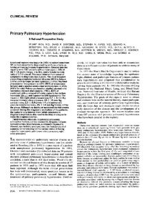
audible tricuspid regurgitation murmur, ascites, and lowerextremity edema. Figure 1 shows the diagnostic process in suspected cases of PH. An electrocardiogram, chest x-ray, and echocardiogram may display signs suggestive of PH (Figs 2-4 and Videos 1 and 2 [supplementary videos are available online]). An echocardiogram should be considered once PH is suspected by history, clinical examination, and risk factors. Possible causes of PH that can be excluded or confirmed by echocardiography are congenital and acquired valvular disease, LV systolic and diastolic dysfunction, large pulmonary embolus (RV dilatation), congenital disease with shunts (ie, atrial septal defect, ventricular septal defect, patent ductus arteriosus, and anomalous pulmonary venous drainage), and other obstructive lesions (coarctation, cor triatriatum, and subaortic valve membrane). Generally, further investigation (RHC) is indicated for patients with unexplained dyspnea, RV systolic pressure (RVSP) ⬎40 mmHg, right atrial enlargement, RV hypertrophy, or interventricular septal flattening. It is important to rule out CTEPH as part of the diagnostic workup. An estimated 3% to 4% of patients that suffer an acute pulmonary embolus (PE) do not fully resolve the thrombus burden, despite anticoagulation, and develop CTEPH.48 Furthermore, 50% of patients with a diagnosis of CTEPH have no
692
STRUMPHER AND JACOBSOHN
Fig 2. This electrocardiogram shows signs of pulmonary hypertension including right atrial and RV hypertrophy: R/S ratio >1 in V1, persistent deep S waves in precordial leads, and signs of right atrial enlargement (peaked P waves in the inferior leads).
prior history of acute pulmonary embolism.49 The screening test of choice to exclude CTEPH is radionuclide perfusion scanning. A normal or very low probability scan essentially excludes CTEPH, whereas a high probability scan warrants further evaluation with a pulmonary angiogram (Fig 5). A spiral computed tomography (CT) scan, although excellent in excluding an acute PE, is less sensitive than perfusion scanning in excluding CTEPH. Although echocardiography may be useful as a screening tool, RHC should be performed to confirm the diagnosis and assess the hemodynamic profile, including (1) oxygen saturation in superior vena cava (SVC), inferior vena cava, right atrium (RA), right ventricle (RV), pulmonary artery (PA), and systemic vessels; (2) pressures in the RA, RV, PA, and PCWP; (3) CO and PVR; and (4) and response to an acute vasodilator therapy. PVR (rather than mPAP) is a more accurate diagnostic criterion to define PH because it reflects the influence of the TPG and CO and only is elevated if the vascular obstruction occurs within the precapillary pulmonary circulation. PVR distinguishes “passive” PAH (elevated mPAP and normal PVR) from PAH because of pulmonary vascular disease (elevated mPAP and elevated PVR). By definition, mPAP and PVR are both elevated in PAH. PH remains a diagnosis of exclusion; after exclusion of airway/lung disease, thromboembolic disease, LV and valvular disease, the diagnostic criteria require both the mPAP ⬎25 mmHg and the PVR ⬎3 Wood units. After noninvasive evaluation with echocardiography, all suspected cases of PH should therefore undergo RHC before the initiation of therapy. During the diagnostic RHC, vasodilator testing should be performed for 2 reasons: (1) acute vasodilator responsiveness identifies patients with a better prognosis, and (2) responders are more likely to have a sustained response to oral calcium channel blockers than nonresponders.50,51 Vasodilator agents most often used are inhaled NO,52 intravenous epoprostenol,53 and intravenous adenosine.54 The definition of a positive vasodilator
response is a decrease in mPAP of ⱖ10 mmHg to an absolute mPAP ⬍40 mmHg, without a decrease in CO. Generally, patients with PH because of causes other than IPAH have a very low rate of long-term responsiveness to calcium channel blockers. THERAPY
Noninvasive Therapy General Measures The following treatment goals should be considered (Fig 6): (1) an improvement in symptoms, (2) an improvement of functional capacity as assessed by the 6-minute walk test or cardiopulmonary exercise testing, (3) lowering mPAP and normalizing CO, (4) reversal of or slowing the rate of progression of the underlying disease, and (5) an improvement in survival. Low-level aerobic exercise (walking) is encouraged, and routine immunizations such as the influenza vaccine and pneumococcal vaccine are advised.55 Avoidance of high altitudes and oxygen supplementation on commercial aircraft for patients with room air saturations ⬍92% are advised.56 Generally, oxygen therapy is indicated if oxygen saturation is ⬍90% on room air. Ideally, pregnancy should be avoided in women with PAH. Routine anticoagulation with coumadin lead to an improvement in survival.57-59 Diuretics and a sodium-restricted diet are indicated in RV dysfunction or overt failure. Digoxin often is added in patients with RV failure, low CO, and atrial arrhythmias although outcome studies are lacking.60 Calcium channel blockers such as long-acting nifedipine, diltiazem, or amlodipine are indicated in a small, select group of patients with IPAH who show acute vasodilator responsiveness to these agents.59 Patients with associated forms of PAH are seldom calcium channel blocker responders.
PULMONARY HYPERTENSION AND RV DYSFUNCTION
693
survival.61-64 Epoprostenol is currently the only therapy for PAH that has definitively been proven to prolong survival. Two large cohort studies showed a survival benefit for epoprostenol-treated patients at 1, 2, and 3 years (88%, 76%, and 63%) versus controls (59%, 46%, and 35%).65 Because of its short half-life (3 minutes), instability at room temperature, and irritating effect on veins, it has to be refrigerated and infused by a syringe pump through a tunneled central venous catheter. The US Food and Drug Administration approved epoprostenol for patients in functional New York Heart Association class 3 and 4 symptomatology with IPAH or PAH because of scleroderma; however, it generally is reserved for those with advanced disease refractory to oral therapy. Although tachyphylaxis with the need for frequent dose adjustments occurs, the beneficial effects of epoprostenol can be sustained for years, and, as a result, many patients have been removed from heart-lung transplantation lists.66 A recent report from the CDC has raised concerns about an increased risk of gram-negative bloodstream infections in patients receiving intravenous prostanoids.67 Alternatively, treprostinil is infused subcutaneously (also
Fig 3. The chest x-ray of a patient with enlarged pulmonary arteries secondary to chronic thromboembolic disease causing pulmonary hypertension: (A) posteroanterior view and (B) lateral view.
Prostanoids In PAH, reduced levels of prostacyclin synthase result in inadequate production of the vasodilator prostacyclin I2. There are currently 3 prostanoids available for treatment of PAH: epoprostenol (ie, prostacyclin and PGI2), treprostinil, and iloprost. Intravenous epoprostenol was the first drug studied. Many studies have shown improved symptomatology, exercise tolerance, hemodynamics, quality of life, and
Fig 4. These transesophageal echocardiographic images show a patient with right ventricular dilatation (A) before and (B) after an ostium secundum atrial septal defect repair. Longstanding left-toright shunting caused chronic right-sided volume overload with resultant RV dilatation. (Color version of figure is available online.)
694
STRUMPHER AND JACOBSOHN
Endothelin Antagonists Endothelin (ET)-1 is an endothelium-derived vasoconstrictor acting on specific endothelin A and B receptors. In the pulmonary circulation, the activation of ET-A receptors causes vasoconstriction and activation of ET-B receptors vasodilatation. Bosentan is a nonselective, competitive ET-A and -B receptor antagonist that promotes pulmonary vasodilatation. Currently, bosentan is used widely in patients with PAH of different causes. In general, oral bosentan improves symptomatology and exercise capacity and increasingly is being used as first-line oral therapy. Abnormal liver enzymes occur frequently at higher doses; hence, the Food and Drug Administration recommendation for monthly liver function tests.73-77 Sitaxsentan is a selective type-A endothelin-receptor blocker, thereby mitigating endothelin-A receptor– dependent vasoconstrictive and proliferative effects while preserving endothelin-B receptor– dependent NO-mediated vasodilation and endothelin clearance. In the STRIDE-1 trial, sitaxsentan improved the 6-minute walk test, New York Heart Association class, CI, and PVR in with IPAH, scleroderma, and CHD associated PAH.78 The warfarin dose should be adjusted after the initiation of sitaxsentan because the inhibition of the CYP2C9 P450 enzyme interferes with the metabolism of warfarin that leads to an increase in international normalized ratio.79 PDE Inhibitors
Fig 5. (A) A computed tomography angiogram of a patient with dilated pulmonary arteries caused by central and peripheral thrombus burden. (B) RV dilatation caused by chronic pressure overload in the same patient.
can be used intravenously) and causes similar symptomatic improvements.68,69 It is a prostacyclin analog with a 3-hour half-life and stability at room temperature delivered by continuous subcutaneous infusion. However, pain and erythema at the infusion site are common. The 3rd prostanoid, iloprost, is given via an ultrasonic nebulizer (2.5-5.0 g) 6 to 9 times a day (half-life of 25 minutes). Airway nebulizations deliver iloprost to resistance pulmonary arteries with minimal effect on systemic blood pressure. In IPAH, short-term inhalation of iloprost resulted in greater pulmonary vasodilatation than NO. It shows similar benefits for symptomatic improvement, and although it lacks the survival benefit of intravenous epoprostenol, there was a trend toward improved survival.70-72
PDE 3 and 5 are enzymes that inactivate 3=, 5= cyclic adenosine monophosphate (cAMP) and cGMP, respectively, the principal second messengers of prostacyclins and NO. Thus, the PDE inhibitors (PDEIs), milrinone and sildenafil, act to augment cAMP- and cGMP-mediated intracellular signaling, leading to vasodilation and decreased PVR. The PDE-5 inhibitor sildenafil at a dose of 20 mg 3 times daily improves exercise capacity, quality of life, and hemodynamics.80-82 Oral sildenafil has been used successfully to manage acute RV dysfunction in heart transplant recipients,83 to wean patients from NO,84 to reduce time on mechanical ventilation, and to prevent the sequelae of CPB on pulmonary endothelial cell function.85,86 More recently, it has been shown that sildenafil is absorbed via the sublingual route.87,88 Although not yet studied definitively, this may have implications for emergent perioperative care. The PDE-3 inhibitor, milrinone, acts similarly but also acts as a positive inotrope (“inodilator”). Milrinone can be administered intravenously or by inhalation; animal CPB studies show that inhaled milrinone causes less systemic hypotension, a smaller decrease in PVR, and a lower heart rate than when given intravenously.86 Combination Therapy Although earlier studies have shown little benefit to combination therapy,89,90 some recent studies have shown symptomatic benefit (without survival benefit) from combination therapies.91,92 The addition of sildenafil to inhaled iloprost or subcutaneous treprostinil is well tolerated and appears effective.93
PULMONARY HYPERTENSION AND RV DYSFUNCTION
695
Fig 6. A therapeutic approach to patients with PAH. CCB, calcium channel blocker; ERA, endothelin-receptor antagonists; PDE5I, phosphodiesterase 5 inhibitors. (Reprinted with permission from McLaughlin et al.12)
Invasive Therapy Atrial Septostomy Observations have shown that patients with Eisenmenger syndrome (right-to-left shunting through an atrial or ventricular septal defect) and PAH generally have superior survival rates compared with IPAH, mainly because of decompression of a pressure-overloaded RV, improved LV filling, and a resulting increase in CO.94,95 Atrial septostomy is considered as a palliative procedure and/or a bridge to lung/heart-lung transplantation in patients with intractable RV failure despite maximal medical therapy.96 The created right-to-left shunt causes a decrease in systemic arterial oxygen saturation that is compensated for by increases in CO and systemic oxygen delivery. Atrial septostomy most commonly is performed by a stepwise balloon dilatation; other techniques include blade septostomy, cutting-edge balloons, and fenestrated Amplatzer devices.97,98 There are no guidelines for the optimal size of the defect; anecdotally, a defect size of 8.5 mm is said to increase CO by 20% to 25%. A decrease in arterial oxygen saturation of 10% and an increase in LV end-diastolic pressure to 18 mmHg preclude further dilatation. Refractory hypoxemia is the most common immediate cause of death, and the overall procedural mortality is 16%. Long-term follow-up after a mean of 2 years generally showed improved symptoms and hemodynamics.99,100 The success rate for bridging patients to transplantation ranged from 30% to 40%.101 Pulmonary Endarterectomy (PEA) CTEPH is underdiagnosed as a cause of PAH and has a poor prognosis if untreated.48,102 Since the first successful report of PEA in 1965, there has been a refinement in patient selection and perioperative techniques, with a reported mortality of as low as 4% to 5% in a single high-volume center.103 The
underlying pathology in CTEPH includes initial thrombus obstruction, thrombus organization, fibrous obstruction of affected proximal arteries, and vascular remodeling in patent distal arteries.104,105 Therefore, it is a disease with a mechanical component (amenable to surgery) and a variable degree of distal small-vessel arteriopathy (amenable to medical therapy). PEA is indicated in patients with positive perfusion scanning, positive pulmonary angiogram, surgically accessible disease, and acceptable surgical risk.106 The goal is to remove sufficient material to lower PVR and increase CO.49,107 In most patients, there is an immediate decrease in PAP as well as an improvement in CO and gas exchange. Postoperative challenges include RV dysfunction caused by residual PAH and/or pulmonary vasoconstriction after extracorporeal circulation and reperfusion pulmonary edema.108 The maximum benefit from surgery may take up to 6 months or longer to become evident. Lung Transplantation Heart-lung transplantation or bilateral lung transplantation is the final option for a minority of patients in whom medical therapy has failed; however, death rates on waiting lists are high because of a global shortage of donor organs. Although effective medical therapy has reduced the rate of transplantation in patients with PAH, approximately 4% of lung and combined heart and lung transplants performed annually worldwide are still for PAH patients with class-4 symptoms or class-3 symptoms despite combination therapy.109-111 The most common indication is for IPAH. Patients with pulmonary venoocclusive disease and pulmonary capillary hemangiomatosis are referred for transplant at the time of diagnosis because medical therapy generally is ineffective.110 Early referral should also take place in patients with scleroderma-associated PAH because of an overall worse prognosis.110,112 On the
696
STRUMPHER AND JACOBSOHN
contrary, patients with congenital left-to-right shunts (ie, atrial septal defects, ventricular septal defects, and patent ductus arteriosus) or Eisenmenger syndrome have higher survival rates.113,114 Single-lung transplants for IPAH have been abandoned because of high rates of perioperative pulmonary edema and poor outcome. However, for patients with CHD and Eisenmenger syndrome (particularly ASD), a single-lung transplant combined with repair of the cardiac defect is possible although heart-lung transplantation has better survival rates in these patients.110,115,116 Although RV afterload is reduced almost immediately after bilateral lung transplantation, hemodynamic instability secondary to RV systolic dysfunction and LV diastolic dysfunction is common in the immediate postoperative period. A retrospective study from Pittsburgh (30 patients who had transplants from 1994 to 2006) showed survival rates of 86% at 1 year, 75% at 5 years, and 66% at 10 years.117 The overall 5-year survival after transplantation for all-cause PAH is 40% to 50%.118 Extracorporeal Support (Extracorporeal Membrane Oxygenation) Possible indications for extracorporeal membrane oxygenation in patients with PAH are acute RV failure and hypoxemia caused by a massive PE, bridge-to-lung transplant, support after lung transplant, treatment of severe reperfusion edema after PEA, and for RV failure unresponsive to conventional medical therapy.119-122 Right Ventricular Assist Devices Patients with end-stage RV failure because of IPAH have fared poorly with ventricular assist devices. The high dP/dT ratio of pulsatile RV assist devices potentially damages the pulmonary microcirculation, causing even greater increases in PVR and PAP, which leads to intraparenchymal pulmonary hemorrhage, hemoptysis, and death. PROGNOSIS
Predictors of a poor prognosis are advanced functional class 3 and 4, rapid symptom progression, poor exercise capacity, significant RV dysfunction, low CO, elevated brain natriuretic peptide, and an associated diagnosis of scleroderma.123,124 Survival also is influenced by etiology, with the best survival rates seen in patients with CHD-associated PAH.125 The natural history and survival of IPAH revealed a median survival of 2.8 years, with 1-, 3-, and 5-year survival rates of 68%, 48%, and 34%, respectively.123 PAH therapy has undergone significant developments over the last 2 decades, but most trials have been of limited size. A recent meta-analysis of 21 trials with 3,140 patients reported improvements in the exercise capacity and a 43% reduction in mortality.126 On the other hand, a metaanalysis of 16 randomized treatment trials of PAH-specific therapies found a nonsignificant reduction in all-cause mortality but a significant improvement in exercise capacity and dyspnea.127
PATHOPHYSIOLOGY AND MANAGEMENT OF PERIOPERATIVE RIGHT VENTRICLE
Decompensation Acute decompensation of patients with PH during the perioperative period is relatively common, frequently lethal, and occurs as a result of acute RV failure.128,129 Patients with Eisenmenger syndrome undergoing a cesarean section have been reported to have a perioperative mortality of up to 70%.130 Patients with PAH undergoing liver transplantation have a mortality as high as 80%.131 The degree of preoperative RV dysfunction and elevation in PVR, together with the type of surgical procedure, are the major predictors of perioperative risk. High-risk surgical procedures include those that could cause significant perioperative systemic inflammatory response; rapid blood loss; high possibility of venous air, CO2, fat, or cement emboli; loss of lung blood vessels; and (reversible) factors leading to acute increases in PVR (eg, acidosis, hypoxia, shivering, pain, and anxiety). All attempts must be made to optimize PVR before surgery, including maximizing medical therapy and preventing conditions that may cause acute deterioration. Patients on chronic intravenous prostacyclin therapy ideally should continue their therapy throughout the perioperative period because discontinuation can precipitate an acute pulmonary hypertensive crisis. Changing to inhaled therapy under controlled conditions may be considered in order to mitigate the potent antiplatelet effect of intravenous prostacyclin. In selected patients not on PAHspecific therapies, a preoperative RHC, vasodilator trial, and PAH-specific therapy may be indicated. Patients with an unacceptable high risk for perioperative decompensation (even after optimization of medical therapy) should not have surgery or should be considered for noninvasive alternatives to surgery. Acute decompensation caused by RV failure frequently is misdiagnosed. Unlike cardiogenic shock from acute LV failure alone (with systemic hypotension, end-organ hypoperfusion, and relatively normal RA pressures), acute RV failure causes increases in RA pressure and systemic venous pressure. The elevated venous (inferior vena cava) pressure (the “outflow pressure” for vitals organs) coupled with a low systemic perfusion pressure (the “inflow pressure” for vital organs) causes a severe reduction in overall perfusion of the vital organs, including kidneys, liver, and the gastrointestinal tract. Therefore, the vital organs have a reduced outflow (venous congestion) and a reduced inflow (low CO). This “double hit” on the vital organs in acute RV failure rapidly can manifest as multiple organ system failure. In addition, the elevated RA pressure may cause hypoxemia in susceptible patients by causing a right-to-left shunt across a patent foramen ovale. It is important to note that tricuspid regurgitation (TR) is common in acute and chronic RV failure, and, hence, thermodilution CO (TdCO) measurements may be misleading.132 TdCO accuracy depends on the severity of the TR (in severe TR, it underestimates CO),133 but there is also a flow dependency of TdCO in TR134 (in acute TR, there is an underestimation of Td CO when the CO is high, an overestimation when the CO is low, and a minimal effect when the CO is midrange). TdCO also may be inaccurate in the presence of anatomic shunts; if there is a left-to-right shunt, thermodilution will measure pul-
PULMONARY HYPERTENSION AND RV DYSFUNCTION
monary rather than systemic blood flow because the cold indicator will be diluted by shunted blood. If there is a right-to-left shunt (Eisenmenger), thermodilution will measure systemic rather than pulmonary blood flow because some of the cold indicator will pass through the shunt. However, the PAP measurements still may be useful to monitor the effect of pulmonary artery vasodilators or vasopressors. Some of the interventions and drugs that improve RV function (ie, 1-adrenergic agonists, phosphodiesterase inhibitors, and calcium sensitizers) may have potentially deleterious effects on the systemic vascular resistance (SVR), particularly if the decrease in SVR is larger than the increase in RV CO. Similarly, systemic vasopressors used to maintain systemic blood pressure and RCA perfusion may cause elevation in PVR. Therefore, the treatment principals of RV failure include the following: 1. Optimization of RV rate and rhythm. Sinus rhythm is important for the optimal filling of a hypertrophied/ dilated right ventricle. Because of the association of RV failure with TR, higher heart rates (80-100 beats/ min) may be desirable to reduce end-diastolic volume. Furthermore, because stroke volume is limited by the increase in RV afterload, it is best to avoid bradycardia (CO ⫽ stroke volume ⫻ heart rate). However, if there is significant RV ischemia, excessive tachycardia may not be tolerated either. A loss of sinus rhythm may lead to acute hemodynamic decompensation, and early synchronized cardioversion should be considered if the changed rhythm exacerbates RV dysfunction. In the event of cardiac pacing, atrial or atrial-ventricular sequential pacing leads to improved RV diastolic filling compared with ventricular pacing alone. 2. Optimization of RV filling. Perioperative CVP monitoring is important; in general, when the CVP is low, the RV must be “coping” even if the PAP and PVR are elevated (ie, the right ventricle must have been “primed” [hypertrophied] and exposed to a progressively higher PAP and PVR over time). On the other hand, an elevated CVP may imply a failing right ventricle with or without TR. The compromised right ventricle will tolerate neither hypovolemia nor overfilling; therefore, an optimal position has to be determined and maintained on the RV Frank-Starling curve (Fig 7). Because the RV is mainly a “volume chamber,” it is less preload dependent than the left ventricle; thus, for a given increase in preload, a smaller increase in stroke volume is expected. However, because it is thin walled, the RV is much more afterload dependent than the LV and the RV CO decreases significantly with an acute increase in mPAP. Past teachings have often suggested that the right ventricle be filled aggressively to passively increase pulmonary blood flow and CO. This may hold true with normal PVR (Fontan physiology) but not in circumstances in which the PVR is high. Injudicious volume loading will result in acute RV distention, increased TR (tricuspid annular dilation), a right-to-left shift of the interventricular septum, impairment of LV end-diastolic filling, reduced stroke
697
Fig 7. The Frank-Starling curve showing the response of the failing right ventricle to an increase in preload.
volume, reduced systemic blood pressure, inadequate RCA perfusion, and an ensuing downward hemodynamic spiral because of progressive RV ischemia and dilation. This is especially true once the CVP reaches values of 15 to 20 mmHg. Optimal RV filling can be assessed with cautious fluid boluses (250 mL of lactated Ringer’s solution) or by an “autotransfusion” maneuver by elevation of the patient’s legs to 45°. Ongoing fluid boluses are indicated if elevation of the legs causes a modest (2-5 mmHg) elevation in CVP and corresponding elevation in PCWP and mean arterial pressure; an elevation of only the CVP (with minimal/no change in PCWP or mean arterial pressure) likely indicates RV distention and precludes further fluid boluses. A relatively underfilled RV is likely the lesser of 2 evils. 3. Maintaining RV myocardial performance includes maintenance of RV coronary perfusion pressure and RV inotropic therapy. Normally, RV coronary perfusion occurs during systole and diastole. However, as the PVR and RVSP rise, RCA flow occurs mainly in diastole (similar to left coronary artery perfusion). RV subendocardial ischemia caused by myocardial oxygen supply-demand imbalance is common in PH. To avoid this, systemic hypotension, excessive increases in RVSP, contractility, and heart rate must be avoided.135 It is important to immediately increase the systemic blood pressure and, subsequently, RCA perfusion pressure when acute RV failure is diagnosed. This can be achieved by optimizing the volume status and use of vasopressors (ie, norepinephrine and vasopressin). Accumulating clinical experiences as well as animal data suggest that vasopressin causes less of an increase in RV afterload than does norepinephrine and reduces the dose of norepinephrine required to maintain the systemic blood pressure.136 Vasopressin has been shown to improve systemic pressures while keeping CO and PAPs stable in patients with persistent postcardiotomy hypotension associated with PH.137 The most important
698
predictor of survival in PH is RV function. Volatile agents depress myocardial function and should be used sparingly in the presence of severe RV failure. Induction doses of propofol and thiopental should be adjusted because both agents decrease both SVR and myocardial contractility. Large doses of opioids should be avoided because it can lead to an acute reduction in sympathetic tone with resultant hypotension and inadequate RV contractility. Etomidate is safe to use because it has little effect on SVR, myocardial contractility and PVR. -Adrenoreceptor agonist therapy (ie, dobutamine and isoprotorenol), PDE-3 inhibitor (milrinone), or a calcium-sensitizer (levosimendan) all improve RV contractility and reduce PVR. However, they also may reduce SVR. If the increase in RV CO does not offset the reduction in SVR, the systemic BP will decrease, and the RCA perfusion will be compromised. This will lead “paradoxically” to a worsening of the RV function because of RV ischemia. Levosimendan is a cardiac inotrope that binds to troponin C, sensitizing the cell to calcium, which leads to increased contractility without increasing intracellular calcium. It has been shown to improve hemodynamics in patients with PH and RV failure while mildly decreasing PVR.138 Because of its ability to decrease afterload, it restores ventricular-pulmonary arterial coupling better than dobutamine.139 Milrinone inhibits the breakdown of 3=, 5= cyclic adenosine monophosphate (cAMP) to adenosine monophosphate, whereas 1-adrenergic agonists stimulate adenylate cyclase, which increases the conversion of adenosine triphosphate to cAMP; hence, milrinone potentiates the inotropic effect of 1-adrenergic agonists in patients with severely decreased myocardial contractility. The 1-adrenergic agonists isoprotorenol and dobutamine are often used in combination with milrinone; together these agents decrease the PVR and have synergistic inotropic effects. Epinephrine, norepinephrine, isoproterenol, and dopamine also have been shown to improve RV contractility.140 PH associated with mild RV dysfunction can be treated with dobutamine alone or with a combination of dobutamine, dopamine, and nitroglycerin. With severe RV dysfunction, the most potent inotropic agents, epinephrine and norepinephrine, with or without milrinone, can be used. It seems that the main advantage in using milrinone is to potentiate the inotropic effect of -agonists (ie, dobutamine, dopamine, epinephrine, and norepinephrine) while adding pulmonary vasodilation. 4. Maintaining transeptal gradient (TSG) and RV geometry. At normal RV systolic pressure (25 mmHg) and LV systolic pressure (125 mmHg), there is a large gradient from the left ventricle to the right ventricle (TSG ⫽ 125-25 mmHg ⫽ 100 mmHg), causing the interventricular septum to bulge into the RV providing a “scaffold” against which the RV-free wall contracts.141 Under normal conditions, this septal position and function account for ⬎50% of RV systolic function.142 In an electrically isolated RV free-wall model, up to 65% of RV function is dependent on proper
STRUMPHER AND JACOBSOHN
functioning of the septum, and the maximum RV developed pressure is reduced by 30% when the septum is inactivated.143,144 Therefore, conditions that reduce the LVSP (systemic hypotension) or increase the RVSP (PH) will reverse the TSG.145 This causes the right ventricle to become more globular (the right ventricle “bulges” into the left ventricle and impairs LV diastolic filling because of increases in RV diastolic volume and pressure), with misalignment of the oblique arrangement of the septal myofibrils leading to an overall decrease in RV function. In order to maintain the TSG, the PVR needs to be reduced, and the systemic blood pressure needs to be maintained. 5. Reduction of PVR. Patients on chronic therapy for PH should continue their established treatment during the perioperative period. Nitrous oxide, ketamine, and protamine increase PVR and should be avoided. During positive-pressure ventilation, PVR increases when lung volumes above the functional residual capacity (FRC) result in the compression of small intra-alveolar vessels (increased transpulmonary pressure); in contrast, extra-alveolar vessel resistance decreases during inspiration. PVR also increases with lung volumes below the FRC because of increased large-vessel resistance through hypoxic pulmonary vasoconstriction. Large tidal volumes and high levels of positive endexpiratory pressure will tend to occlude the intra-alveolar capillaries in well-ventilated areas and divert flow to poorly ventilated areas with a resultant increase in ventilation/perfusion (V/Q) mismatching, a decrease in oxygen saturation, and an increase in PVR. The aforementioned contribution of the intra- and extra-alveolar vessels accounts for the characteristic U-shaped relationship between lung volume and PVR, which is minimal at the FRC and increased at high or low lung volumes (Fig 8). In contrast to systemic arteries, pulmonary vessels constrict with hypoxia (Euler-Liljestrand reflex) and dilate with hyperoxia. Thus, perioperative hypoxemia, hypercarbia, atelectasis, pleural effusions, hypothermia, fluid overload, pain, and anxiety can all cause acute rises in PVR with resultant RV decompensation. Therefore, perioperative ventilation strategies of patients with PAH should incorporate high concentrations of oxygen, low tidal volumes (6 mL/kg of predicted body weight), a respiratory rate sufficient to achieve mild hypocarbia, and optimum levels of positive end-expiratory pressure (5-10 cmH2O). Early drainage of pleural effusions and recruitment maneuvers should be considered. Intravenous air or particulate material (precipitated drugs) should be avoided because of the potential for right-to-left embolization through an opened foramen ovale. Apart from the previously mentioned physiologic considerations, the PVR also can be reduced with the following agents. Intravenous Agents Unfortunately, none of the available intravenous PA vasodilators is selective enough not to cause accompanying systemic vasodilation, which potentially could lead to reductions in the RV coronary perfusion and RV ischemia. The PVR/SVR ratio determines the degree of pulmonary vasodilatation; if the PVR is elevated more than the SVR, intravenous PA vasodilators will produce relatively more pulmonary than systemic vasodi-
PULMONARY HYPERTENSION AND RV DYSFUNCTION
699
vasodilation. The timing of milrinone administration is also important; initiating the milrinone infusion before surgery leads to lower PAPs after CPB compared with initiating the infusion during CPB.150 Nesiritide, a recombinant form of B-type natriuretic peptide, binds to a particulate guanylate cyclase receptor, activating the conversion of guanosine triphosphate to cGMP, causing decreases in mPAP, PVR, PCWP, and CVP. However, the intraoperative use of nesiritide is hampered by its effect of lowering SVR.151 Inhaled Agents
Fig 8. The relationship between lung volume and pulmonary vascular resistance. TLC, total lung capacity; RV, residual volume; FRC, functional residual capacity. (Color version of figure is available online.)
lation. If the decrease in PVR leads to a high enough increase in RV CO, it could minimize the effect of the decreased SVR and, in fact, even increase systemic blood pressure. In contrast, if pulmonary vasodilatation does not increase RV CO, a decrease in systemic blood pressure may not be avoided. PA vasodilators also inhibit hypoxic pulmonary vasoconstriction, which could lead to V/Q mismatching and hypoxemia. The use of intravenous calcium channel blockers is not recommended in the acute setting because these agents may decrease RV contractility, CO, and systemic blood pressure. Pulmonary artery vasodilation with intravenous nitroglycerin or sodium nitroprusside can be useful in patients with PH with/without accompanying RV failure provided simultaneous systemic hypotension is managed appropriately.146 Nitroglycerin has the added advantage of increasing the coronary perfusion to ischemic myocardium. Intravenous nitroglycerin, nitroprusside, and citrulline are all metabolized to NO, which binds to soluble guanylate cyclase, catalyzing the conversion of guanosine triphosphate to cGMP. Increased levels of cGMP set off a cascade that ultimately leads to decreased phosphorylated myosin light chains and vasodilation. Citrulline (NO precursor) has been shown to prevent PH in children with CHD and is well tolerated during cardiac surgery147 and, therefore, represents a possible future therapy in adults. Intravenous epoprostenol (PGI2) has been used successfully in the treatment of protamine-induced PAH and to facilitate weaning patients with severe PAH from CPB.148 However, it has significant systemic vasodilating properties and generally is not recommended for treating acute RV failure. PGE1 is also a potent pulmonary vasodilator that has been shown to improve RV function in cardiac surgical patients.149 The PDE-3 inhibitor milrinone lowers PVR and has positive inotropic properties (“inodilator”) through the inhibition of the breakdown of cAMP that leads to
Inhaled vasodilators cause preferential vasodilation in wellventilated lung zones, with resultant improvement in V/Q matching and arterial oxygen saturation. A higher arterial oxygen partial pressure in itself will decrease PVR. Inhaled agents decrease mPAP, but in contrast to intravenous vasodilators (which decrease CVP, mPAP, PAWP, and systemic BP), inhaled agents have minimal effects on LV preload and systemic BP. Thus, inhaled agents, by increasing pulmonary blood flow could potentially precipitate pulmonary edema in the presence of LV dysfunction.152 Inhaled NO (iNO) is a selective pulmonary artery vasodilator (increased cGMP production) that reduces PVR without decreasing SVR because of immediate inactivation by binding to hemoglobin.153,154 iNO has been proven to be particularly beneficial in patients with PH and associated RV failure in whom the combination of iNO and intravenous dobutamine caused increases in CO and arterial oxygen partial pressure while mPAP remained stable. iNO may be lifesaving in weaning patients with severe PH and RV failure from CPB.155 When used for an acute rise in PVR in association with high-inspired oxygen levels, it may be associated with toxicity from free radical, peroxynitrite, nitrogen dioxide, and methemoglobin production. iNO is expensive and administered via a specialized delivery system. Inhaled epoprostenol (iPGI2) has been found suitable in the management of patients with PAH undergoing mitral valve surgery, resulting in a greater ease of weaning from CPB, shorter intubation times, and a shorter intensive care unit stay.148,156 iPGI2 increases smooth muscle cell cAMP, which leads to pulmonary artery vasodilation; it is hydrolyzed before producing systemic effects. It is a cost-effective alternative to iNO but can be cumbersome to use because of it being administered in glycine, which makes the solution sticky and requires ventilator filters to be changed frequently to avoid malfunction of ventilator valves.157 iNO and iPGI2 are equally effective in decreasing mPAP and PVR.158 Both have been proven to selectively vasodilate the pulmonary circulation and improve RV function in pediatric and adult cardiac surgical patients, patients with acute respiratory distress syndrome, and newborns with persistent PH.159,160 These 2 inhaled agents also may be useful in patients with PH who are hypoxemic secondary to V/Q abnormalities. Because their prolonged use may downregulate endogenous vasodilator pathways, both may cause rebound PH upon withdrawal. Inhaled iloprost, a stable analog of prostacyclin, has been beneficial in the management of intraoperative PH and RV dysfunction in animal and human heart transplant recipients and can be used to help wean patients with PH from CPB.161,162 It is water
700
STRUMPHER AND JACOBSOHN
soluble, has a longer half-life than inhaled epoprostenol, and easily is administered by intermittent nebulization. Inhaled milrinone can be used to decrease PVR perioperatively; it is associated with less systemic hypotension, a smaller decrease in PVR, and a lower heart rate compared with intravenous milrinone.49 Similar findings have been reported among patients with PH undergoing mitral valve replacement.163 Several other inhaled agents have shown selective vasodilation comparable to iNO and iPGI2; these include inhaled PGE1, NO donors (NONO-ates), sodium nitroprusside, and nitroglycerin. Diethylenetriamine (NO donor) release NO at a stochiometrically predictable rate has a slow half-life (24 hours) and reverses PH without systemic vasodilation.164 Nebulized diethylenetriamine (150 g) showed selective pulmonary vasodilation in acute respiratory distress syndrome.165 In children with PH caused by CHD, inhaled nitroglycerin decreases PAP and PVR without much effect on heart rate, PCWP, and SVR.166 The phosphodiesterase-5 inhibitor sildenafil inhibits the breakdown of cGMP to GMP, leading to vasodilation and a decrease in PVR. Oral sildenafil has been used successfully to
manage acute RV dysfunction in heart transplant recipients, wean patients from NO, prevent rebound PH in patients on NO, reduce the duration required for mechanical ventilation, and prevent pulmonary endothelial cell dysfunction.83-85 Oral sildenafil also potentiates and prolongs the effects of iNO.167 Although sublingually administered sildenafil has not been studied, this route should be considered when acute perioperative increases in PVR compromise RV function. SUMMARY
In summary, PH is a rare but lethal disease. The perioperative management of patients with PH and associated RV dysfunction is complex and requires a thorough understanding of the pathophysiology involved. Failure to make an early diagnosis and institute the correct therapy will lead to high perioperative morbidity and mortality. The anesthesiologist has to be aware of the potential treatment strategies including optimizing physiologic parameters, the use of selective pulmonary artery vasodilators, and inotropic and systemic blood pressure support.
REFERENCES 1. McLaughlin V, McGoon M: Pulmonary arterial hypertension. Circulation 114:1417-1431, 2006 2. Farber H, Loscalzo J: Pulmonary arterial hypertension. N Engl J Med 351:655–166, 2004 3. Fishman AP: A century of pulmonary hemodynamics. Am J Respir Crit Care Med 170:109-113, 2004 4. Taichman D, Mandel J: Epidemiology of pulmonary hypertension. Clin Chest Med 28:1-22, 2007 5. Dresdale DT, Schultz M, Michtom RJ: Primary pulmonary hypertension I, clinical and hemodynamic study. Am J Med 11:686-705, 1951 6. Riedel B: The pathophysiology and management of perioperative pulmonary hypertension with specific emphasis on the period following cardiac surgery. Int Anesthesiol Clin 37:55-79, 1999 7. Wagenvoort CA, Wagenvoort H: Primary pulmonary hypertension: A pathologic study of the lung vessels in 156 classically diagnosed cases. Circulation 42:1163-1184, 1970 8. Simonneau G, Robbins I, Beghetti M, et al: Updated clinical classification of pulmonary hypertension. J Am Coll Cardiol 54:S43S53, 2009 (suppl) 9. Galiè N, Hoeper MM, Humbert M, et al: Guidelines for the diagnosis and treatment of pulmonary hypertension: ESC/ERS GUIDELINES. Eur Heart J 30:2493-2537, 2009 10. Kovacs G, Berghold A, Scheidl S, et al: Pulmonary arterial pressure during rest and exercise in healthy subjects. A systematic review. Eur Respir J 34:888-894, 2009 11. Badesch BD, Champion HC, Gomez-Sanchez MA, et al. Diagnosis and assessment of pulmonary arterial hypertension. J Am Coll Cardiol 54:S55-S56, 2009 12. McLaughlin V, Archer SL, Badesch BD, et al, for the Writing Committee: CCF/AHA 2009 Expert Consensus Document on Pulmonary Hypertension: A Report of the American College of Cardiology Foundation Task Force on Expert Consensus Documents and the American Heart Association Developed in Collaboration With the American College of Chest Physicians; American Thoracic Society, Inc.; and the Pulmonary Hypertension Association. J Am Coll Cardiol 53:1573-1619, 2009 13. Hatano S, Strasser T: Primary Pulmonary Hypertension. Report on a WHO Meeting. October 15-17, 1973. Geneva: World Health Organization; 1975
14. Fishman AP: Clinical classification of pulmonary hypertension. Clin Chest Med 22:385-391, 2001 15. Simonneau G, Galiè N, Rubin LJ, et al: Clinical classification of pulmonary hypertension. J Am Coll Cardiol 43:5S-12S, 2004 16. Humbert M, Sitbon O, Chaouat A, et al: Pulmonary arterial hypertension in France: Results from a national registry. Am J Respir Crit Care Med 173:1023-1030, 2006 17. Walker AM, Langleben D, Korelitz JJ, et al: Temporal trends and drug exposures in pulmonary hypertension: An American experience. Am Heart J 152:521-526, 2006 18. Benza RL, Miller DP, Gomberg-Maitland M, et al: Predicting survival in pulmonary arterial hypertension. Insights from the Registry to Evaluate Early and Long-Term Pulmonary Arterial Hypertension Disease Management (REVEAL). Circulation 122:1644 –172, 2010 19. Herget J, Wilhelm J, Novotna J, et al: A possible role of the oxidant tissue injury in the development of hypoxic pulmonary hypertension. Physiol Res 49:493-501, 2000 20. Archer S, Rich S: Primary pulmonary hypertension: A vascular biology and translational research “work in progress.” Circulation 102:2781-2791, 2000 21. Rich S, Dantzker DR, Ayres SM, et al: Primary pulmonary hypertension. A national prospective study. Ann Intern Med 107:216223, 1987 22. Trembath RC, Thomson JR, Machado RD, et al: Clinical and molecular genetic features of pulmonary hypertension in patients with hereditary hemorrhagic telangiectasia. N Engl J Med 345:325-334, 2001 23. Christman BW, McPherson CD, Newman JH, et al: An imbalance between the excretion of thromboxane and prostacyclin metabolites in pulmonary hypertension. N Engl J Med 327:70-75, 1992 24. Rubens C, Ewert R, Halank M, et al: Big endothelin-1 and endothelin-1 plasma levels are correlated with the severity of primary pulmonary hypertension. Chest 120:1562-1569, 2001 25. Stewart DJ, Levy RD, Cernacek P, et al: Increased plasma endothelin-1 in pulmonary hypertension: Marker or mediator of disease? Ann Intern Med 114:464-469, 1991 26. Abenhaim L, Moride Y, Brenot F, et al: Appetite-suppressant drugs and the risk of primary pulmonary hypertension. N Engl J Med 335:609-616, 1996
PULMONARY HYPERTENSION AND RV DYSFUNCTION
27. Eddahibi S, Humbert M, Fadel E, et al: Serotonin transporter overexpression is responsible for pulmonary artery smooth muscle hyperplasia in primary pulmonary hypertension. J Clin Invest 108: 1141-1150, 2001 28. Marcos E, Adnot S, Pham MH, et al: Serotonin transporter inhibitors protect against hypoxic pulmonary hypertension. Am J Respir Crit Care Med 168:487-493, 2003 29. Launay JM, Herve P, Peoc’h K, et al: Function of the serotonin 5-hydroxytryptamine 2B receptor in pulmonary hypertension. Nat Med 8:1129-1135, 2002 30. Giaid A, Yanagisawa M, Langleben D, et al: Expression of endothelin-1 in the lungs of patients with pulmonary hypertension. N Engl J Med 328:1732-1739, 1993 31. Steudel W, Ichinose F, Huang PL, et al: Pulmonary vasoconstriction and hypertension in mice with targeted disruption of the endothelial nitric oxide synthase (NOS 3) gene. Circ Res 81:34-41, 1997 32. Petkov V, Mosgoeller W, Ziesche R, et al: Vasoactive intestinal peptide as a new drug for treatment of primary pulmonary hypertension. J Clin Invest 111:1339-1346, 2003 33. Seeney M, Yuan JX: Hypoxic pulmonary vasoconstriction: role of voltage-gated potassium channels. Respir Res 1:40-48, 2000 34. Simonneau G, Fartoukh M, Sitbon O, et al: Primary pulmonary hypertension associated with the use of fenfluramines derivatives. Chest 114:195S-199S, 1998 (suppl 3) 35. Schaiberger PH, Kennedy TC, Miller FC, et al: Pulmonary hypertension associated with long-term inhalation of “crank methamphetamine. Chest 104:614-616, 1993 36. Silver RM: Scleroderma—Clinical problems: The lungs. Rheum Dis Clin North Am 22:825-840, 1996 37. Speich R, Jenni R, Opravil M, et al: Primary pulmonary hypertension in HIV infection. Chest 100:1268-1271, 1991 38. Cool CD, Rai PR, Yeager ME, et al: Expression of human herpes virus 8 in primary pulmonary hypertension. N Engl J Med 349:1113-1122, 2003 39. McDonnell PJ, Toye PA, Hutchins GM: Primary pulmonary hypertension and cirrhosis: Are they related? Am Rev Respir Dis 127:437-441, 1983 40. Yang YY, Lin HC, Lee WC, et al: Portopulmonary hypertension: Distinctive hemodynamic and clinical manifestations. J Gastroenterol 36:181-186, 2001 41. Castro M, Krowka MJ, Schroeder DR, et al: Frequency and clinical implications of increased pulmonary artery pressures in liver transplant patients. Mayo Clin Proc 71:543-551, 1996 42. Molden D, Abraham JL: Pulmonary hypertension: its association with hepatic cirrhosis and iron accumulation. Arch Pathol Lab Med 106:328-331, 1982 43. Dingli D, Utz JP, Krowka MJ, Oberg AL, Tefferi A. Unexplained pulmonary hypertension in chronic myeloproliferative disorders. Chest 120:801-8; 2001. 44. Marvin KS, Spellberg RD: Pulmonary hypertension secondary to thrombocytosis in a patient with myeloid metaplasia. Chest 103:642644, 1993 45. Hill G, McClean D, Fraser R, et al: Pulmonary hypertension as a consequence of alveolar capillary plugging by malignant megakaryocytes in essential thrombocythaemia. Aust N Z J Med 26:852-853, 1996 46. Grisaru D, Rachmilewitz EA, Mosseri M, et al: Cardiopulmonary assessment in betathalassemia major. Chest 98:1138-1142, 1990 47. Klings ES, Farber HW: Role of free radicals in the pathogenesis of acute chest syndrome in sickle cell disease. Respir Res 2:280-285, 2001
701
48. Pengo V, Lensing AW, Prins MH, et al: Incidence of chronic thromboembolic pulmonary hypertension after pulmonary embolism. N Engl J Med 350:2257-2264, 2004 49. Hoeper MM, Mayer E, Simonneau G, et al: Chronic thromboembolic pulmonary hypertension. Circulation 113:2011-2020, 2006 50. Sitbon O, Humbert M, Jais X, et al: Long-term response to calcium channel blockers in idiopathic pulmonary arterial hypertension. Circulation 111:3105-3111, 2005 51. Morales-Blanhir J, Santos S, de Jover L, et al: Clinical value of vasodilator test with inhaled nitric oxide for predicting long-term response to oral vasodilators in pulmonary hypertension. Respir Med 98:225-234, 2004 52. Pepke-Zaba J, Higenbottam TW, Dinh’Xuan AT, et al: Inhaled nitric oxide as a cause of selective pulmonary vasodilatation in pulmonary hypertension. Lancet 338:1173-1174, 1991 53. Rubin LJ, Groves BM, Reeves JT, et al: Prostacyclin-induced acute pulmonary vasodilation in primary pulmonary hypertension. Circulation 66:334-338, 1982 54. Schrader BJ, Inbar S, Kaufmann L, et al: Comparison of the effects of adenosine and nifedipine in pulmonary hypertension. J Am Coll Cardiol 19:1060-1064, 1992 55. Mereles D, Ehlken N, Kreuscher S, et al: Exercise and respiratory training improve exercise capacity and quality of life in patients with severe chronic pulmonary hypertension. Circulation 114:14821489, 2006 56. Mohr LC: Hypoxia during air travel in adults with pulmonary disease. Am J Med Sci 335:71-79, 2008 57. Frank H, Mlczoch J, Huber K, et al: The effect of anticoagulant therapy in primary and anorectic drug-induced pulmonary hypertension. Chest 112:714-721, 1997 58. Fuster V, Steele PM, Edwards WD, et al: Primary pulmonary hypertension: Natural history and the importance of thrombosis. Circulation 70:580-587, 1984 59. Rich S, Kaufmann E, Levy PS: The effect of high doses of calcium-channel blockers on survival in primary pulmonary hypertension. N Engl J Med 327:76-81, 1992 60. Rich S, Seidlitz M, Dodin E, et al: The short-term effects of digoxin in patients with right ventricular dysfunction from pulmonary hypertension. Chest 114:787-792, 1998 61. Barst RJ, Rubin LJ, Long WA, et al: A comparison of continuous intravenous epoprostenol (prostacyclin) with conventional therapy for primary pulmonary hypertension. The Primary Pulmonary Hypertension Study Group. N Engl J Med 334:296-302, 1996 62. Sitbon O, Humbert M, Nunes H, et al: Long-term intravenous epoprostenol infusion in primary pulmonary hypertension: Prognostic factors and survival. J Am Coll Cardiol 40:780-788, 2002 63. Badesch DB, Tapson VF, McGoon MD, et al: Continuous intravenous epoprostenol for pulmonary hypertension due to the scleroderma spectrum of disease. A randomized, controlled trial. Ann Intern Med 132:425-434, 2000 64. Cabrol S, Souza R, Jais X, et al: Intravenous epoprostenol in inoperable chronic thromboembolic pulmonary hypertension. J Heart Lung Transplant 26:357-362, 2007. 65. McLaughlin VV, Shillington A, Rich S: Survival in primary pulmonary hypertension: the impact of epoprostenol therapy. Circulation 106:1477-1482, 2002 66. Shapiro SM, Oudiz RJ, Cao T, et al: Primary pulmonary hypertension: improved long-term effects and survival with continuous intravenous epoprostenol infusion. J Am Coll Cardiol 30:343-349, 1997 67. Centers for Disease Control and Prevention: Bloodstream infections among patients treated with intravenous epoprostenol or intravenous treprostinil for pulmonary arterial hypertension—Seven sites, United States, 2003-2006. MMWR Morb Mortal Wkly Rep 56:170172, 2007
702
68. Laliberte K, Arneson C, Jeffs R, et al: Pharmacokinetics and steady-state bioequivalence of treprostinil sodium (Remodulin) administered by the intravenous and subcutaneous route to normal volunteers. J Cardiovasc Pharmacol 44:209-214, 2004 69. Simonneau G, Barst RJ, Galie N, et al: Continuous subcutaneous infusion of treprostinil, a prostacyclin analogue, in patients with pulmonary arterial hypertension: A double-blind, randomized, placebocontrolled trial. Am J Respir Crit Care Med 165:800-804, 2002 70. Olschewski H, Simonneau G, Galie N, et al: Inhaled iloprost for severe pulmonary hypertension. N Engl J Med 347:322-329, 2002 71. Opitz CF, Wensel R, Winkler J, et al: Clinical efficacy and survival with first-line inhaled iloprost therapy in patients with idiopathic pulmonary arterial hypertension. Eur Heart J 26:1895-1902, 2005 72. Hoeper MM, Schwarze M, Ehlerding S, et al: Long-term treatment of primary pulmonary hypertension with aerosolized iloprost, a prostacyclin analogue. N Engl J Med 342:1866-1870, 2000 73. Channick RN, Simonneau G, Sitbon O, et al: Effects of the dual endothelin-receptor antagonist bosentan in patients with pulmonary hypertension: A randomised placebo-controlled study. Lancet 358: 1119-1123, 2001 74. Rubin LJ, Badesch DB, Barst RJ, et al: Bosentan therapy for pulmonary arterial hypertension. N Engl J Med 346:896-903, 2002 75. McLaughlin VV, Sitbon O, Badesch DB, et al: Survival with first-line bosentan in patients with primary pulmonary hypertension. Eur Respir J 25:244-249, 2005 76. Galie N, Beghetti M, Gatzoulis MA, et al: Bosentan therapy in patients with Eisenmenger syndrome: A multicenter, double-blind, randomized, placebo-controlled study. Circulation 114:48-54, 2006 77. Jais X, D’Armini AM, Jansa P, et al: Bosentan for treatment of inoperable chronic thromboembolic pulmonary hypertension: BENEFit (Bosentan Effects in iNopErable Forms of chronic Thromboembolic pulmonary hypertension), a randomized, placebo- controlled trial. J Am Coll Cardiol 52:2127-2134, 2008 78. Barst RJ, Langleben D, Frost A, et al: Sitaxsentan therapy for pulmonary arterial hypertension. Am J Respir Crit Care Med 169:441447, 2004 79. Widlitz AC, Barst RJ, Horn EM: Sitaxsentan: A novel endothelin-A receptor antagonist for pulmonary arterial hypertension. Expert Rev Cardiovasc Ther 3:985-991, 2005 80. Lewis GD, Shah R, Shahzad K, et al: Sildenafil improves exercise capacity and quality of life in patients with systolic heart failure and secondary pulmonary hypertension. Circulation 116:15551562, 2007 81. Galie N, Ghofrani HA, Torbicki A, et al: Sildenafil citrate therapy for pulmonary arterial hypertension. N Engl J Med 353:21482157, 2005 82. Hoeper MM, Welte T: Sildenafil citrate therapy for pulmonary arterial hypertension. N Engl J Med 354:1091-1093, 2006 83. De Santo LS, Mastroianni C, Romano G, et al: Role of sildenafil in acute posttransplant right ventricular dysfunction: Successful experience in 13 consecutive patients. Transplant Proc 40:2015-2018, 2008 84. Lee JE, Hillier SC, Knoderer CA: Use of sildenafil to facilitate weaning from inhaled nitric oxide in children with pulmonary hypertension following surgery for congenital heart disease. J Intensive Care Med 23:329-334, 2008 85. Aubin MC, Laurendeau S, Mommerot A, et al: Differential effects of inhaled and intravenous sildenafil in the prevention of the pulmonary endothelial dysfunction due to cardiopulmonary bypass. J Cardiovasc Pharmacol 51:11-17, 2008 86. Lamarche Y, Malo O, Thorin E, et al: Inhaled but not intravenous milrinone prevents pulmonary endothelial dysfunction after cardiopulmonary bypass. J Thorac Cardiovasc Surg 130:83-92, 2005
STRUMPHER AND JACOBSOHN
87. Freitas AF Jr, Bacal F, Jr, et al: Impact of sublingual sildenafil on pulmonary hypertension in patients with heart failure. Arq Bras Cardiol 92:116-126, 2009 88. Deveci S, Pes¸kirciog˘lu L, Aygün C, et al: Sublingual sildenafil in the treatment of erectile dysfunction: Faster onset of action with less dose. Int J Urol 11:989-992, 2004 89. Humbert M, Barst RJ, Robbins IM, et al: Combination of bosentan with epoprostenol in pulmonary arterial hypertension: BREATHE-2. Eur Respir J 24:353-359, 2004 90. Hoeper MM, Leuchte H, Halank M, et al: Combining inhaled iloprost with bosentan in patients with idiopathic pulmonary arterial hypertension. Eur Respir J 28:691-694, 2006 91. McLaughlin VV, Oudiz RJ, Frost A, et al: Randomized study of adding inhaled iloprost to existing bosentan in pulmonary arterial hypertension. Am J Respir Crit Care Med 174:1257-1263, 2006 92. Simonneau G, Rubin LJ, Galie N, et al: Addition of sildenafil to long-term intravenous epoprostenol therapy in patients with pulmonary arterial hypertension: A randomized trial. Ann Intern Med 149:521530, 2008 93. Gomberg-Maitland M, McLaughlin V, Gulati M, et al: Efficacy and safety of sildenafil added to treprostinil in pulmonary hypertension. Am J Cardiol 96:1334-1336, 2005 94. Hopkins WE, Ochoa LL, Richardson GW, et al: Comparison of the hemodynamics and survival of adults with severe primary pulmonary hypertension or Eisenmenger syndrome. J Heart Lung Transplant 15:100-105, 1996 95. Hopkins WE: The remarkable right ventricle of patients with Eisenmenger syndrome. Coron Artery Dis 16:19-25, 2005 96. Doyle RL, McCrory D, Channick RN, et al: Surgical treatments/ interventions for pulmonary arterial hypertension: ACCP evidencebased clinical practice guidelines. Chest 126:63S-71S, 2004 97. Micheletti A, Hislop AA, Lammers A, et al: Role of atrial septostomy in the treatment of children with pulmonary arterial hypertension. Heart 92:969-972, 2006 98. Prieto LR, Latson LA, Jennings C: Atrial septostomy using a butterfly stent in a patient with severe pulmonary arterial hypertension. Catheter Cardiovasc Interv 68:642-647, 2006 99. Espı’nola-Zavaleta N, Vargas-Barro’n J, Tazar JI, et al: Echocardiographic evaluation of patients with primary pulmonary hypertension before and after atrial septostomy. Echocardiography 16:625-634, 1999 100. Reichenberger F, Pepke-Zaba J, McNeil K, et al: Atrial septostomy in the treatment of severe pulmonary arterial hypertension. Thorax 58:797-800, 2003 101. Rothman A, Sklansky MS, Lucas VW, et al: Atrial septostomy as a bridge to lung transplantation in patients with severe pulmonary hypertension. Am J Cardiol 84:682-686, 1999 102. Lewczuk J, Piszko P, Jagas J, et al: Prognostic factors in medically treated patients with chronic pulmonary embolism. Chest 119:818-823, 2001 103. Thistlethwaite PA, Kaneko K, Madani MM, et al: Techniques and outcomes of pulmonary endarterectomy surgery. Ann Thorac Cardiovasc Surg 14:274-282, 2008 104. Moser KM, Bloor CM: Pulmonary vascular lesions occurring in patients with chronic major vessel thromboembolic pulmonary hypertension. Chest 103:685-692, 1993 105. Yi ES, Kim H, Ahn H, et al: Distribution of obstructive intimal lesions and their cellular phenotypes in chronic pulmonary hypertension: A morphometric and immunohistochemical study. Am J Respir Crit Care Med 162:1577-1586, 2000 106. Kim NH: Assessment of operability in chronic thromboembolic pulmonary hypertension. Proc Am Thorac Soc 3:584-588, 2006
PULMONARY HYPERTENSION AND RV DYSFUNCTION
107. Jamieson SW, Kapelanski DP, Sakakibara N, et al: Pulmonary endarterectomy: Experience and lessons learned in 1,500 cases. Ann Thorac Surg 76:1457-1464, 2003 108. Mayer E, Klepetko W: Techniques and outcomes of pulmonary endarterectomy for chronic thromboembolic pulmonary hypertension. Proc Am Thorac Soc 3:589-593, 2006 109. Christie JD, Edwards LB, Aurora P, et al: Registry of the International Society for Heart and Lung Transplantation: Twenty-fifth official adult lung and heart/lung transplantation report—2008. J Heart Lung Transplant 27:957-969, 2008 110. Orens JB, Estenne M, Arcasoy S, et al, for the Pulmonary Scientific Council of the International Society for Heart and Lung Transplantation: International guidelines for the selection of lung transplant candidates: 2006 update—A consensus report from the Pulmonary Scientific Council of the International Society for Heart and Lung Transplantation. J Heart Lung Transplant 25:745-755, 2006 111. Toyoda Y, Thacker J, Santos R, et al: Long-term outcome of lung and heart-lung transplantation for idiopathic pulmonary arterial hypertension. Ann Thorac Surg 86:1116-1122, 2008 112. Kawut SM, Taichman DB, Archer-Chicko CL, et al: Hemodynamics and survival in patients with pulmonary arterial hypertension related to systemic sclerosis. Chest 123:344-350, 2003 113. Nihill MR, O’Laughlin MP, Mullins CE: Effects of atrial septostomy in patients with terminal cor pulmonale due to pulmonary vascular disease. Catheter Cardiovasc Diagn 24:166-172, 1991 114. Kerstein D, Levy PS, Hsu DT, et al: Blade balloon atrial septostomy in patients with severe primary pulmonary hypertension. Circulation 91:2028-2035, 1995 115. Waddell TK, Bennett L, Kennedy R, et al: Heart-lung or lung transplantation for Eisenmenger syndrome. J Heart Lung Transplant 21:731-737, 2002 116. Cottini SR, Lerch N, de Perrot M, et al: Risk factors for reperfusion injury after lung transplantation. Intensive Care Med 32: 557-563, 2006 117. Toyoda Y, Thacker J, Santos R, et al: Long-term outcome of lung and heart-lung transplantation for idiopathic pulmonary arterial hypertension. Ann Thorac Surg 86:1116-1122, 2008 118. Trulock EP, Edwards LB, Taylor DO, et al, for the International Society for Heart and Lung Transplantation: Registry of the International Society for Heart and Lung Transplantation: Twenty third official adult lung and heart lung transplantation report. J Heart Lung Transplant 25:880-892, 2006 119. Maggio P, Hemmila M, Haft J, et al: Extracorporeal life support for massive pulmonary embolism. J Trauma 62:570-576, 2007 120. Vlasselaers D, Verleden GM, Meyns B, et al: Femoral venoarterial extracorporeal membrane oxygenation for severe reimplantation response after lung transplantation. Chest 118:559-561, 2000 121. Thistlethwaite PA, Madani MM, Kemp AD, et al: Venovenous extracorporeal life support after pulmonary endarterectomy: indications, techniques, and outcomes. Ann Thorac Surg 82:2139-2145, 2006 122. Conrad SA, Rycus PT, Dalton H: Extracorporeal Life Support Registry Report 2004. ASAIO J 51:4-10, 2005 123. D’Alonzo GE, Barst RJ, Ayres SM, et al: Survival in patients with primary pulmonary hypertension. Results from a national prospective registry. Ann Intern Med 115:343-349, 1991 124. Stupi AM, Steen VD, Owens GR, et al: Pulmonary hypertension in the CREST syndrome variant of systemic sclerosis. Arthritis Rheum 29:515-524, 1986 125. Kuhn KP, Byrne DW, Arbogast PG, et al: Outcome in 91 consecutive patients with pulmonary arterial hypertension receiving epoprostenol. Am J Respir Crit Care Med 167:580-586, 2003 126. Galie N, Manes A, Negro L, et al: A meta-analysis of randomized controlled trials in pulmonary arterial hypertension. Eur Heart J 30:394-403, 2009
703
127. Macchia A, Marchioli R, Marfisi R, et al: A meta-analysis of trials of pulmonary hypertension: A clinical condition looking for drugs and research methodology. Am Heart J 153:1037-1047, 2007 128. Ramakrishna G, Sprung J, Ravi BS, et al: Impact of pulmonary hypertension on the outcomes of noncardiac surgery: Predictors of perioperative morbidity and mortality. J Am Coll Cardiol 45:16911699, 2005 129. Blaise G, Langleben D, Hubert B: Pulmonary arterial hypertension: Pathophysiology and anesthetic approach. Anesthesiology 99: 1415-1432, 2003 130. Martin JT, Tautz TJ, Antognini JF: Safety of regional anesthesia in Eisenmenger’s syndrome. Reg Anesth Pain Med 27:509-513, 2002 131. Krowka MJ, Mandell MS, Ramsay MA, et al: Hepatopulmonary syndrome and portopulmonary hypertension: A report of the multicenter liver transplant database. Liver Transpl 10:174-182, 2004 132. Nishikawa T, Dohi S: Errors in the measurement of cardiac output by thermodilution. Can J Anaesth 40:142-153, 1993 133. Balik M, Pachl J, Hendl J: Effect of the degree of tricuspid regurgitation on cardiac output measurements by thermodilution. Intensive Care Med 8:1117-1121, 2002 134. Heerdt PM, Blessios GA, Beach ML, et al: Flow dependency of error in thermodilution measurement of cardiac output during acute tricuspid regurgitation. Cardiothorac Vasc Anesth 15:183-187, 2001 135. Szabo G, Soos P, Bahrle S, et al: Adaptation of the right ventricle to an increased afterload in the chronically volume overloaded heart. Ann Thorac Surg 82:989-995, 2006 136. Braun EB, Palin CA, Hogue CW: Vasopressin during spinal anesthesia in a patient with primary pulmonary hypertension treated with intravenous epoprostenol. Anesth Analg 99:36-37, 2004 137. Price LC, Forrest P, Sodhi V, et al: Use of vasopressin after caesarean section in idiopathic pulmonary arterial hypertension. Br J Anaesth 99:552-555, 2007 138. Poelzl G, Zwick RH, Grander W, et al: Safety and effectiveness of levosimendan in patients with predominant right heart failure. Herz 33:368-373, 2008 139. Kerbaul F, Rondelet B, Demester JP, et al: Effects of levosimendan versus dobutamine on pressure load–induced right ventricular failure. Crit Care Med 34:2814-2819, 2006 140. Calvin JE Jr: Inotropic therapy for the failed heart: Picking the right drug for the job. Cardiothorac Vasc Anesth Update 10:1-13, 1991 141. Damiano RJ, Asano T, Smith PK, et al: Functional consequences of right ventricular isolation procedure. J Thorac Cardiovasc Surg 100:569-579, 1990 142. Saleh S, Liakopoulos OJ, Buckberg GD: The septal motor of biventricular function. Eur J Cardiothorac Surg 29:S126-S138, 2006 (suppl 1) 143. Damiano RJ, La Follette P Jr, Cox JL, et al: Significant left ventricular contribution to right ventricular systolic function. Am J Physiol 261:H1514-H1524, 1991 144. Belenkie I, Horne SG, Dani R, et al: Effects of aortic constriction during experimental acute right ventricular pressure loading. Further insights into diastolic and systolic ventricular interaction. Circulation 92:546-554, 1995 145. Damiano RJ, Cox JL, Lowe JE, et al: Left ventricular pressure effects on right ventricular pressure and volume outflow. Cathet Cardiovasc Diagn 19:269-278, 1990 146. Ziskind Z, Pohoryles L, Mohr R, et al: The effect of low-dose intravenous nitroglycerine on pulmonary hypertension immediately after replacement of a stenotic mitral valve. Circulation 72:164-169, 1985 147. Barr FE, Tirona RG, Taylor MB, et al: Pharmacokinetics and safety of intravenously administered citrulline in children undergoing
704
congenital heart surgery: Potential therapy for postoperative pulmonary hypertension. J Thorac Cardiovasc Surg 134:319-326, 2007 148. Lowson SM: Alternatives to nitric oxide. Br Med Bull 70:119131, 2004 149. D’Ambra M, LaRaia PJ, Philbin DM, et al: Prostaglandin E1—A new therapy for refractory right ventricular failure and pulmonary hypertension after mitral valve replacement. J Thorac Cardiovasc Surg 89:567-572, 1985 150. Lamarche Y, Perrault LP, Maltais S, et al: Preliminary experience with inhaled milrinone in cardiac surgery. Eur J Cardiothorac Surg 31:1081-1087, 2007 151. Salzberg SP, Filsoufi F, Anyanwu A, et al: High-risk mitral valve surgery: Perioperative hemodynamic optimization with nesiritide (BNP). Ann Thorac Surg 80:502-506, 2005 152. Semigran MJ: Hemodynamic effects of inhaled mitric oxide in heart failure. Am J Coll Cardiol 24:982-988, 1994 153. Oz MC, Ardehali A: Collective review: Perioperative uses of inhaled nitric oxide in adults. Heart Surg Forum 7:E584-E589, 2004 154. Fujita Y, Nishida O, Sobue K, et al : Nitric oxide inhalation is useful in the management of right ventricular failure caused by myocardial infarction. Crit Care Med 30:1379-1381, 2002 155. Inglessis I: Hemodynamic effects of inhaled nitric oxide in right ventricular infarction and cardiogenic shock. J Am Coll Cardiol 44:793-798, 2004 156. Fattouch K, Sbraga F, Sampognaro R, et al: Treatment of pulmonary hypertension in patients undergoing cardiac surgery with cardiopulmonary bypass: a randomized, prospective, double-blind study. J Cardiovasc Med (Hagerstown) 7:119-123, 2006 157. DeWet CJ, Affleck DG, Jacobsohn E, et al: Inhaled prostacyclin is safe, effective, and affordable in patients with pulmonary hypertension, right heart dysfunction, and refractory hypoxemia after cardiothoracic surgery. J Thorac Cardiovasc Surg 127:1058-1067, 2004 158. Haraldsson A: Comparison of inhaled nitric oxide and inhaled aerosolized prostacyclin in the evaluation of heart transplant candidates with elevated pulmonary vascular resistance. Chest 114:780-786, 1998
STRUMPHER AND JACOBSOHN
159. Steudel W, Hurford WE, Zapol WM, et al: Inhaled nitric oxide: Basic biology and clinical application. Anesthesiology 91:1090-1121, 1999 160. Walmrath D, Schneider T, Schermuly R, et al: Direct comparison of nitric oxide and aerosolized prostacyclin in acute respiratory distress syndrome. Am J Respir Crit Care Med 153:991-996, 1996 161. Rex S, Missant C, Claus P, et al: Effects of inhaled iloprost on right ventricular contractility, right ventriculo-vascular coupling and ventricular interdependence: A randomized placebo-controlled trial in an experimental model of acute pulmonary hypertension. Crit Care 12:R113, 2008 162. Winterhalter M, Simon A, Fischer S, et al: Comparison of inhaled iloprost and nitric oxide in patients with pulmonary hypertension during weaning from cardiopulmonary bypass in cardiac surgery: A prospective randomized trial. J Cardiothorac Vasc Anesth 22:406413, 2008 163. Wang H, Gong M, Zhou B, et al: Comparison of inhaled and intravenous milrinone in patients with pulmonary hypertension undergoing mitral valve surgery. Adv Ther 26:462-468, 2009 164. Cornfield DN, Martin EB, Hampl V, et al: Aerosol delivery of diethylene-triamine/nitric oxide, a nitric oxide adduct, causes selective pulmonary vasodilation in perinatal lambs. J Lab Clin Med 134:419425, 1999 165. Lam CF, Van Heerden PV, Blott J, et al: The selective pulmonary vasodilatory effect of inhaled DETA/NO, a novel nitric oxide donor, in ARDS—A pilot human trial. J Crit Care 19:48-53, 2004 166. Goyal P, Kiran U, Chauhan S, et al: Efficacy of nitroglycerin inhalation in reducing pulmonary arterial hypertension in children with congenital heart disease. Br J Anaesth 97:208-214, 2006 167. Lepore JJ: Hemodynamic effects of sildenafil in patients with congestive heart failure and pulmonary hypertension: Combined administration with inhaled nitric oxide. Chest 127:16471653, 2005