Pulmonary Hypertension, Secondary: [Print] - eMedicine Pulmonology
Seite 1 von 32
emedicine.medscape.com eMedicine Specialties > Pulmonology > Pulmonary Hypertension
Pulmonary Hypertension, Secondary Nader Kamangar, MD, FACP, FCCP, FAASM, Associate Professor of Clinical Medicine, Division of Pulmonary, Critical Care and Sleep Medicine, Multicampus Pulmonary and Critical Care Fellowship Program, University of California, Los Angeles, David Geffen School of Medicine; Medical Director, Hospitalist/Intensivist Program, Olive View-UCLA Medical Center; Associate Program Director, Combined Pulmonary and Critical Care Fellowship Program, Cedars-Sinai/Olive View-UCLA Medical Center/West Los Angeles Veterans Affairs Medical Center Shahriar Pirouz, MD, Resident Physician, Department of Internal Medicine, Olive View University of California Los Angeles Medical Center; Sat Sharma, MD, FRCPC, Professor and Head, Division of Pulmonary Medicine, Department of Internal Medicine, University of Manitoba; Site Director, Respiratory Medicine, St Boniface General Hospital Updated: Jun 1, 2010
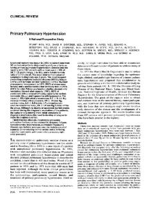
Introduction Background Pulmonary hypertension (PH) defined as a mean pulmonary arterial (PA) pressure of greater than 25 mm Hg at rest or greater than 30 mm Hg during exercise, is characterized by a progressive and sustained increase in pulmonary vascular resistance that eventually leads to right ventricular (RV) failure. Pulmonary hypertension is a life-threatening condition if untreated; treatment success rates vary based on etiology. See the images below.
Gross pathology on a patient who died from severe pulmonary hypertension secondary to persistent patent ductus arteriosus.
http://emedicine.medscape.com/article/303098-print
04.10.2010
Pulmonary Hypertension, Secondary: [Print] - eMedicine Pulmonology
Seite 2 von 32
Another view (of picture in Media File 1) of gross pathology on a patient who died from severe pulmonary hypertension secondary to persistent patent ductus arteriosus.
During a pulmonary arterial thromboendarterectomy, a bilateral proximal thrombus was carefully dissected and extracted, leading to the resolution of secondary pulmonary artery hypertension.
Cardiac disorders, pulmonary disorders, or both in combination are the most common causes of secondary pulmonary hypertension. Cardiac diseases produce pulmonary hypertension via volume or pressure overload, although subsequent intimal proliferation of pulmonary resistance vessels adds an obstructive element. Perivascular parenchymal changes along with pulmonary vasoconstriction are the mechanism of pulmonary hypertension in respiratory diseases. Therapy for pulmonary hypertension is targeted at the underlying cause and its effects on the cardiovascular system. Novel therapeutic agents such as prostacyclin and others undergoing clinical trials have led to the possibility of specific therapies for these once untreatable disorders.
http://emedicine.medscape.com/article/303098-print
04.10.2010
Pulmonary Hypertension, Secondary: [Print] - eMedicine Pulmonology
Seite 3 von 32
The following clinical guideline summaries are available: z
(1) Medical therapy for pulmonary arterial hypertension: ACCP evidence-based clinical practice guidelines. (2) 2007 addendum [1,2 ]
z
Guidelines on diagnosis and treatment of pulmonary arterial hypertension. The Task Force on Diagnosis and Treatment of Pulmonary Arterial Hypertension of the European Society of Cardiology[3 ]
z
ACCF/AHA 2009 expert consensus document on pulmonary hypertension: a report of the American College of Cardiology Foundation Task Force on Expert Consensus Documents and the American Heart Association[4 ]
Frequency United States
The overall prevalence of pulmonary hypertension in the general population is unknown, owing to the heterogeneity of the disease. In specific subgroups of pulmonary hypertension patients, studies have estimated the prevalence as follows: z
In an observational study of 277 patients with HIV infection, .46% of patients had pulmonary hypertension.[5 ]In comparison with prior studies, no change in prevalence rate was seen with modern highly active antiretroviral treatment (HAART).[6 ]
z
A systematic review of several studies of patients with obstructive sleep apnea (OSA) estimated the prevalence of pulmonary hypertension at 15-20%.[7 ]
z
A systematic review of several studies among patients with chronic obstructive pulmonary disease (COPD) estimated the prevalence of pulmonary hypertension at 10-30%.[8 ]
z
In scleroderma patients, the incidence has been estimated to be 6-60% of all patients, with the variance based on the extent of disease.[9 ]
Mortality/Morbidity Based on the US Centers for Disease Control and Prevention (CDC) Pulmonary Hypertension Surveillance from 1980-2002, the following was reported:[10 ] z
The age-standardized death rates for the total US population increased from 5.2 deaths to 5.4 deaths per 100,000 population.
z
The main increase in death rates was seen among women, with 3.3 deaths to 5.5 deaths per 100,000 population, and blacks, with 4.6 deaths to 7.3 deaths per 100,000 population.
z
The death rate in males decreased over this time, from 8.2 deaths to 5.4 deaths per 100,000 population.
Clinical History The clinical manifestations of secondary pulmonary arterial hypertension (SPAH) are frequently masked by the underlying etiology. Obtaining a careful history may help exclude some of the numerous causes of secondary pulmonary hypertension. Important clues to a specific secondary cause include past history of heart murmur, deep venous thrombosis or pulmonary embolism, Raynaud phenomenon, arthritis or arthralgias, rash, heavy alcohol consumption, hepatitis, heavy snoring, daytime hypersomnolence, morning headaches, morbid obesity, and a family history of hypertension. Patients with SPAH often have nonspecific symptoms that reflect the underlying etiology. Other symptoms include the following: z
Dyspnea upon exertion
z
Fatigue
z
Lethargy
z
Syncope with exertion
z
Chest pain
Less common symptoms include the following:
http://emedicine.medscape.com/article/303098-print
04.10.2010
Pulmonary Hypertension, Secondary: [Print] - eMedicine Pulmonology
z
Cough
z
Hemoptysis
z
Hoarseness (due to compression of the recurrent laryngeal nerve by the distended pulmonary artery)
Seite 4 von 32
Typical exertional angina has been reported in as many as 8.5% of patients with SPAH secondary to mitral stenosis. This most likely occurs because of the pulmonary artery distension, right ventricular ischemia, or both in combination.
Physical Physical examination findings may include the following: z
The intensity of the pulmonic component of the second heart sound (P2) may be increased, and a systolic ejection murmur may be heard over the left sternal border. The P2 may demonstrate fixed or paradoxic splitting. A right ventricular heave may be palpated.
z
A prominent a wave may be observed in the jugular venous pulse, and a right-sided fourth heart sound (S4) with a left parasternal heave may be heard.
z
Right ventricular failure leads to systemic venous hypertension and cor pulmonale. Signs are the high-pitched systolic murmur of tricuspid regurgitation, hepatomegaly, a pulsatile liver, ascites, and peripheral edema. In this scenario, a right ventricular third heart (S3) sound is also heard.
z
Signs of underlying cardiac, pulmonary, liver, or collagen-vascular disease are often present.
Causes Pulmonary hypertension was previously divided into 2 categories: primary pulmonary hypertension and secondary pulmonary hypertension, based on identifiable etiology. In 1998, the World Health Organization (WHO) proposed a clinical classification of pulmonary hypertension based on similarities in pathophysiology, clinical presentation, and therapeutic options. Group 1, pulmonary arterial hypertension (PAH), has 2 subgroups: z
Subgroup 1 - Patients with sporadic and familial idiopathic pulmonary arterial hypertension (IPAH)
z
Subgroup 2 - Conditions with known localization of lesions the small pulmonary arterioles, including collagen-vascular disease (scleroderma/CREST syndrome), congenital left-to-right shunts, portopulmonary hypertension, HIV-associated pulmonary hypertension, newborn pulmonary hypertension, and drug-induced (eg. anorexigens) pulmonary hypertension
Group 2, pulmonary venous hypertension, consists of left-sided myocardial and valvular diseases and extrinsic compression of the pulmonary veins (eg tumors) and pulmonary veno-occlusive disease. Group 3, pulmonary hypertension associated with lung diseases and/or hypoxemia, consists of diseases causing inadequate arterial oxygenation. The 3 main groups are those due to lung disease (eg, COPD, interstitial lung disease), impaired respiration (eg, OSA,[11 ] alveolar hypoventilation disorders), and long-term exposure to high altitude. Group 4, pulmonary hypertension due to chronic thrombotic and/or embolic disease, has 2 subgroups: z
Subgroup 1 - Chronic thromboembolic pulmonary hypertension (CTEPH) of proximal arteries
z
Subgroup 2 - Pulmonary embolisms within distal pulmonary arteries, which may be due to thrombosis, tumor, parasites, in situ thrombosis, or sickle cell disease
Group 5, pulmonary hypertension due to direct effect of pulmonary vasculature, consists of inflammatory diseases effecting pulmonary vasculature including, schistosomiasis, sarcoidosis, histocytosis X, and fibrosing mediastinitis. Functional classifications of PAH The classes listed below are based on information adapted from the executive summary of the world symposium on Primary Pulmonary Hypertension in Evian, France in 1998. z
Class I: These are patients with pulmonary hypertension but without resulting limitation of physical activity. Ordinary physical activity does not cause undue dyspnea or fatigue, chest pain, or near syncope.
z
Class II: These are patients with pulmonary hypertension resulting in slight limitation of physical activity. They are comfortable at
http://emedicine.medscape.com/article/303098-print
04.10.2010
Pulmonary Hypertension, Secondary: [Print] - eMedicine Pulmonology
Seite 5 von 32
rest. Ordinary physical activity causes undue dyspnea or fatigue, chest pain, or near syncope. z
Class III: These are patients with pulmonary hypertension resulting in marked limitation of physical activity. They are comfortable at rest. Less than ordinary activity causes undue dyspnea or fatigue, chest pain, or near syncope.
z
Class IV: These are patients with pulmonary hypertension with an inability to perform any physical activity without symptoms. These patients manifest signs of right-sided heart failure. Dyspnea and/or fatigue may even be present at rest. Discomfort is increased by any physical activity.
Differential Diagnoses Apnea, Sleep
Emphysema
Arteriovenous Malformations
Mitral Regurgitation
Atrial Myxoma
Mitral Stenosis
Atrial Septal Defect
Pulmonary Hypertension, Primary
Cardiomyopathy, Dilated
Restrictive Lung Disease
Cardiomyopathy, Hypertrophic
Systemic Lupus Erythematosus
Cardiomyopathy, Restrictive Chronic Obstructive Pulmonary Disease
Workup Laboratory Studies Arterial blood gas determinations should be performed in pulmonary hypertension (PH) patients to assess for hypoxemia. A collagen-vascular disease screening should be performed. This includes measuring the erythrocyte sedimentation rate, rheumatoid factor levels, and antinuclear antibody levels. Synthetic liver function test results (ie, albumin levels, prothrombin time, bilirubin levels) may indicate liver disease associated with portal hypertension. HIV testing and hepatology serology tests should be performed on patients at risk. A complete blood cell count, biochemistry panel, prothrombin time, and activated partial thromboplastin time should be performed at baseline.
Imaging Studies Chest radiography The classic finding on a chest radiograph from a patient with pulmonary arterial hypertension is enlargement of central pulmonary arteries, attenuation of peripheral vessels, and oligemic lung fields. Findings of right ventricular and right atrial dilatation are possible. See the images below:
http://emedicine.medscape.com/article/303098-print
04.10.2010
Pulmonary Hypertension, Secondary: [Print] - eMedicine Pulmonology
Seite 6 von 32
Chest radiograph of a patient with secondary pulmonary hypertension shows enlarged pulmonary arteries. This patient had an atrial septal defect.
A 54-year-old woman with history of scleroderma (CREST variety, ie, calcinosis cutis, Raynaud phenomenon, esophageal motility disorder, sclerodactyly, and telangiectasia) developed dyspnea that worsened upon exertion. Images from a high-resolution CT scan of the lungs showed no parenchymal disease. The patient was found to have severe pulmonary arterial hypertension.
Two-dimensional echocardiography Signs of chronic right ventricular pressure overload are present, which include increased thickness of the right ventricle with paradoxical bulging of the septum into the left ventricle during systole. In later stages, right ventricular dilatation occurs, leading to right ventricular hypokinesis. Right atrial dilatation and tricuspid regurgitation are also present. Doppler echocardiography Doppler echocardiography is the most reliable noninvasive method to estimate pulmonary arterial pressure. Tricuspid regurgitation is usually present in patients with pulmonary arterial hypertension, which aids measurement of pulmonary artery pressure when using the modified Bernoulli equation. The efficacy of Doppler echocardiography depends on the ability to adequately locate the tricuspid regurgitant jet. Furthermore, acoustic windows may be limited in patients who have other diseases (eg, chronic obstructive pulmonary disease [COPD]) or in those who are obese. Tricuspid regurgitation is generally detected in more than 90% of patients with severe SPAH, and a correlation of greater than 95% is
http://emedicine.medscape.com/article/303098-print
04.10.2010
Pulmonary Hypertension, Secondary: [Print] - eMedicine Pulmonology
Seite 7 von 32
observed when the pressure is measured using catheterization. Doppler echocardiography is a useful noninvasive test for long-term follow-up.
Other Tests Electrocardiography Signs of right ventricular hypertrophy or strain may be observed. These include right axis deviation, an R-to-S wave ratio greater than 1 in lead V1, increased P-wave amplitude, and an incomplete or complete right bundle-branch block pattern. Ventilation perfusion lung scanning Ventilation perfusion scan should be performed to exclude chronic thromboembolic pulmonary hypertension. A high- or low-probability scan result is most useful, whereas intermediate-probability results should lead to performing pulmonary angiography. Diffuse mottled perfusion can be observed in patients with primary pulmonary hypertension, as opposed to segmental or subsegmental mismatched defects observed in patients with SPAH. See the image below:
A ventilation/perfusion scan of bilateral mismatched segmental and subsegmental defects, suggesting chronic thromboembolic hypertension.
Pulmonary function testing
http://emedicine.medscape.com/article/303098-print
04.10.2010
Pulmonary Hypertension, Secondary: [Print] - eMedicine Pulmonology
Seite 8 von 32
Pulmonary function tests (ie, spirometry and diffusing capacity for carbon monoxide) should be performed in patients with SPAH to exclude an underlying pulmonary disorder. Diffusing capacity is universally reduced in patients with pulmonary hypertension. These tests may show an obstructive pattern suggestive of COPD or a restrictive pattern suggestive of an interstitial lung disease. Furthermore, the severity of the lung disorder may be established by pulmonary function test findings because they provide both the qualitative and quantitative data.
Procedures Right-sided heart catheterization Right-sided heart catheterization is the criterion standard test for the diagnosis, quantification, and characterization of pulmonary arterial hypertension. Left-sided heart dysfunction and intracardiac shunts can be excluded, and the cardiac output can be measured. The indications for this procedure are (1) difficulty with the accurate measurement of pulmonary arterial hypertension with Doppler echocardiography and (2) the need for a precise measurement of pulmonary vascular resistance to conduct a vasodilator trial to assess the acute response to vasodilators. Acute vasoreactivity is determined by administering a short-acting vasodilator such as prostacyclin, inhaled nitric oxide, or adenosine. An acute response often predicts a beneficial effect from oral agents, such as calcium channel blockers.[12 ] See the images below:
This left pulmonary arterial angiogram shows large central pulmonary arteries and attenuation of peripheral vessels, but thrombosis cannot be identified because it has organized along the vessel walls.
http://emedicine.medscape.com/article/303098-print
04.10.2010
Pulmonary Hypertension, Secondary: [Print] - eMedicine Pulmonology
Seite 9 von 32
Bilateral angiogram should be performed in patients suggested to have chronic thromboembolic pulmonary arterial hypertension. This right pulmonary arterial angiogram from the patient in Media File 8 again shows no evidence of a filling defect, therefore excluding acute thrombosis. Angioscopy is a potentially useful procedure in this setting.
Histologic Findings The histopathologic lesions in patients with secondary pulmonary hypertension are similar to those observed in patients with primary pulmonary hypertension. These pathological changes are the result of long-standing hypertension rather than a consequence of different causes. The plexiform lesion is observed in patients with all types of pulmonary arterial hypertension. These lesions consist of medial hypertrophy, eccentric or concentric laminar intimal proliferation and fibrosis, fibrinoid degeneration, and thrombotic lesions. Fresh or organized and recanalized thrombi may also be present. Diverse types of intimal and muscular lesions of the small muscular arteries may cause the clinical syndrome of pulmonary hypertension, and a plexiform lesion reflecting the abrupt onset of pulmonary hypertension is likely, rather than the lesion being a distinctive cause.
Treatment Medical Care The treatment of pulmonary hypertension (PH) is primarily directed at treatment of the underlying disease. Effective therapy should be instituted in the early stages, before irreversible changes in pulmonary vasculature occur. Once the cause of secondary pulmonary arterial hypertension (SPAH) has been established, the management consists of specific interventional therapy, specific medical therapy, or general supportive therapy. Oxygen supplementation Oxygen has a proven benefit in reducing patient mortality in selected patients with pulmonary arterial hypertension (PAH). Two large trials have demonstrated a definite mortality benefit for patients with COPD, the most common cause of pulmonary arterial hypertension. Survival rates are highest in patients who have less severe SPAH, patients in whom the pulmonary arterial pressure decreases, or patients in whom exercise capacity improves with oxygen therapy.
http://emedicine.medscape.com/article/303098-print
04.10.2010
Pulmonary Hypertension, Secondary: [Print] - eMedicine Pulmonology
Seite 10 von 32
Although long-term study results are not available, oxygen administration may also benefit other groups of patients with SPAH. Therefore, patients who have PaO2 of less than 55 mm Hg at rest from any cause, those who have desaturation during exercise, and those who perform better on oxygen therapy should be prescribed long-term oxygen therapy. Medicare indications for continuous long-term oxygen therapy include the following: z
Arterial PaO2 of less than or equal to 55 mm Hg or an arterial oxygen saturation (SaO2) of less than or equal to 88%
z
PaO2 of 56-59 mm Hg or an SaO2 of 89%, in the presence of evidence of cor pulmonale, right-sided heart failure, or erythrocytosis (hematocrit >55%)
Calcium channel blocker[13,14 ] In a controlled study of 70 patients treated with calcium channel blockers (CCBs), approximately 50% maintained actual long-term New York Heart Association (NYHA) functional class improvement at 1 year, without the need for another treatment The most commonly observed adverse effects with the CCBs are systemic hypotension and lower limb edema. In one study, 10-1414% of idiopathic pulmonary arterial hypertension patients were seen to develop Raynaud syndrome. Sildenafil[15,16,17,18 ] In one controlled study evaluating 278 patients with group 1 pulmonary arterial hypertension treated with sildenafil for a 12-week period, the trial showed improvements in 6-minute exercise capacity, decrease in mean pulmonary artery pressures, and decrease in World Health Organization (WHO) functional class for a 12-month period. One uncontrolled study of 104 patients with chronic thromboembolic pulmonary hypertension (CTEPH) treated with sildenafil for a 12month period showed significant improvements in WHO functional class and pulmonary vascular resistance. Bosentan[19,20,21,22 ] Endothelin-1 exerts a direct vasoconstrictor effect and leads to the proliferation of vascular smooth muscle cells and is a proinflammatory mediator. The effects of endothelin-1 are mediated through the EtA and EtB endothelin receptors. EtA receptors mediate sustained vasoconstriction and proliferation of vascular smooth muscle cells. EtB receptors result in clearance of endothelin and induce the production of nitric oxide and prostacyclin by endothelial cells. Bosentan is an orally active dual (EtA and EtB) endothelin-receptor antagonist. The efficacy of oral bosentan in patients with pulmonary arterial hypertension that was either primary or associated with scleroderma was demonstrated in terms of a significant increase in walking distance. Bosentan also improved the cardiac index, right ventricular systolic function, and function of the left ventricle. Less clinical worsening, defined as death, lung transplantation, or hospitalization for pulmonary hypertension, was present. Combination therapy In one controlled study of 25 patients with idiopathic pulmonary arterial hypertension and scleroderma-associated pulmonary hypertension in whom monotherapy with bosentan had failed, a significant improvement in WHO functional status and exercise capacity was observed in patients with idiopathic pulmonary arterial hypertension, but not in the patients with scleroderma-associated pulmonary hypertension.[23 ] In one controlled trial, sildenafil at 80 mg was added to patients already receiving intravenous epoprostenol, and insufficient improvement was observed. This trial proved that the combination was more effective than the placebo for improving exercise capacity and pulmonary arterial pressure. It also demonstrated a significant reduction in the number of patients showing clinical worsening and an improvement of survival among the patients with the most severe disease.[24 ] Prostacyclin therapy[25 ] Epoprostenol[26,27,28 ] Intravenous prostacyclin (epoprostenol) induces relaxation of vascular smooth muscle and inhibits its growth and platelet aggregation through the increase in intracellular cyclic adenosine monophosphate A prospective, randomized, open-label trial was conducted on 81 patients with primary pulmonary hypertension. After 12 weeks, epoprostenol therapy led to functional improvement, as shown by an improved 6-minute walk test and a decrease of 8% in mean pulmonary artery pressure. However, no long-term randomized trial of epoprostenol in patients with pulmonary arterial hypertension has been conducted.
http://emedicine.medscape.com/article/303098-print
04.10.2010
Pulmonary Hypertension, Secondary: [Print] - eMedicine Pulmonology
Seite 11 von 32
Intravenous epoprostenol improved exercise tolerance, hemodynamics, and long-term survival in a cohort of 178 patients with primary pulmonary hypertension as compared with historical controls. Another trial, in which a cohort of 162 patients was studied after 1 year of receiving epoprostenol therapy, confirmed that the patients' clinical function improved significantly, even though improvements in hemodynamic measures were modest. Improvement with epoprostenol has also been reported in patients who had primary pulmonary hypertension associated with congenital left-to-right cardiac shunts, portal hypertension, and HIV infection.[29 ] Epoprostenol is administered only by continuous intravenous infusion with the use of a portable infusion pump connected to a permanent catheter. Common adverse effects of epoprostenol include jaw pain, headache, diarrhea, flushing, leg pain, and nausea, although they are generally mild and dose related. Other complications include catheter-related sepsis, pump failure, or dislocation of the central venous catheter. Sudden drug interruption may be life threatening. Treprostinil[30,31 ] Treprostinil is a stable prostacyclin analogue administered as a continuous subcutaneous infusion delivered by a minipump. A multicentric randomized trial evaluated treprostinil versus placebo over 12 weeks in 470 patients. The study showed that patients with pulmonary arterial hypertension had increases in 6-minute walk distances, dyspnea, and hemodynamic measurements. A subsequent multicenter retrospective study of 122 patients with pulmonary arterial hypertension or CTEPH treated over a 3-year period showed significant improvement in long-term survival rates. A randomized controlled trial by McLaughlin et al demonstrated the addition of inhaled treprostinil improved exercise capacity and quality of life among patients with PAH (n = 212) who remained symptomatic despite therapy with bosentan or sildenafil.[32 ] Iloprost[33,34 ] Iloprost is a chemically stable prostacyclin analogue that can be delivered by inhaler by producing aerosol particles that deposit in the alveoli. The disadvantage of iloprost is its short duration of action; therefore, it must be inhaled as many as 6 times a day. One 12-week trial involving 203 patients showed an increase in patient scores on a 6-minute walk test and improvement in NYHA functional class, as well as improved hemodynamics. Adverse effects included cough, hypotension, and syncope associated with vasodilation. The long-term efficacy of inhaled iloprost remains disappointing because the only trials performed show a high dropout rate and no improvement in survival compared with conventional therapy. Digoxin[35 ] Digoxin has been shown to be beneficial for patients with supraventricular tachycardia–associated left ventricular dysfunction, but verapamil has been proven to be better than digoxin for controlling the heart rate. Anticoagulation[36,37 ] Current evidence suggests that in patients with idiopathic pulmonary arterial hypertension (IPAH), thrombotic arteriopathy (abnormalities of blood coagulation factors, antithrombotic factors, and the fibrinolytic system) forms, contributing to a prothrombotic state. In a review of 7 observational studies evaluating anticoagulation in pulmonary arterial hypertension, 5 showed a mortality benefit.
Surgical Care Patients with an atrial septal defect, mitral stenosis, or chronic pulmonary thromboembolic disease should be considered for surgical management. Pulmonary arterial hypertension resolves following successful surgical procedures, unless it is too far advanced. Although lung transplantation is reserved for patients with severe primary pulmonary hypertension, a subset of patients with secondary pulmonary arterial hypertension (SPAH) has undergone successful transplantation at several centers. These patients had SPAH due to collagen-vascular disease, drug-induced pulmonary arterial hypertension, or pulmonary venous obstruction. Stability of the underlying causative disorder and the patient's ability to tolerate an extensive surgical procedure are prerequisites. Heart-lung transplantation has been performed in patients with SPAH due to congenital cardiac disease or severe left ventricular dysfunction. Lung transplantation, which has historically been the treatment of choice for severe pulmonary arterial hypertension, in recent years has only been needed for patients who are still in NYHA functional class IV after 3 months of therapy with epoprostenol. Long-term benefits of lung transplantation remain disappointing, with 50% survival at 5 years. Chronic pulmonary hypertension from thromboembolism is much more prevalent than is generally appreciated. Pulmonary endarterectomy offers a cure for the condition, and wider recognition of the efficacy of the operation and the entity are important.[38 ] Pulmonary endarterectomy is a technically demanding procedure, now performed with success at only selected centers. However, excellent results can be obtained with proper patient selection, meticulous surgical technique, and careful postoperative management. An endarterectomy (not an embolectomy) of all affected parts of the lung is performed, and cardiopulmonary bypass, systemic cooling, and circulatory arrest are essential to clear all affected areas of the pulmonary vasculature. Pulmonary endarterectomy has proven to be
http://emedicine.medscape.com/article/303098-print
04.10.2010
Pulmonary Hypertension, Secondary: [Print] - eMedicine Pulmonology
Seite 12 von 32
permanently curative, although an inferior vena caval filter should be placed in all patients to prevent recurrence, and the patients must have life-long anticoagulation. The largest of the case series for thromboendarterectomy operations for thromboembolic pulmonary hypertension reviews the outcomes of 743 patients who underwent this operation between 1999 and 2004. The outcomes showed that the procedure can be performed safely in patients with severe thromboembolic pulmonary hypertension, regardless of the magnitude of the preoperative pulmonary artery systolic pressure.[39 ]
Medication Although treatment of secondary pulmonary hypertension consists primarily of that necessary for the underlying disease, several medications and oxygen are used in different clinical settings. Currently, definite proof of effectiveness is lacking for several of these treatments.
Anticoagulants Long-term anticoagulation with warfarin should be considered in selected patients with SPAH. These include patients with chronic pulmonary emboli, pulmonary venoocclusive disease, and atrial fibrillation induced by left- or right-sided heart failure who are at high risk for developing venous thromboembolism (eg, those with cor pulmonale or immobility secondary to severe dyspnea).
Warfarin (Coumadin)
Interferes with hepatic synthesis of vitamin K–dependent coagulation factors. Used for prophylaxis and treatment of venous thrombosis, pulmonary embolism, and thromboembolic disorders. Tailor dose to maintain an INR in the range of 2-3. Tailor dose to maintain desired INR. Recurrence of DVT and PE increases dramatically when INR drops to 5. Procoagulant vitamin K–dependent proteins are responsible for a transient hypercoagulable state when warfarin is first started and when it is stopped. This phenomenon occasionally causes warfarin-induced necrosis of large areas of skin or of distal appendages. Heparin is always used to protect against this hypercoagulability when warfarin is started; however, when warfarin is stopped, the problem resurfaces, causing an abrupt temporary rise in the rate of recurrent venous thromboembolism. At least 186 different foods and drugs have been reported to interact with warfarin. Clinically significant interactions have been verified for a total of 26 common drugs and foods, including 6 antibiotics and 5 cardiac drugs. Every effort should be made to keep the patient adequately anticoagulated at all times because procoagulant factors recover first when warfarin therapy is inadequate. Patients who have difficulty maintaining adequate anticoagulation while taking warfarin may be asked to limit their intake of foods that contain vitamin K. Foods that have moderate-to-high amounts of vitamin K include Brussels sprouts, kale, green tea, asparagus, avocado, broccoli, cabbage, cauliflower, collard greens, liver, soybean oil, soybeans, certain beans, mustard greens, peas (black-eyed peas, split peas, chick peas), turnip greens, parsley, green onions, spinach, and lettuce. Dosing Adult
5-15 mg/d PO; adjust dose according to desired INR Pediatric
Administer weight-based dose of 0.05-0.34 mg/kg/d PO; adjust dose according to desired INR Infants may require doses at high end of range Interactions
Drugs that may decrease anticoagulant effects include griseofulvin, carbamazepine, glutethimide, estrogens, nafcillin, phenytoin, rifampin, barbiturates, cholestyramine, colestipol, vitamin K, spironolactone, oral contraceptives, and sucralfate Medications that may increase anticoagulant effects of warfarin include oral antibiotics, phenylbutazone, salicylates, sulfonamides, chloral hydrate, clofibrate, diazoxide, anabolic steroids, ketoconazole, ethacrynic acid, miconazole, nalidixic acid, sulfonylureas, allopurinol, chloramphenicol, cimetidine, disulfiram, metronidazole, phenylbutazone, phenytoin, propoxyphene, sulfonamides, gemfibrozil, acetaminophen, and sulindac Contraindications
Documented hypersensitivity; severe liver or kidney disease; open wounds or GI ulcers Precautions Pregnancy
D - Fetal risk shown in humans; use only if benefits outweigh risk to fetus Precautions
http://emedicine.medscape.com/article/303098-print
04.10.2010
Pulmonary Hypertension, Secondary: [Print] - eMedicine Pulmonology
Seite 13 von 32
Do not switch brands after achieving therapeutic response; caution in patients with active tuberculosis or diabetes; patients with protein C or protein S deficiency are at risk of developing skin necrosis
Calcium channel blockers Efficacy has been evaluated primarily in patients with primary pulmonary hypertension. Efficacy of these agents is unclear in patients with SPAH. In selected patients (ie, patients with scleroderma), these agents may be tried only after a vasodilator response is demonstrated. Act by inhibiting calcium ions from entering slow channels or select voltage-sensitive areas of vascular smooth muscle.
Nifedipine (Adalat, Procardia)
Vasodilator that dilates both systematic and pulmonary vascular beds. Higher than usual doses are required for optimal vasodilation of pulmonary arteries. Dosing Adult
10-20 mg IR cap PO tid initially; gradually increase as BP allows (not