0021-972X/00/$03.00/0 The Journal of Clinical Endocrinology & Metabolism Copyright © 2000 by The Endocrine Society
Vol. 85, No. 3 Printed in U.S.A.
The ret/PTC Mutations Are Common in Sporadic Papillary Thyroid Carcinoma of Children and Young Adults* CYDNEY L. FENTON, YVONNE LUKES, DIARMUID NICHOLSON, CATHERINE A. DINAUER, GARY L. FRANCIS, AND R. MICHAEL TUTTLE Departments of Pediatrics (C.L.F., C.A.D., G.L.F.) and Clinical Investigation (Y.L., D.N.), Walter Reed Army Medical Center, Washington, D.C. 20307; F. Edward Hebe´rt School of Medicine, Uniformed Services University of Health Sciences (C.L.F., C.A.D., G.L.F.), Bethesda, Maryland 20814; and Endocrine Service, Memorial Sloan-Kettering Cancer Center (R.M.T.), New York, New York 10021 ABSTRACT The ret/PTC rearrangements (PTC-1, PTC-2, and PTC-3) are characteristic of papillary thyroid cancer (PTC). In adults, PTC-1 is common and may be associated with an aggressive clinical course. The incidence and significance of ret/PTC mutations are less well understood in children. We examined spontaneous PTC from 33 patients (23 females and 10 males) with a median age of 18 yr (range, 6 –21 yr) and a median follow-up of 3.5 yr (range, 0 –13.4 yr). The ret/PTC mutations were identified in 15 tumors (45%), including 8 PTC-1 (8 of 15, 53%), 2 PTC-2 (2 of 15, 13%), 2 PTC-3 (2 of 15, 13%), and 3 (3 of 15, 20%)
combined PTC mutations (PTC-1 and PTC-2). This distribution is significantly different (P ⫽ 0.001, by 2 analysis) from that reported for children with radiation-induced PTC. There was no correlation between the presence or type of ret/PTC mutation and patient age, tumor size, focality, extent of disease at diagnosis, or recurrence. We conclude that ret/PTC mutations are 1) common in sporadic childhood PTC, 2) predominantly PTC-1, 3) frequently multiple, and 4) of different distribution than that reported for children with radiationinduced PTC. (J Clin Endocrinol Metab 85: 1170 –1175, 2000)
T
The majority of their cases (n ⫽ 57) were children with radiation-induced PTC that were found to contain predominantly PTC-3 mutations. There were only 17 children with spontaneous PTC, and in these PTC-1 was predominant. No correlation with clinical outcome was reported for either group, although it has been generally recognized that radiation-induced PTC following the Chernobyl nuclear accident is more aggressive (4, 6). These data indicate that mutations producing ret/PTC-3 rearrangements are common events in radiation-induced PTC in children (8). However, none of these previous studies correlated the presence of ret/PTC mutations to the clinical outcome of individual patients. Furthermore, the optimal control group of children who developed nonradiation-related, sporadic PTC remains small (n ⫽ 17). For these reasons, the current study was undertaken to examine the incidence of ret/PTC mutations, the type of ret/PTC mutations, and the clinical outcome for a cohort of 33 sporadic PTC that presented during childhood or adolescence.
HE THYROID-SPECIFIC oncogene ret/PTC was first described in 1987, when a rearranged form of the RET protooncogene was shown to transform NIH-3T3 cells (1, 2). RET rearrangements were subsequently identified in a subset of adult papillary thyroid carcinoma (PTC) and were recently identified as important features of PTC following the Chernobyl nuclear accident (3). The frequency of ret/PTC rearrangements has varied according to the geographic area. The highest prevalence was reported in Italy (33–35%) and the lowest in Saudi Arabia (2.5%) and Japan (0 –9%) (2, 4 – 8). However, most of these studies examined only a small number of adult cases and made no reference to childhood tumors (5). Motomura et al. recently reported that 71% of sporadic PTC from children in the United States and 87% of PTC from children living in radiation-contaminated areas of Belarus contain rearrangements of the ret oncogene (2, 9). Received July 16, 1999. Revision received October 20, 1999. Accepted December 2, 1999. Address all correspondence and requests for reprints to: R. Michael Tuttle, M.D., Memorial Sloan-Kettering Cancer Center, Box 419, 1275 York Avenue, New York, New York 10021. E-mail: rmtuttle@hotmail. com. * This work was supported by an intramural research grant (WU 6414) from the Department of Clinical Investigation, Walter Reed Army Medical Center (Washington DC). The opinions or assertions contained herein are the private views of the authors and are not to be construed as official or as reflecting the opinions of the Uniformed Services University of the Health Sciences, the Department of the Army, or the Department of Defense. A portion of this study was presented at the Annual Meeting of the Pediatric Academic Societies, San Francisco, California, May 1– 4, 1999, and was published in abstract form in Pediatric Research 45:89A, 1999.
Materials and Methods This study received prior approval from the human use committee, Department of Clinical Investigation, Walter Reed Army Medical Center (Washington, DC).
Clinical data The centralized tumor registry for the Department of Defense (ACTUR) was searched for all individuals diagnosed with thyroid cancer before 21 yr of age. Original medical records were abstracted to construct a clinical database maintained by the principal investigator (10). The extent of disease at diagnosis was defined according to the classification system of DeGroot et al. (11). Disease class I was confined to the thyroid gland. Disease class II involved the regional lymph nodes.
1170
The Endocrine Society. Downloaded from press.endocrine.org by [${individualUser.displayName}] on 23 January 2017. at 06:38 For personal use only. No other uses without permission. . All rights reserved.
ret/PTC IN SPORADIC CHILDHOOD THYROID CANCER Disease class III had either direct extension beyond the thyroid capsule or inadequate surgical resection. Disease class IV involved distant metastasis to bone or lung. Recurrence was defined as the appearance of new disease (new radioactive iodine uptake or biopsy-proven disease) in any patient who had been free of disease (no disease detected by palpation or radioactive iodine scan) for at least 4 months. The clinical data for several of these patients [137 with PTC and 33 with follicular thyroid cancer (FTC)] have been previously reported (10). The corresponding original paraffin-embedded tissue blocks were available for 53 of these patients. Slides were stained with hematoxylin and eosin and were reviewed to establish the diagnoses. We were unable to extract ribonucleic acid (RNA) from 9 tissue blocks, leaving a final number of 44 cases in this study. This included 33 patients (75%) with sporadic PTC, 1 patient (2.3%) with radiation-associated PTC, 8 patients (18.1%) with FTC, 1 patient (2.3%) with medullary thyroid cancer (MTC), and 1 patient (2.3%) with intrathyroidal lymphoma (LYM). Only 1 patient (no. 10) had a history of previous radiation exposure. This patient was treated with external beam radiation for a diagnosis of Hodgkin’s lymphoma at the age of 8 yr. His thyroid cancer was first detected 11 yr later. A thorough review of the remaining cases revealed nothing to suggest radiation exposure in any other patient. Because the emphasis in this study is on sporadic PTC, the data from the single patient with radiation exposure are presented separately.
RNA extraction and complementary DNA (cDNA) preparation Total RNA was isolated from five sections (1 m thick) immediately adjacent to the diagnostic slides. Sections were pooled, deparaffinized using three extractions with xylene (1 ml, 65 C, 10 min), and collected by centrifugation (16,000 ⫻ g, room temperature, 5 min). The tissue was then rehydrated with graded ethanol washes (100%, 80%, and 50%, room temperature, 10 min) and diethyl pyrocarbonate (DEPC)-treated water. The DEPC-treated water was removed (16,000 ⫻ g, room temperature, 5 min), and the pellet was resuspended in 0.5 ml 0.1 mol/L Tris-HCl, 25 mol/L ethylenediamine tetraacetate, and 1% SDS (pH 7.3) containing 500 g proteinase K (Sigma, St. Louis, MO) and incubated with vigorous shaking (52 C, 24 h), with an additional 250 g proteinase K added after 24 h (52 C, additional 24 h). After solubilization, 0.5 ml solution D [4 mol/L guanidinium isothiocyanate, 25 mmol/L sodium citrate (pH 7.0), 0.5% N-lauroylsarcosine, and 0.1 mol/L -mercaptoethanol], 25 L sodium acetate (pH 4.0), 0.25 mL DEPC-treated water-saturated phenol, and 50 L chloroform/isoamyl alcohol (49:1) were added. The aqueous phase was removed, and the RNA was precipitated (100% ethanol, ⫺20 C, 1 h). The pellet was redissolved in 0.3 mL solution D, precipitated with 100% ethanol (⫺70 C, 30 min), and washed (70% ethanol). The RNA was then treated with deoxyribonuclease I, ribonuclease free (20 U/200 L incubation volume; Roche Molecular Biochemical, Indianapolis, IN) and extracted with phenol followed by chloroform/isoamyl alcohol (24:1), and precipitated (100% ethanol, ⫺70 C, 1 h). The pellet was finally resuspended in DEPC-treated water containing RNase inhibitor (1 U/22 L; Ambion, Inc., Austin, TX). One microgram of total RNA was reverse transcribed with random hexamers (2.5 mol/L) using Moloney murine leukemia virus reverse transcriptase at a final concentration of 2.5 U/L, deoxy-NTPs (1 mmol/L each), MgCl2 (5 mmol/L), PCR buffer II (1⫻), and RNase inhibitor (1 U) in a final volume of 20 L and thermocycled (22 C for 10 min, 42 C for 15 min, 99 C for 5 min, and a 5 C soak) for one cycle (PE Applied Biosystems, Foster City, CA). PCR amplification for each ret/ PTC rearrangement contained 1 L cDNA template in a final volume of 5 L of a Perkin-Elmer Corp. (Foster City, CA) PCR reaction buffer mixture using TaqGold polymerase. The reaction was cycled for 10 min
1171
at 95 C followed by 15 s at 95 C and 1 min at 60 C for a total of 43 cycles in a PE 9600 thermocyler (PE Applied Biosystems). The amplified product was separated either in a 4%, 3:1 NuSieve agarose gel (FMC, Rockland, ME) and detected using a 1:10,000 dilution of Syber Green stain (FMC) or a Novex (San Diego, CA) 20% Tris/borate/EDTA polyacrylamide gel and silver stained. The primer pairs were designed to span the known breakpoints specific for each of the ret/PTC mutations in addition to the internal housekeeping gene glyceraldehyde-3-phosphate dehydrogenase gene (GAPDH). Template negative controls and RNA extracted from nonpapillary thyroid cancers were included as negative controls along with each amplification. Reverse transcriptase-negative controls were included as well. Positive control clones for ret/PTC-1, ret/PTC-2, and ret/PTC-3 were provided by Dr. C. Jhiang, Ohio State University (Columbus, OH). Table 1 shows the primer sequences for ret/PTC-1, ret/PTC-2, ret/ PTC-3, and GAPDH in both the sense and antisense directions. The three primer pairs for the different ret/PTC mutations shared a common antisense primer, but unique sense primers. These were synthesized by Biotechnologies Ltd. (Laurel, MD) and were designed to amplify the region across the fusion point of the chimera using information reported by Klugbauer et al. (11). Based on this ret/PTC primer design, the amplified sequence for ret/PTC-1 would be 81 bp, that for ret/PTC-2 would be 108 bp, and that for ret/PTC-3 would be 134 bp. The GAPDH primers were purchased from Perkin-Elmer Corp. Three of the samples (no. 1, 3, and 13) that were found to contain ret/PTC mutations by gel electrophoresis were sequenced to confirm the identity of the chimeric products for each of the ret/PTC rearrangements. Amplified PCR products of the appropriate size were excised from the agarose gel. The DNA was eluted by two successive freezethaw cycles (⫺70 C, room temperature) and centrifuged at 10,000 ⫻ g for 10 min, followed by a purification step (30-m pore size filter; Millipore Corp., Bedford, MA) of the supernatant. Approximately 10 ng DNA template and 1.5 pmol of the appropriate template were added to BigDye Terminator Ready Reaction Mix (PE Applied Biosystems). Reactions were run according to the kit protocol for 25 cycles. Extension products were purified by ethanol precipitation and were separated by electrophoresis in a 4% acrylamide gel on an ABI Prism 377 DNA Sequencer (PE Applied Biosystems). Sequences from the tumors were aligned and compared to the reported sequences for ret/PTC-1, ret/ PTC-2, and ret/PTC-3 using the Sequencer program (Genecode, Ann Arbor, MI). Sequences were confirmed with more than 99% probability for all three samples.
Data analysis and statistical significance After RT-PCR amplification, data were only included if at least GAPDH or one of the three ret/PTC mutations was successfully amplified (44 of 53 attempted samples). Statistical analysis was performed using SPSS for Windows 95 (SPSS, Inc., Chicago, IL). Correlation was performed using Pearson correlation, and nonparametric analysis was performed using the 2 test.
Results
Sporadic PTC from 33 patients (23 females and 10 males) and 1 radiation-associated PTC (1 male) were examined. The details of tumor class, treatment, adjunctive therapy, and recurrence are shown in Table 2. The median age at diagnosis was 18 yr (range, 6 –21 yr). Thirty-five percent of the patients were less than 17 yr of age, 18% were between the ages of 17–19 yr, and 47% were between 19 –21 yr. The majority of
TABLE 1. RET/PTC primer design
ret/PTC-1 ret/PTC-2 ret/PTC-3 GAPDH
Sense
Antisense
Expected size (bp)
5⬘-CAAAGCCAGCGTTACCATCG-3⬘ 5⬘-GAAATTGTGGGGCATCGACC-3⬘ 5⬘-CAAGCTCCTTACATACC-3⬘ 5⬘-GAAGGTGAAGGTCGGAGTC-3⬘
5⬘-CCTTCTCCTAGAGTTTTTCC-3⬘ 5⬘-CCTTCTCCTAGAGTTTTTCC-3⬘ 5⬘-CCTTCTCCTAGAGTTTTTCC-3⬘ 5⬘-GAAGATGGTGATGGGATTTC-3⬘
81 108 134 226
The Endocrine Society. Downloaded from press.endocrine.org by [${individualUser.displayName}] on 23 January 2017. at 06:38 For personal use only. No other uses without permission. . All rights reserved.
1172
JCE & M • 2000 Vol 85 • No 3
FENTON ET AL.
TABLE 2. Demographics of patients with papillary thyroid carcinoma Case no.
131 I post-op
Tumor size (cm)/focality
Recurrence
F/U (months)
Clinical status
ret/PTC rearrangement
Yes Yes
2.0/Unifocal 1.2/Multifocal
Yes/11 months Yes/12 months
63 107
NED Alive, N/A
Total Lobectomy Subtotal Total Total Total Total Subtotal
N/A N/A No No No Yes Yes N/A
2.6/Unifocal 2.0/Unifocal 1.5/Unifocal 0.5/Multifocal 2.4/Unifocal 1.5/Unifocal 2.9/Unifocal 1.1/Unifocal
No No No No No No No No
0 17 18 10 50 2 100 77
N/A Alive, N/A NED NED NED NED NED Alive, N/A
ret/PTC-1 ret/PTC-1 ret/PTC-2 ret/PTC-2
I
Subtotal
Yes
0.7/Unifocal
No
106
NED
20/M 20/F 21/F 21/F
I I I I
Total Total Total Total
Yes Yes N/A N/A
5.0/multifocal 0.7/unifocal 2.3/multifocal 1.8/unifocal
No No No No
24 39 18 68
NED NED NED NED
13/M 14/F 10/F 13/F 15/F 17/F 18/F 19/M 19/F 19/M 20/F 20/F 21/F 19/F 15/M 10/M 17/M 20/M 20/F
II II II II II II II II II II II II II III III III III III IV
Subtotal Total Total Subtotal Total Total Total Total Total N/A Total Total Total Total Total Subtotal Total Total Total
Yes Yes Yes N/A Yes Yes Yes Yes Yes N/A Yes Yes N/A Yes Yes Yes Yes Yes Yes
4.2/multifocal 3.0/multifocal 0.8/unifocal 5.5/multifocal 0.7/multifocal 2.0/multifocal 1.8/multifocal 0.5/unifocal 2.3/multifocal 1/multifocal N/A 1/unifocal 1/multifocal 2.0/multifocal N/A 5.0/unifocal 2.6/multifocal N/A 0.5/multifocal
Yes/159 months Yes/67 months No N/A No No No No No No No No No No Yes/40 months Persistent No No No
161 86 15 0 85 116 27 36 37 104 92 101 22 86 60 27 3 46 63
AD NED NED N/A NED NED Alive, N/A NED NED Alive, N/A NED NED Alive, N/A AD AD AD AD Alive, N/A NED
Age (yr)/sex
Class
Surgery
1 2
14 /M 21/F
I I
Total Total
3 4 5 6 7 8 9 10
6/F 11/F 11/F 14/M 17/F 18/F 18/F 19/Ma
I I I I I I I I
11
19/F
12 13 14 15 16 17 18 19 20 21 22 23 24 25 26 27 28 29 30 31 32 33 34
ret/PTC-3 ret/PTC-2 ret/PTC-1 ret/PTC-3 ret/PTC-1 ret/PTC-2 ret/PTC-3 ret/PTC-1 ret/PTC-1 ret/PTC-2 ret/PTC-1 ret/PTC-1 ret/PTC-1 ret/PTC-1 ret/PTC-1 ret/PTC-1
Class, Disease class according to DeGroot et al. (10); Surgery, extent of initial operative procedure; 131I, documented patient received 131I after initial surgery; Tumor size, greatest diameter of the primary lesion as noted on pathology report at diagnosis; Focality, multifocal vs. unifocal lesions at diagnosis; Recurrence, recurrence and/months after initial therapy when recurrence was first detected; F/U, length of follow-up in months. Clinical status: N/A, not available; AD, alive with disease; NED, no evidence of disease. a Single patient with radiation exposure.
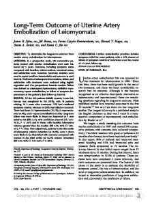
patients (82%) were defined as having DeGroot disease class I or disease class II (12). The median postoperative follow-up time was 3.6 yr (range, 0 –13.4 yr). Forty-four percent were followed for 5 yr or more. This cohort is similar to that of most series, in which the majority of young patients with PTC are female and have either class I or class II disease (9). FTC from eight patients (seven females and one male) were also examined. The median age at diagnosis was 19.5 yr (range, 16 –21 yr). MTC from one patient and an intrathyroidal LYM from one patient were also examined. Figure 1 shows the results of the PCR amplification. The expected 226-bp GAPDH product was identified in 42 samples (Fig. 1A). Figure 1, B, C, and D, shows representative gel electrophoretograms for the 3 different ret/PTC oncogenes. We sequenced three cases to confirm the identity of the ret/PTC mutation identified on gel. In case 1, gel electrophoresis identified ret/PTC-1 mutation, which was confirmed by the sequence data. In case 3, ret/PTC-2 was identified by gel electrophoresis and confirmed by sequence
determination. In case 13, ret/PTC-3 was identified by gel electrophoresis and sequence confirmation. As shown in Table 3, 15 subjects had ret/PTC mutations (15 of 33, 45%). Eight of the 15 mutations (53%) were identified as ret/PTC-1, 2 (13%) were ret/PTC-2, 2 (13%) were ret/PTC-3, and 3 (20%) contained both ret/PTC-1 and ret/PTC-2. Nine patients with ret/PTC mutations were classified as having disease class I, and 6 were classified as having disease class II. None was identified with either disease class III or IV. Two of the 15 patients with ret/PTC mutations (13.3%) developed recurrent PTC within 12 months. The single patient (case 10) with radiation-associated PTC contained both ret/PTC-1 and ret/PTC-3, was classified as disease class I, and did not develop recurrence. As expected, none of the patients with FTC or MTC or the single patient with LYM was positive for ret/PTC mutations. In contrast, of the 18 patients who were negative for ret/ PTC mutations, 3 (of 18, 16.6%) developed recurrent PTC.
The Endocrine Society. Downloaded from press.endocrine.org by [${individualUser.displayName}] on 23 January 2017. at 06:38 For personal use only. No other uses without permission. . All rights reserved.
ret/PTC IN SPORADIC CHILDHOOD THYROID CANCER
1173
FIG. 1. Results of PCR amplification. RNA was reverse transcribed, and the cDNA products were separated by electrophoresis. A, GAPDH amplification products from 7 representative samples. The 226-bp GAPDH product was identified on 4% agarose gel stained with Syber Green. B, ret/PTC-1 amplification products from 10 subjects and a positive control clone resolved over a 4% agarose gel and stained with Syber Green. The positive control (lane 11) and 3 tumor samples (cases 1, 14, and 24; lanes 4, 6, and 9, respectively) reveal the expected 81-bp amplification product. C, ret/PTC-2 amplification products from 15 subjects and a positive control resolved over polyacrylamide gel and stained with silver. The positive control (lane 16) and 2 tumor samples (case 3, lane 5; case 8, lane 10) reveal the expected 108-bp product. D, ret/PTC-3 amplification products from 14 subjects and a positive control resolved over polyacrylamide gel and stained with silver. The positive control (lane 15) and 1 tumor sample (case 13, lane 8) reveal the expected 134-bp product.
TABLE 3. Distribution of RET/PTC mutations Number of patients (n ⫽ 34)
ret/PTC-positive patients PTC-1 PTC-2 PTC-3 PTC-1/PTC-2 PTC-1/PTC-3a
Disease class I (n ⫽ 16)
Disease class II (n ⫽ 13)
8
2
6
2 2 3 1
2 2 3 1
18
6
Disease class III (n ⫽ 4)
Disease class IV (n ⫽ 1)
Recurrence (n ⫽ 5)
Time to recur (months)
1
11
1
12
3
40,67,159
Median follow-up (yr)
4.7
ret/PTC-negative patients ret/PTC (⫺) a
7
4
1
3.4
Patient previously treated with external beam radiation therapy.
Recurrence developed slightly later in the patients who were ret/PTC negative (40 –160 months); however, this difference only approached statistical significance (P ⫽ 0.08, by independent sample two-tail t test). There was no correlation between the presence of ret/PTC mutations and patient age, tumor size, tumor focality (unifocal vs. multifocal disease), extent of disease at diagnosis, treatment (extent of surgery and/or use of adjunctive radioactive iodine ablation), recurrence, or disease-free survival.
Discussion
We amplified GAPDH and the ret/PTC oncogenes (ret/ PTC-1, ret/PTC-2, and ret/PTC-3) from 34 PTC removed from children and young adults. Only 1 patient had a history of prior radiation exposure. Fifteen of the 33 spontaneous PTCs (45.5%) contained at least 1 ret/PTC mutation. The prevalence of ret/PTC rearrangements in childhood thyroid cancer is not well known. Several researchers have
The Endocrine Society. Downloaded from press.endocrine.org by [${individualUser.displayName}] on 23 January 2017. at 06:38 For personal use only. No other uses without permission. . All rights reserved.
1174
JCE & M • 2000 Vol 85 • No 3
FENTON ET AL.
studied ret/PTC rearrangements in childhood thyroid cancers that developed after the Chernobyl nuclear disaster (13). However, reports of ret/PTC rearrangements in sporadic pediatric thyroid cancers are rare. As shown in Table 4, previous studies reported variable prevalence of ret/PTC rearrangements in young patients with sporadic PTC. Bongazone et al. examined 9 PTC from Italian children (4 –19 yr of age) and found that 67% contained ret/PTC mutations (2, 14). Motomura et al. examined 8 Japanese children (9 –14 yr of age) and found that 37.5% contained ret/PTC mutations (2). Finally, Nikiforov et al. examined 17 children (5–18 yr of age) from the United States and found that 71% contained ret/PTC rearrangements (2, 9). In all 3 studies ret/PTC-1 was predominant. This pattern is similar to that reported in adult populations (2, 15). None of these studies, however, correlated the presence of ret/PTC rearrangements with the clinical outcome for individual children. The overall frequency of ret/PTC mutations in our series (45.5%) is similar to that determined by all 3 previous studies and almost identical to that found by Williams et al., who studied children in the United Kingdom with spontaneous PTC. In their study, 10 of 21 (47%) tumors had ret/PTC mutations (5). However, they did not identify the different types of ret/PTC rearrangements, nor did they provide any clinical correlation. The distribution of ret/PTC rearrangements in childhood thyroid cancer is based predominantly on PTC that developed after exposure to the Chernobyl nuclear accident. Initial studies of Chernobyl-associated PTC identified ret/PTC-3 as the most common form of RET rearrangement in radiationinduced childhood PTC (3, 10 –12, 14). However, Pisarchik et al. found that 29% of adult and childhood PTC in Belarus (n ⫽ 31) actually contained ret/PTC-1 rearrangements (3). Pisarchik hypothesized the increase in frequency of the ret/ PTC-1 rearrangements could be related to the longer latency period in their cases, which developed PTC later than those in the initial reports. The patients who had ret/PTC-3 rearrangements had been detected much earlier after the Chernobyl incident (3). The distribution of ret/PTC mutations in our series (53% ret/PTC-1, 13% ret/PTC-2, and 13% ret/PTC-3) is significantly different from the distribution reported for radiationassociated PTC (P ⫽ 0.001, by 2 analysis) (8, 9, 11). There are
several possible reasons for this difference. First, it is possible that ret/PTC-3 could be specifically induced by radiation exposure. Only 1 patient in our series had prior radiation exposure, and his tumor was found to contain both ret/ PTC-1 and ret/PTC-3. Second, previous reports contained a greater number of children with extensive disease. For example, the series by Pisarchik et al. included 15 children with disease class III (3). In contrast, only 6 patients in our series had disease class III or IV. It is therefore possible that ret/ PTC-3 mutations could be associated with more invasive disease. Finally, the children in this study were all from the United States, and the differences seen in the distribution of ret/PTC mutations may reflect a difference in either ethnic background or geographic regions. Overall, our data are most consistent with previous studies of spontaneous adult PTC, in which ret/PTC-1 mutations have been most common (2, 15). Sugg et al. found that ret/ PTC-1 mutations were most frequent in adult PTC, followed by ret/PTC-3 and finally by ret/PTC-2 (1). The clinical significance of ret/PTC mutations is debated, even in adults. Sugg et al. correlated the presence of several oncogenes with clinical outcome for 20 adults with PTC (16). They found ret/PTC mutations in 7 of 20 (35%) of their cases. Five of the 7 patients with metastatic disease had ret/PTC rearrangements. The researchers hypothesize that ret/PTC rearrangements may have a potential role in metastasis. In a follow-up to their initial study, Bongarzone et al. examined the clinicopathological features of adults and children with ret/NTRK1 rearrangements (6). They examined 76 patients (4 – 80 yr of age), of whom 34.2% (26 of 76) were positive for ret/PTC mutations. With respect to children less than 20 yr of age, 60% (6 of 10) had a ret/NTRK1 rearrangement. There was a positive correlation between ret/NTRK1 mutations and young age (⬍30 yr) as well as locally advanced disease (6). However, the study did not separate ret/PTC rearrangements from NTRK1 rearrangements for all age groups. In contrast, Mayr et al. examined 58 PTC from patients aged 17– 84 yr of age and found ret/PTC mutations in 13.8%, with ret/PTC-1 being most frequent (17). The patients with ret/PTC mutations commonly had lymph node involvement at diagnosis. In contrast, distant metastatic spread and death from disease were not observed in pa-
TABLE 4. RET/PTC rearrangements in pediatric papillary thyroid cancer Authors (ref.)
Age range of patients (yr)
No. of children
Radiation exposure
Geographic area
% with ret/PTC rearrangements
Predominant type of ret/PTC rearrangement
Bongarzone (14) Motomura (2) Williams (5) Nikiforov (9) Lee (4) Pisarchik (3) Fuggazzola (8) Nikiforov (9) Klugbauer (10) Pisarchik (13) Delvincourt (7)
4–19 9–14 7–14 5–18 31– 81 12–21 6–14 5–18 8–39 10–28 19–70
9 8 21 23 0 14 6 38 12 35 2
No No No No No Yes Yes Yes Yes Yes N/A
Italy Japan United Kingdom United States China Belarus Belarus Belarus Belarus Belarus France
67 38 47 48 54 43 67 76 67a 23b 50a
ret/PTC-1 ret/PTC-1 ret/PTC-1 ret/PTC-1 ret/PTC-2 ret/PTC-1 ret/PTC-3 ret/PTC-3 ret/PTC-3 ret/PTC-1 N/A
a
Percentage of children with ret/PTC rearrangements. Percentage of all patients with ret/PTC rearrangements. N/A, Information not available. b
The Endocrine Society. Downloaded from press.endocrine.org by [${individualUser.displayName}] on 23 January 2017. at 06:38 For personal use only. No other uses without permission. . All rights reserved.
ret/PTC IN SPORADIC CHILDHOOD THYROID CANCER
tients with ret/PTC mutations. Delvincourt et al. examined 16 PTC from patients aged 19 – 68 yr and found ret/PTC mutations in 12.5% (2 of 16). They found no correlation between the presence or absence of ret/PTC mutations and distant metastasis (7). The data from our study reveal no correlation between the presence or absence of ret/PTC mutations and clinical outcome. Furthermore, there was no relationship between any specific type of ret/PTC mutation and clinical outcome. However, our median follow-up for this cohort is relatively short (3.6 yr) and may not be long enough to detect all recurrences. This is in direct contrast to the results reported by Sugg et al. (1), but is consistent with the findings of Mayr et al. (17). Our study found double ret/PTC mutations in 12% of the PTC examined. This frequency is similar to that identified by Sugg et al. (1). They examined 36 PTC and found a total of 16 ret/PTC mutations. Two of the 16 samples contained multiple ret/PTC rearrangements (12.5%). In addition, 9 of 39 occult cancers (23%) contained 2 forms of ret/PTC mutations. In summary, we found that ret/PTC mutations are present in spontaneous childhood PTC and that they occur more frequently than in adults. The distribution of mutations is similar to that seen in adults, with ret/PTC1 being the most common. However, the distribution is significantly different from that seen in PTC from the Chernobyl population. Double ret/PTC mutations were common, occurring in 12% of the samples examined. The presence of ret/PTC mutations does not correlate to the extent of disease at diagnosis or short term clinical outcome for children and young adults. References 1. Sugg SL, Ezzat S, Rosen I, Freeman JL, Asa S. 1998 Distinct multiple RET/PTC gene rearrangements in multifocal papillary thyroid neoplasia. J Clin Endocrinol Metab. 83:4116 – 4122. 2. Motomura T, Nikiforov YE, Namba H, et al. 1998 ret Rearrangements in Japanese pediatric and adult papillary thyroid cancers. Thyroid. 8:485– 489.
1175
3. Pisarchik AV, Ermak G, Demidchik EP, Mikhalevich LS, Kartel NA, Figge J. 1998 Low prevalence of the ret/PTC3r1 rearrangement in a series of papillary thyroid carcinomas presenting in Belarus ten years post-Chernobyl. Thyroid. 8:1003–1008. 4. Lee CH, Hsu LS, Chi CW, Chen GD, Yang AH, Chen JY. 1998 High frequency of rearrangement of the RET protooncogene (RET/PTC) in Chinese papillary thyroid carcinomas. J Clin Endocrinol Metab. 83:1629 –1632. 5. Williams H, Rooney S, Thomas GA, Cummins G, Williams E. 1996 RET activation in adult and childhood papillary thyroid carcinoma using a reverse transcriptase-n-polymerase chain reaction approach on archival-nested material. Br J Cancer. 74:585–589. 6. Bongarzone I, Vigneri P, Mariani L, Collini P, Pilotti S, Pierotti MA. 1998 RET/NTRK1 rearrangements in thyroid gland tumors of the papillary carcinoma family: correlation with clinicopathological features. Clin Cancer Res. 4:223–228. 7. Delvincourt C, Patey M, Flament JB, et al. 1996 Ret and trk proto-oncogene activation in thyroid papillary carcinomas in French patients from the Champagne-Ardenne Region. Clin Biochem. 29:267–271. 8. Fugazzola L, Pilotti S, Pinchera, et al. 1995 Oncogenic rearrangements of the RET proto-oncogene in papillary thyroid carcinomas from children exposed to the Chernobyl nuclear accident. Cancer Res. 55:5617–5620. 9. Nikiforov E, Rowland JM, Bove KE, Monforte-Munoz H, Fagin JA. 1997 Distinct pattern of ret oncogene rearrangements in morphological variants of radiation-induced and sporadic thyroid papillary carcinomas in children. Cancer Res. 57:1690 –1694. 10. Dinauer CAW, Tuttle RM, Robie DK, et al. 1998 Clinical features associated with metastasis and recurrence of differentiated thyroid cancer in children. Clin Endocrinol (Oxf). 49:619 – 628. 11. Klugbauer S, Lengfelder E, Demidchik EP, Rabes HM. 1995 High prevalence of RET rearrangement in thyroid tumors of children after the Chernobyl reactor accident. Oncogene. 11:2459 –2467. 12. DeGroot L, Kaplan E, McCormick M, Strauss F. 1990 Natural history, treatment, and course of papillary thyroid carcinoma. J Clin Endocrinol Metab. 71:414 – 424. 13. Pisarchik AV, Ermak G, Fomicheva V, Kartel NA, Figge J. 1998 The ret/PTC rearrangement is a common feature of Chernobyl-associated papillary thyroid carcinomas from Belarus. Thyroid. 8:133–139. 14. Bongarzone I, Fugazzola L, Vigneri P. 1996 Age-related activation of the tyrosine kinase receptor protooncogenes RET and NTRK1 in papillary thyroid carcinoma. J Clin Endocrinol Metab. 81:2006 –2009. 15. Learoyd DL, Messina M, Zedenius J, Guinea AI, Delbridge LW, Robinson BG. 1998 RET/PTC and RET tyrosine kinase expression in adult papillary thyroid carcinomas. J Clin Endocrinol Metab. 83:3631–3635. 16. Sugg SL, Ezzat S, Zheng L, Freeman JL, Rosen IB, Asa SL. 1999 Oncogene profile of papillary thyroid carcinoma. Surgery. 125:46 –52. 17. Mayr B, Brabant G, Goretzki P, Ru¨schoff J, Dietmaier W, Dralle H. 1997 ret/PTC-1, -2, and -3 oncogene rearrangements in human thyroid carcinomas: implications for metastatic potential? J Clin Endocrinol Metab. 82:1306.
The Endocrine Society. Downloaded from press.endocrine.org by [${individualUser.displayName}] on 23 January 2017. at 06:38 For personal use only. No other uses without permission. . All rights reserved.