Global Veterinaria 8 (1): 76-82, 2012 ISSN 1992-6197 © IDOSI Publications, 2012
Prevalence of Some Milk Borne Bacterial Pathogens Threatening Camel Milk Consumers in Egypt 1
1
Abeer, A. AlAll, 2Azza S.A. Gouda, 1H.A. Dardir and 3A.K. Ibrahim
Department of Food Hygiene and Control, Faculty of Veterinary Medicine, Cairo University 2 Department of Microbiology, Desert Research Center, Cairo 3 Department of Clinical Pathology, Faculty of Veterinary Medicine, Cairo University
Abstract: Camel milk represents the principal milk for consumers in the arid and sub-arid areas where camels present the main animal source. Regular consumption of camel’s milk in those areas is mainly occurred in raw state. So the present work was planned to investigate the possibility of transmission of 3 milk borne pathogens including Salmonella spp., E. coli and Listeria monocytogenes in a total of 185 camel’s milk samples collected from Sinai, Aswan and Sharqia Governorates. Conventional diagnosis revealed isolation and identification of 5 Salmnella spp. with special interest to presence of S. enteritidis, S. typhi S. typhimurium and S. anatum and 12 E. coli isolates, with special consideration to presence of E. coli O157:H7 and E. coli O26:H11. Two isolates of Listeria monocytogenes had been detected. Multiplex PCR assay found to be rapid, economic and sensitive tool for accurate detection of the three organisms concurrently. In addition, this selected multiplex PCR assay detected virulence genes (InvA, Eae and ActA) of Salmonella spp., E. coli and Liseria monocytogenes, respectively enhancing evaluation of the pathogenicity of these pathogenic strains present in milk samples. Finally, it was concluded that for improving quality of raw camel’s milk, enhancing the milking protocols and sanitizing programs associated with camel’s milk production should be carried out. Key words: Camel milk
Listeria monocytogenes
E. coli spp. and Salmonella spp.
INTRODUCTION
Escherichia coli and it is pathogenic [6]. Contamination of raw milk by pathogenic bacteria from source external to the udder may be caused by salmonellae strains, which produce many outbreaks of enteritis [7]. Listeria monocytogenes, Shiga toxin-producing E. coli (STEC) and serotypes of Salmonella are considered as important food borne pathogens [8, 9]. Listeria monocytogenes has been recognized as a cause of disease in humans and animals and has been responsible for listeriosis outbreaks in past years [10, 11]. Various reports showed that Listeria spp. can be found in dairy products [12], meat and poultry [13]. Raw milk and dairy products made from unpasteurized milk have been responsible for E. coli outbreaks including strain O157:H7 [14, 15]. The detection methods for milk borne pathogens generally involve: (a) colony isolation on selective media, (b) use of biochemical tests and (c) serotyping using antibodies against specific bacterial antigens [15, 16]. These procedures are cumbersome and time consuming.
Camel milk is one of the most valuable food resources for pastoral people in arid and semiarid areas. In the last years milk consumption among urban population has been increased [1, 2]. Nowadays, public health concern associated with microbial food safety has arisen [3]. Numerous epidemiological reports proved that, non-heat treated milk and raw-milk products represent the major factors responsible for illnesses caused by food borne pathogens [4]. Camel milk and meat are the principal animal foods in arid and semi-arid areas of the African and Asian countries [3]. FAO [5] has reported that, more than 18 million camels around the world support the survival of millions of people. Raw camel milk may contain microorganisms pathogenic for man and their source may lie either within or outside the udder. Pathogenic bacteria may present in raw milk as a direct consequence of udder disease. Among the organisms commonly producing mastitis is
Corresponding Author: Abeer, A. AlAll, Department of Food Hygiene, Faculty of Veterinary Medicine, Cairo University.
76
Global Veterinaria, 8 (1): 76-82, 2012
Isolation of Salmonella, E. coli and L. monocytogenes: It was carried out according to protocols described by APHA [31]. Strains presenting a biochemical profile suggestive of Salmonella were submitted to additional biochemical tests [32]. The strains confirmed as Salmonella spp. in the Central Laboratory of Egyptian Ministry of Health were differentiated serologically into species and subspecies as described by Popoff [33]. Colonies suspected to be E. coli were examined according to Ewing [32] and Orskov and Orskov [34]. E. coli were selected for subculture and sero-grouping. Determination of the EPEC sero-groups (O antigens and H antigens) was performed by agglutination tests using polyvalent and monovalent sera against O antigens (O26, O55, O82, O111, O113, O119, O126, O125, O126, O127, O128, O142 and O157) and flagellar H antigens (H1 to H 56) according to the instructions of the manufacturer (Bio-Rad Co and Statens Serum institute, Copenhagen, Denmark), respectively. For testing samples for Listeria spp., 25 ml milk of each sample was homogenized with 225 ml of enrichment broth (Oxoid, Hampshire, United Kingdom) for 2 min. The enrichments were incubated at 37°C for 48 hrs. A loopful of the enrichment culture was streaked onto Oxford Listeria spp. selective agar (Oxoid) and incubated for 48 hrs at 37°C and examined for typical Listeria spp. colonies.
In certain cases, it takes several days to establish the identity of particular bacteria. Therefore, new approaches in milk safety are needed for fast and efficient detection of low numbers of bacteria likely to be present in milk. Several methods were tested in recent years to facilitate the identification of bacteria in foods. Molecular techniques, such as PCR, have been used extensively for several years for identification and characterization of bacteria in food samples including meat and dairy products [17, 18]. However, these assays used selective enrichment techniques to recover bacteria in food samples and they take 48-72 hrs before the identity of bacteria can be established. The detection of pathogenic bacteria is a fundamental objective of food microbiology ensuring food quality. Regarding this, PCR technology has successfully shortened analysis time and has been widely applied for the detection of food borne pathogens [19]. Several of these PCR- based methods were developed for the detection of L. monocytogenes involving a pre-enrichment step [20, 21]. E. coli frequently contaminates food and it is a good indicator of fecal pollution [25-27]. Presence of E. coli in milk products indicates the presence of enteropathogenic microorganisms, which constitute a public health hazard. Enteropathogenic E. coli can cause severe diarrhea and vomiting in infants and young children [28]. Salmonella is one of the main causes of food borne diseases worldwide, in humans and animals [29]. The infective dose of Salmonella can be as low as 15 to 20 cells, depending upon age and health of host. Although most outbreaks are associated with the consumption of egg products, there are also reports of outbreaks related to the consumption of milk and ice cream [30]. The study was planned to elucidate the safety status of camel’s milk for Egyptian consumers. The study was directed to detect specific pathogens including Salmonella spp., E. coli spp. and L. monocytogenes by conventional and molecular assays.
Molecular Investigations: A volume of 500 ul of each milk sample was extracted using Sambrook method [35] to obtain purified DNA. The extracted samples were amplified by multiplex PCR assay for detection of Salmonella spp., E. coli spp. and Listeria monocytogenes using the primers listed in Table (1). The reaction mixture (50 µl) contained 5 µl of extracted DNA, 1 µl of each primer (20 pmol/µl), 0.6 µl of deoxynucleosidetriphosphate (10 mmol/L), 3 µl of 10 X thermophilic buffer (Promega), 1.8 µl of MgCl2 (25 mmol/L), 0.2 µl of Taq DNA polymerase (5 U/µl) and complete the reaction volume using distilled water in a 0.2-ml reaction tube. Primers were synthesized by (Metabion. GmbH, Germany) for each gene. Amplification protocol included initial denaturation, at 95°C for 5 min followed by 93°C for one min, 55°C for 1 min and 72°C for one min the three steps were repeated for 39 cycle and finally kept at 72 for 10 min. The presence of PCR products was determined by electrophoresis of 10 µl of
MATERIALS AND METHODS Collection of Samples: A total of 185 raw camel milk samples was collected from Sinai (42 samples), Aswan (73 samples) and Sharquia (70 samples), the samples were transported to the laboratory in an insulated ice box without delay until being examined. 77
Global Veterinaria, 8 (1): 76-82, 2012 Table 1: List of the oligonucleotides primers used for multiplex PCR assay Organism
Gene
Primer
S. enterica
InvA
E. coli*
Intemin
L. monocytogenes
Act A
Sal-f: Sal-r: Eae-f Eae-r Acta-f: Acta-r:
Band Size Bp Aattatcgccacgttcgggcaa Tcgcaccgtcaaaggaacc Tgcggcacaacaggcggcga Cggtcgccgcaccaggattc gctgatttaagagatagaggaaca Tttatgtggttatttgctgtc
Reference
284
Rahn et al. [36]
629
Heuvelink et al. [37]
827
Zhou and Jiao [38]
*Enteropathogenic attachment & effacement
the DNA product in a 1.5 % agarose gel with 1 X TAE buffer (40 mM Tris-HCl, 1 mM EDTA/L, 1.14 ml/L glacial acetic acid, pH 7.8) at a voltage of 4 volts /cm and stained with 0.5 mg/ml ethidium bromide and the fluorescent bands were visualized with a UV transilluminator and photographed. A 100-bp DNA ladder (Gibco BRL) was used as a molecular marker.
Regarding to serotypes of bacterial isolates (Table 3) Salmonella spp., infection in camels has been reported in various countries, including Sudan [46], USA [47] and, more recently, from Somalia [48], Ethiopia [49], Egypt [50-52] and UAE [53] with similar prevalence rate and varied serotypes. While Omer and Eltinay [54], reported that the examined samples were free of Salmonella spp. Regarding to E. coli isolates and serovars E. coli isolates with variable antigenic structure had been reported by Obied and Bagadi [55] in Saudi camel’s milk. Both Benkerroum et al. [27] and Semereab and Molla [56], demonstrated high E. coli count for Moroccan and Ethiopian camel’s milk, respectively. The high incidence of E. coli serovars was detected to declare that both E. coli O157:H7 and E. coli O26:H11 to be of high prevalence regarding to the rest of E. coli isolates. Similar results were demonstrated by Hajian et al. [57]. Listeria monocytogenes was found to be less in spread but had a high fatality rate. Many authors stated that L. monocytogenes may enter the food chain through carrier animals that shed the organism in the milk and feces due to the microorganism resistance to adverse environmental conditions [58, 59]. Our results revealed a low incidence of L. monocytogenes in Sharqia milk samples while L monocytogenes isolation was negative in milk samples collected from both Sinai and Aswan. In addition, camel herds usually present in scarcity of veterinary care [25] and lack of using appropriate sanitizers between milking intervals, which could enhance the microbial colonization. Multiplex PCR assays are found to be less labor and save time and reduce risk of manipulation with pathogenic organisms for long time. Rapid and sensitive PCR give a chance of covering many microorganisms in a short time for accurate detection [37]. In the present study, multiplex PCR assay (Table 4, Photo 1) based on detection of DNA revealed an overall prevalence of Salmonella spp. based on detection of InvA gene responsible for attachment of Salmonella spp. [36] was 3.24%, regarding to the locality the highest prevalence was in Sharquia 4.2%, followed by Aswan 2.7% and finally Sinai milk samples 2.3 %. In case
RESULTS AND DISCUSSION The main objectives of present study were to assess the possible hazards that might occur as a result of consuming raw camel’s milk (fresh milk, un-pasteurized). So, investigations were directed with the main interest to declare the microbial eminence of Egyptian camel’s milk collected from three different ecological areas, that including Sinai, Aswan and Sharqia governorates. As well as the present study was directed to embrace detection of 3 main food borne pathogenic bacteria, Salmonella spp., E. coli and Listeria monocytogenes. Although many authors described the ability of camel milk to inhibit the growth of many bacterial spp. due to the lytic action of lysozyme and lactoferrin contained in camel’s milk [39, 42, 43]. Camel milk still represents a significant source of infection for human [3, 44, 45]. Regarding to the overall prevalence of tested samples by using bacteriological isolation and biochemical identification, results revealed that an overall prevalence of Salmonella Spp. (5) 2.7%, E. coli spp. (12) 6.48% and Listeria spp. (2) 1.08 %. Regarding to locality Salmonella Spp. was detected in a prevalence rate ranging from 2.38- 2.85% where the lowest rate was detected in Sinai milk samples while the highest rate was detected in Sharqia milk samples. E. coli spp. was detected in a prevalence rate ranging from 5.71 - 7.14% where the lowest rate was detected in Sharqia milk samples while the highest rate was detected in Sinai milk samples. Listeria Spp. was detected in a prevalence rate ranging from 0.00 - 2.85% where the negative results were detected in Sinai and Aswan milk samples while the highest rate was detected in Sharqia milk samples. 78
Global Veterinaria, 8 (1): 76-82, 2012 Table 2: Prevalence of pathogenic bacteria isolated from camel’s milk samples Total (185) -----------------------------Positive % Salmonella E. coli Listeria
5 12 2
2.703 6.486 1.081
Sinai (42) ----------------------------Positive % 1 3 0
Aswan (73) ----------------------------Positive %
2.381 7.143 0.000
2 5 0
Sharquia (70) ----------------------------Positive %
2.740 6.849 0.000
2 4 2
2.857 5.714 2.857
Table 3: The specific incidence of pathogens isolated from camel’s milk samples Salmonella Spp.
E coli Spp.
Subspecies
Number
Type
Antigenic structure
Origin
S. enteridis
2
S,typhi S,typhiurium S,anatum
1 1 1
E1 E1 B B E1
1,9,12 1,9,12 9,12,VI 1,4,5,12 3,10
Sinai Aswan Sharqia Aswan Sharqia
Number
L monocytogenes
O antigens
H antigens
Origin
4
157
7
Sinai Sharqia Aswan Sharqia Sharqia Sharqia Sinai Aswan Sinai Aswan Aswan
3
26
11
1 1 1 1
113 126 126 82
21 20 20 8
Number
----------------------- Serotypes-------------------------
Origin
2
-------------
Aswan Sinai
-----------
Table 4: Detection of virulence genes using multiplex PCR assay Total (185) -----------------------------Positive % Salmonella E. coli Listeria
6 14 4
3.243 7.568 2.162
Sinai (42) ----------------------------Positive % 1 2 0
Aswan (73) ----------------------------Positive %
2.381 4.762 0.00
2 7 2
2.740 9.589 2.740
Sharquia (70) ----------------------------Positive % 3 5 2
4.286 7.143 2.857
of E. coli, the overall prevalence of intemin gene (Enteropathogenic attachment and effacement) responsible for E. coli adherence and attachment to intestinal epithelial cells [37] was 7.56%. Aswan milk samples recorded the highest prevalence (7) 9.5%, then Sharquia milk samples (5) 7.1% and Sinai milk samples (2) 4.7%. The overall prevalence of Acta A gene responsible for cell to cell spread enhancing and establishing the infection by Listeria monocytogenes [38], was (4) 2.16% and the prevalence was ranging from (2) 2.8% to (2) 2.7% in Sharquia and Aswan milk samples, respectively, while it was negative in Sinai milk samples. The increased level of detection using multiplex PCR assay when compared with conventional methods may be attributed to the high sensitivity of PCR in addition to inhibitory effects of camel milk components on recovery of bacteria during isolation which is due to
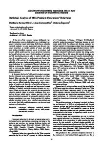
Photo 1: Electrophoretic pattern of multiplex PCR assay where M lane showed 100-1000 bp ladder, lane +ve con: positive control for L. monocytogenes ActA gene 829 bp, E. coli. Eae gene 629 bp and Salmonella spp InvA gene 284 bp. Lanes 1-9 showed results of some camel’s milk samples where 2&7 negative samples while 1, 3, 4, 5, 6, 8 and 9 positive sample with corresponding bands size to each of the three organisms 79
Global Veterinaria, 8 (1): 76-82, 2012
presence of high level of lactoferrin [39] and lysozyme that may cause direct lysis of bacteria [60]. But Polymerase chain reaction can detect DNA when either present in low level or fragmented DNA [61, 62]. Detection of such virulence genes by using multiplex PCR give attention to the feeding persons in raw camel’s milk that may develop risk specially toward infant, immune-compromised persons and aged man so they express sever illness, So planning for pathogen detection in foods necessitate applying a reliable methods which is obligatory role to keep food safe for consumers. However, peculiar characteristics of certain foods, such as milk and dairy products can directly influence pathogens recovery by isolation. With respect to milk in general and camel milk specifically significant interferences in the recovery of L. monocytogenes, Salmonella spp and E. coli [61-63] may occur. Based on the study findings: Almost of raw camel milk samples were produced and handled under poor hygienic conditions with high health risk to the consumers. So, improving quality of raw camel milk, require enhancing the milking protocols and sanitizing programs should be conducted. As the camel milk may be responsible for transmission of some health hazard microorganisms that increase the need for highly effective diagnostic procedures to maintain the health of milk consumers.
7.
8.
9.
10.
11.
12.
REFERENCES 1.
2. 3.
4.
5. 6.
13.
Farah, Z., 2004. An introduction to the camel. In: Milk and meat from the camel - Handbook on products and processing. Eds., FARAH, Z. and A. FISCHER, vdfHochschulverlag, Zürich, Switzerland, pp: 15-28. Chaibou, M., 2005. Productivité zootechnique du désert: le cas du basin laitier d'Agadez au Niger. Diss. Med. Vet. Montpellier, France. El-Ziney, M.G. and A.I. Al-Turki, 2007. Microbiological quality and safety assessment of camel milk (Camlus Dormedaries) in Saudi Arabia (Qassim Region). Appl. Ecology and Environ. Res., 5: 115-122. Make References like this Style. De Buyser, M.L., B. Dufour, M. Maire and V. Lafarge, 2001. Implication of milk and milk products in foodborne disease in France and different industrialized countries- Int. J. Food Microbiol., 67: 1-17. FAO, 2003. Report on FAO workshop on camel milk. Sinell, H.J., 1973. Food Infections, from Animals. In: The Microbiological Safety of Foods. Eds., B.C. Hobbs and J.H. B. Christian. Academic Press, London and New York.
14. 15. 16. 17. 18.
19.
80
Robinson, D.A., W.J. Edgar, G.L. Gibson, A.A. Matcheit and A.A. Robertson, 1979. Campylobacter enteritis associated with consumption of unpasteurized milk L. Brit.Medical J., 1: 1171. McClell, R.G. and A.C. Pinder, 1994. Detection of Salmonella typhimurium in dairy products with flow cytometry and monoclonal antibodies. Appl. Environ. Microbiol., 60: 4255-4262. Olsen, J.E., S. Aabo, W. Hill, S. Notermans, K. Wernars, P.E. Granum, T. Popovic, H.N. Rasmussen and O. Olsvik, 1995. Probes and polymerase chain reaction for detection of food-borne bacterial pathogens. Int. J. Food Microbiol., 28: 1-78. Bille, J., 1990. Epidemiology of human listeriosis in Europe, with special reference to the Swissoutbreak. In: A.L. Miller, J.L. Smith and G.A. Somkuti, (Eds.), Foodborne Listeriosis. Elsevier Science Publishers B.V. (Biomedical Division), Amsterdam, pp: 71-74. Broome, C., B. Gellin and B. Schwartz, 1990. Epidemiology of listeriosis in the United States. In: A.L. Miller, J.L. Smith and G.A. Somkuti, (Eds.), Food-borne Listeriosis. Elsevier Science Publishers B.V. (Biomedical Division), Amsterdam, pp: 61-65. Marth, E.H. and E.T. Ryser, 1990. Occurrence of Listeria in foods: milk and dairy foods. In: A.L. Miller, J.L. Smith and G.A. Somkuti, (Eds.), Food-borne Listeriosis. Elsevier Science Publishers B.V. (Biomedical Division), Amsterdam, pp: 151-164. Carosella, J.M., 1990. Occurrence of Listeria monocytogenes in meat and poultry. In: A.L. Miller, J.L. Smith and G.A. Somkuti, (Eds.), Food-borne Listeriosis. Elsevier Science Publishers B.V. (Biomedical Division), Amsterdam, pp: 165-173. Farber, J.M. and P.I. Peterkin, 1991. Listeria monocytogenes, a food borne pathogen. Microbiol. Rev., 55: 476-511. McClure, P.J. and S. Hall, 2000. Survival of Escherichia coli in foods. J. Appl. Microbiol., 88: 61S-70S. Knabel, S.J., 1995. Food borne illness: role of home food handling practices. Food Technol. 49: 119-131. Hill, W.E., 1996. The polymerase chain reaction: application for the detection of foodborne pathogen. Crit. Rev. Food Sci. Nutr., 36: 123-173. Wang, R.F., W.W. Cao and C.E. Ceniglia, 1997. A universal protocol for PCR detection of 13 Spp. Of food borne pathogens in food. J. Appl. Microbiol., 83: 727-736. Bruce, I.J., 1994. Nucleic acid amplification mediated microbial identification. Sci. Prog., 77: 183-206.
Global Veterinaria, 8 (1): 76-82, 2012
20. Bansal, N.S., 1996. Development of a polymerase chain reaction for the detection of Listeria monocytogenes in foods. Lett. Appl. Microbiol., 22: 353-356. 21. Manzano, M., L. Cocolin, P. Ferroni, C. Cantoni and G. Comi, 1997. A simple and fast PCR protocol to detect Listeria monocytogenes from meat. J. Sci. Food. Agric., 73: 25-30. 22. Fluit, A.C., R. Torensma, M.J. Visser, C.J. Aarsman, M.J. Poppelier, B.H. Keller, P. Klapwijk and J. Verhoef, 1993. Detection of Listeria monocytogenes in cheese with magnetic immunopolymerasechainreactionassay. Appl. Environ. Microbiol., 59: 1289-1293. 23. Nogva, H.K., K. Rudi, K. Naterstad, A. Holck and D. Lillehaug, 2000. Application of 5’-nuclease PCR for quantitative detection of Listeria monocytogenes in pure cultures, water, skim milk and unpasteurized whole milk. Appl. Environ. Microbiol., 66: 4266-4271. 24. Herman, L.M.F., J.H.G.E. DeBlock and R.J.B. Moermans, 1995. Direct detection of Listeria monocytogenes in 25ml of raw milk by a two-step PCR with nested primers. Appl. Environ. Microbiol., 61: 817-819. 25. Dilielo, L.R., 1982. Methods in Food and Dairy Microbiology. AVI publishing Co. Inc. Westport Connt. USA, pp: 39. 26. Soomro, A., M. Arain, M. Khaskheli and B. Bhutto, 2002. Isolation of Escherichia coli from raw milk and milk products in relation to public health sold under market condition at Tandojam. Pak. J. Nutr., 1(3): 151-152. 27. Benkerroum, N., Y. Bouhal, A. EI Attar and A. Marhaben, 2004. Occurrence of Shiga toxinproducing E. coli 0157:H7 in selected diary and meat products marketed in the city of Rabat, Morocco. J. Food. Prot., 67: 1234-1237. Make References like this Style. 28. Anon, Y., 1975. E. coli Enteritis. Lancet, pp: 1131-1132. 29. ICMSF (International Commission On Microbiological Specifications For Foods), 1996. Salmonelas. In: Microorganismos de los alimentos: Características de los patógenosmicrobianos. Ed: T.A. Roberts, A.C. Baird-Parker and R.B. Tompkin, Editorial Acribia, S.A. Zaragoza, España, pp: 255-305. 30. FDA, 2007. FDA and CDC re-mind consumers of the Dangers of Drinking Raw Milk.
31. American Public Health Association (APHA), 1992. Compendium of methods for the microbiological examination of foods. 3rd ed. Ed. Vanderzant, C. and D.F. Splittoesser, Washington, DC 2005. USA. 32. Ewing, W.H., 1986. Edward and Ewing’s Identification of Enterobacteriaceae. 4th ed. NY: Elsevier Science. 33. Popoff, M.Y., 2001. Antigenic Formulas of the Salmonella Serovars, 8th ed., WHO Collaborating Centre for Reference and Research on Salmonella, Institut Pasteur, Paris, France. 34. Orskov, F. and I. Orskov, 1992. Escherichia coli serotyping and disease in man and animals. Can J. Microbiol., 38(7): 699-704. 35. Sambrook, J.E.F., E. Fritsch and T. Maniatis, 1989. Molecular cloning: A Laboratory Manual, 2nd ed., Cold Spring Harbor Laboratory Press, New York. 36. Rahn, K., S.A. De Grandis, R.C. Clarke, S.A. McEwen, J.E. Galan, C. Ginocchio R. Curtiss III and C.L. Gyles, 1992. Amplification of an invA gene sequence of Salmonella enterica by polymerase chain reaction as a specific method of detection of Salmonella. Molecular and Cellular Probes, 6: 271-279. 37. Heuvelink, A.E., N.C.A.J. Van De Kar, J.F.G.M. Meis, L.A.H. Monnens and W.J.G. Melchers, 1995. Characterization of verocytotoxin-producing Escherichia coli O157 isolates from patients with hemolytic uremic syndrome in Western Europe. Epidemiology and Infection, 115: 1-14. 38. Zhou, Z. and X. Jiao, 2005. Molecular grouping and pathogenic analysis of Listeria monocytogenes of clinical and food origin Food Control, 16: 867-872. 39. Al-Majali, A.M., Z.B. Isamail, Y. Al-hami and A.Y. Nour, 2007. Lactoferrin concentration in milk from camels (CamelusDromedarius) with and without sub-clinical mastitis. Intern. J. Appl. Res. Vet. Med., 5(3): 120-124. 40. Farah, Z., 1993. A review article. Composition and characteristics of camel milk. J. Dairy Res., 60: 603-626. 41. Duhaiman, A.S., 1988. Purification of camel’s milk lysozyme and its lytic effect on E. coli and Micrococcus lysodeikticus. Comp. Biochem. Physiol., 91b: 793-796. 42. Wernery, U., 2003. New Observations on Camels and their Milk, Dar Al Fajr pub. Abu Dhabi, United Arab Emirates, pp: 41-42. 43. Elagamy, E.I., R. Ruppanner, A. Ismail, C.P. Champagene and R. Assaf, 1992. Antimicrobial and antiviral activity of camel milk protective proteins. J. Dairy Res., 59: 169-175. 81
Global Veterinaria, 8 (1): 76-82, 2012
44. Matofari, J.W., A. Shitandi, P.L. Shalo, N.J. Nanua and M. Younan, 2007. A survey of Salmonella enterica contamination of camel milk in Kenya. African J. Microbiol. Res., 1(4): 046-050. 45. Vanegas, M.C., E. Vasquez, A.J. Marthinez and A.M. Rueda, 2009. Detection of Listeria monocytogenes in raw whole milk for human consumption in Colombia by real time PCR. food Control, 20: 430-432. 46. Curason, G., 1998. Unemaladie du dromadaire analogue au farcin du boeuf. Bull. Soc. Cent. Med. Vet. Supplement to Rec. Med. Vet., 94(71): 491-496. 47. Bruner D. Moran, 1949. Salmonella infections of domestic animals. Conell Vet., 39(1): 53-63. 48. Cheyne, A., R. Pegram and C. Cartright, 1977. An outbreak of Salmonellosis in camels in North East of the Somali Democratic Republic. Tropical Animal Health Production, 9(4): 238-240. 49. Pegram, R.G. and F. Tareke, 1981. Observation on the health of Afar livestock. Ethopian Vet. J., 5: 11-15. 50. Refai, M., W. EL-Said, K. Osman, Z. Lofti, E.E. Safwat and S. Elias, 1984. Salmonella in slaughtered camels in Egypt. ZagaZig Vet. J., 9: 266-267. 51. Yassien, N.A., 1985. Salmonella in slaughtered camels. M.V. Sci. Thesis. Faculty of Veterinary Medicine, Cairo University. 52. Osman, A.R., 1995. Path. Study on intestinal infections in camels. Thesis. Faculty of Veterinary Medicine Cairo University. 53. Wernery, U., 1992. The prevalence of Salmonella infections in camels (Camelus dromedarius) in the UAE. Br. Vet. J., 148(5): 445-450. 54. Omar, R.H. and A.H. Eltinay, 2008. Microbial quality of camel's raw milk in central southern region of united Arab Emirates. Emir. J. Food Agric., 20(1): 76-83.
55. Obied, A.I. and H.O. Bagadi, 1996. Mastitis in camelus dromedaries and the somatic content of camel's milk. Research in Veterinary Sci., 61: 55-58. 56. Semereab, T. and B. Molla, 2001. Bacteriological quality of raw milk of camel (Camelus dromedarius) in AFAR region (Ethiopia). J. Camel Res., 8: 51-54. 57. Hajian, S., E. Rahimi and H. Mommtaz, 2011. A three years study of E. coli O157 H7 in cattle, camel, sheep, goats, chicken and beef minced meat. IPCBEE, Volume 9. Singpooare. 58. Chan, Y.C., K.J. Boor and M. Wiedmann, 2007. SigmaB-dependent and sigmaBindependent mechanisms contribute to transcription of Listeria monocytogenes cold stress genes during cold shock and cold growth. Appl. and Environ. Microbiol., 73(19): 6019-6029. 59. Wiedmann, M., T.J. Arvik, R.J. Hurley and K.J. Boor, 1998. General stress transcription factor sigmaB and its role in acid tolerance and virulence of Listeria monocytogenes. J. Bacteriol., 180(14): 3650-3656. 60. Al-Haj, O.A. and H.A. Al-Kanhal, 2010. Compositional, technological and nutritional aspect of dromedary camel milk. Intern. Dairy J., 20: 811-821. 61. Jay, J.M., 1995. Foods with low numbers of microorganisms may not be the safest foods OR, why did human listeriosis and hemorrhagic colitis become food borne diseases? Dairy Food Environ. Sanit., 15: 674-7. 62. Jay, J.M., 1996. Microorganisms in fresh ground meats: the relative safety of products with low versus high numbers. Meat Sci., 43: S59-66. 63. De Boer, E., 1998, Update on media for isolation of Enterobacteriaceae from foods. Int. J. Food Microbiol., 45: 43-53.
82