Review Articles
Subinvolution of the Placental Site as an Anatomic Cause of Postpartum Uterine Bleeding A Review Jamie A. Weydert, MD; Jo Ann Benda, MD
● Context.—Subinvolution of the placental site is an anatomic cause of delayed postpartum uterine bleeding that may be underrecognized by general surgical pathologists. Objective.—To review the physiology of uteroplacental arterial development and normal postpartum involution, and to present the characteristic clinical and histopathologic features of subinvolution. Data Sources.—Literature review (MEDLINE via PubMed and Ovid) regarding the pathology and pathophysiology of placental site subinvolution. Review of the clinical and pathologic characteristics of our own institution’s previ-
ously diagnosed cases of subinvolution from hysterectomy and endomyometrial curettage specimens. Conclusions.—Surgical pathologists must be aware of the cardinal histopathologic findings of subinvolution, and this diagnosis must be considered in every postpartum curettage or hysterectomy specimen presented to the surgical pathologist. Subinvolution of the placental site is an important diagnosis, as this process implies an idiopathic cause, rather than an iatrogenic cause, of postpartum uterine bleeding. The etiology of placental site subinvolution remains poorly characterized. (Arch Pathol Lab Med. 2006;130:1538–1542)
P
tis, and gestational trophoblastic disease in this clinical setting is insufficient evaluation. Though possibly underrecognized by general surgical pathologists, subinvolution of the placental site (placental site vascular subinvolution or subinvolution of uteroplacental arteries) is an important contributor to secondary postpartum bleeding. Subinvolution of the placental site refers to delayed or inadequate physiologic closure and sloughing of the superficial modified spiral arteries at the placental attachment site. Subinvolution can be identified and documented by the typical clinical features and the histologic findings in postpartum endometrial curettings and hysterectomy specimens. Although detailed studies have been performed by several authors to determine the normal and abnormal anatomy and histology of the uterine placental site,5–12 there is a relative dearth of practice-oriented literature on the topic of subinvolution. This article briefly reviews the physiology of normal uteroplacental arterial development and involution and the proposed pathophysiology of subinvolution. Comparisons to another disease of the uteroplacental vasculature, preeclampsia, are also considered. Using case material from our own institution and descriptions offered in the literature, the essential histologic findings of subinvolution are illustrated. Establishing a diagnosis of subinvolution is critical, as this process implies an idiopathic, and not iatrogenic, cause of delayed postpartum uterine bleeding.
ostpartum uterine bleeding is defined as uterine blood loss greater than 500 mL in the case of a vaginal delivery and greater than 1000 mL after cesarean birth. Primary postpartum bleeding occurs within the first 24 hours after completion of the third stage of labor, while secondary (delayed) postpartum bleeding occurs after this time frame.1 The risk factors and associated uterine pathology of primary postpartum bleeding are well known and include uterine atony, retained placenta, inherited disorders of coagulation, consumptive coagulopathy, endometrial infection, and perineal lacerations. Uterine atony is regarded as the most common physiologic abnormality responsible for primary postpartum bleeding; therefore, obstetricians are well informed with regard to the diagnosis and treatment of uterine atony.2,3 Secondary postpartum bleeding, in contrast, is less frequently addressed; however, it is estimated to complicate about 1% of all pregnancies. Although primary and secondary postpartum bleeding may share common causes, the etiology of secondary postpartum bleeding often remains unknown if the patient can be managed conservatively.4 If endometrial curettage is performed, surgical pathologists examine uterine curettings in order to establish an etiology of the bleeding. Occasionally, pathologists are confronted with a postpartum hysterectomy specimen in severe cases. However, excluding the presence of chorionic villi, endometriAccepted for publication March 24, 2006. From the Department of Pathology, University of Iowa, Iowa City. The authors have no relevant financial interest in the products or companies described in this article. Reprints: Jamie A. Weydert, MD, Department of Pathology, 200 Hawkins Dr, 5239C RCP, Iowa City, IA 52242-1009 (e-mail:
[email protected]). 1538 Arch Pathol Lab Med—Vol 130, October 2006
DEVELOPMENT OF UTEROPLACENTAL BLOOD FLOW: TRANSFORMATION OF THE SPIRAL ARTERIES The developing fetus ultimately derives its nutrition from the maternal blood supplied to the placenta via the maternal uterine spiral arteries. Early in gestation (6–10 Subinvolution of the Placental Site—Weydert & Benda
wk), placenta-derived extravillous cytotrophoblasts (EVTs) invade the decidual portion of the maternal spiral arteries, initially forming endovascular ‘‘plugs’’ that actually impede blood flow to the developing placenta.7 As gestation moves into the second trimester, the endovascular EVTs replace the normal endothelial lining of the artery and express endothelial-type markers and angiogenic factors.13,14 The EVTs also take residence within and around the walls of the spiral arteries (interstitial EVTs). This vascular colonization process extends as deep as the inner third of the myometrium.5–7 The interstitial EVTs work to remodel the arterial walls, and a histological correlate of this remodeling is the observation of hyaline fibrinoid material that replaces the arterial media, with loss of elastic fibers.12 Trophoblast-independent mechanisms of vascular alteration are also at work during this time but are beyond the scope of this discussion. The net physiologic effect of this remodeling and colonization process is the creation of large-caliber, high-flow, but low-resistance arteries sufficient to meet the demands of the growing placenta and fetus. NORMAL INVOLUTION OF THE UTEROPLACENTAL ARTERIES After delivery, a physiologic mechanism of uteroplacental arterial involution is required to eliminate these altered vessels. In the third trimester, this process begins modestly as the endovascular EVTs are replaced by maternalderived endothelial cells. The exact temporal relationships are not fully elucidated, but several involution events occur within the uteroplacental vascular bed in the few days after delivery. These changes include occlusive fibrointimal thickening, endarteritis, thrombosis, regeneration of the internal elastic lamina, and disappearance of both endovascular and interstitial EVTs (Figure 1, A and B). Although the histomorphology of involutional events is well described, the exact molecular basis that triggers these changes remains poorly understood.15–17 Necrosis and sloughing of the decidua and superficial endomyometrium occur in tandem with the involutional vascular changes. Contraction of the uterine smooth muscle also contributes to mechanical shrinkage and involution of the placental site as a whole.9 Taken together, one very important consequence of these changes is to limit the loss of blood at the placental site after delivery. PATHOPHYSIOLOGY OF SUBINVOLUTION The EVTs obviously play an important role in the development of adequate uteroplacental arterial flow, and an abnormal interaction between these cells and the maternal tissue is proposed as the mechanism for subinvolution.16 An immunologic basis for this type of altered fetomaternal interaction has been suggested and is an especially attractive idea when compared to the findings in preeclampsia. In preeclampsia, poor investment and remodeling of the spiral arteries by EVTs is found. Mounting epidemiologic and molecular evidence suggests that poor interaction between EVTs and maternal decidual tissue may be due to an abnormal immunologic recognition process.8,18–20 The respective pathophysiology of preeclampsia and subinvolution may represent opposite ends of a spectrum of abnormal cellular interactions at the fetomaternal tissue interface. Other evidence supporting an abnormal immunologic basis for subinvolution includes the observations by Andrew et al15 of differential immunoglobulin and Arch Pathol Lab Med—Vol 130, October 2006
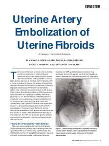
Figure 1. Normal involution. A, Low-power view showing clusters of normally involuted uteroplacental arteries. B, Medium-power view showing fibrointimal obliteration of the uteroplacental artery lumen (hematoxylin-eosin, original magnifications ⫻6.7 [A] and ⫻16.7 [B]).
complement deposition in involuted and subinvoluted vessels. In this study, the deposition of immunoglobulins (IgG, IgA, IgM) and complement proteins (C1q, C3d, C4) in the walls of normally involuted vessels is described; these components are absent in subinvoluted vessels. Another insight into the mechanism of subinvolution is related to EVT apoptosis. While the EVTs in preeclampsia appear to lose expression of the antiapoptosis protein Bcl2, increased expression of this protein has been reported in subinvolution.20,21 Perhaps persistent expression of this antiapoptosis protein plays a role in maintaining the uteroplacental vessels in a state similar to gestation in cases of subinvolution. Despite these interesting observations, there remains no uniform consensus as to a defined pathophysiologic sequence of events in subinvolution. DIAGNOSTIC SURGICAL PATHOLOGY OF SUBINVOLUTION The diagnosis of subinvolution can be made by pathologic examination of postpartum endometrial curettage material, provided the placental site is sampled. Vascular subinvolution may be detected in the presence of rare degenerating chorionic villi, which is a valuable observation when the amount of villous tissue present does not acSubinvolution of the Placental Site—Weydert & Benda
1539
Figure 2. Subinvolution. A, Low-power view of clusters of patent uteroplacental arteries filled with red cells and loose fibrin (arrow). B, Mediumpower view of a partially thrombosed uteroplacental artery. C, Low-power view of subinvoluted vessels (arrow) adjacent to normally involuted vessels (arrowhead). D, High-power view showing extravillous trophoblasts (thin arrow) within the vessel wall (hematoxylin-eosin, original magnifications ⫻6.7 [A], ⫻16.7 [B], ⫻6.7 [C], and ⫻100 [D], respectively).
count for bleeding. Therefore, the diagnosis of subinvolution should be carefully excluded in all cases, and the temptation to only record the presence or absence of villous tissue should be avoided. A number of microscopic features have been described in the literature.11,12,15–17 The essential findings identifiable on hematoxylin-eosin staining include large, patent, and dilated superficial myometrial vessels with pink hyaline material replacing the media and partial intravascular thrombosis of variably aged thrombi (Figure 2, A and B). These abnormal vessels are typically clustered with little intervening myometrial tissue and may be located adjacent to normally involuted vessels (Figure 2, C). Extravillous interstitial and/or endovascular trophoblasts should also be identified. The cytologic features of EVT cells include polygonal shape, abundant amphophilic cytoplasm, and vesicular nuclei (Figure 2, D). Immunohistochemistry may be helpful to identify these cells, as they stain readily with commercially available antibodies directed at lowmolecular-weight cytokeratin, inhibin-␣, Mel-CAM, and human placental lactogen (Figure 3, A and B).22 Rare cells may also stain with  human chorionic gonadotrophin. Other, though less constant, microscopic features have 1540 Arch Pathol Lab Med—Vol 130, October 2006
been described in subinvolution. These include lack of internal elastic lamina duplication, partial or complete absence of a true endothelial lining, and immunohistochemical expression of the antiapoptosis protein Bcl-2. Histochemical stains for elastin (elastic von Gieson or elastochrome) can be very helpful in delineating the vessel wall anatomy. Immunoperoxidase staining for vascular endothelial markers may also be helpful in defining the extent of endothelial regeneration, particularly anti-CD31. The constellation of histologic findings in cases of subinvolution likely reflects the nature of this process as a biologic continuum of changes, lying somewhere between appropriate involution and complete lack of it. The extent of subinvolution varies from case to case, and the diagnostic findings may be quantitatively limited in any given specimen. In cases where the complete set of essential findings is not observed, subinvolution may be suggested as a diagnostic possibility. A diagnosis of subinvolution should only be rendered in the appropriate clinical setting, which is secondary (delayed) postpartum bleeding. Physiologic patency of uteroplacental arteries will be seen in the immediate (⬍24 h) postpartum period and should not be unequivocally interpreted as subinvolution. Subinvolution of the Placental Site—Weydert & Benda
for presentation is within the second week after delivery.16 In our case files, we identified 8 patients since 1997 with a pathologic diagnosis of subinvolution (3 hysterectomies, 5 curettings). As can be seen in the Table, 3 of our patients presented within the second week postpartum, 3 presented between the third and seventh weeks postpartum, and time of presentation was not available for the remaining 2 patients. Subinvolution has also been reported to cause bleeding after induced abortion23 and after molar pregnancy evacuation.24 The severity of the bleeding is also variable, but typically there is an abrupt onset of increased uterine bleeding, which prompts the patient to seek medical attention. Endometrial curettage may not be curative, and continued brisk bleeding may require hysterectomy in spite of fluid and coagulation maintenance. Clinical associations cited in the literature include multiparity and older age.16 Four of the patients in our case file were multiparous, and the age range was 19 to 34 years, but 7 of the 8 patients were 27 years of age or older. SUMMARY Subinvolution of the placental site is a cause of delayed postpartum bleeding. This process is likely underrecognized by general surgical pathologists. The histologic findings of dilated, clustered, myometrial arteries partially occluded by thrombi of various ages and invested with extravillous trophoblasts are diagnostic of subinvolution in the appropriate clinical setting. The cause of subinvolution is not known, but this process may be a manifestation of an abnormal interaction between fetal-derived trophoblasts and maternal tissue. Subinvolution is an important process to recognize, as it implies an idiopathic, and not an iatrogenic, cause of delayed postpartum bleeding. References
Figure 3. Subinvolution. A, Medium-power view of a patent uteroplacental artery. B, Same artery immunostained using antibodies against low-molecular-weight cytokeratin (DakoCytomation, Carpinteria, Calif) showing numerous persistent extravillous trophoblasts within the vessel wall (hematoxylin-eosin and immunoperoxidase, original magnifications ⫻16.7).
CLINICAL FEATURES AND ASSOCIATIONS Delayed postpartum bleeding should always raise the possibility of subinvolution. Although subinvolution may cause bleeding anytime between 1 week and several months postpartum, the most common reported timeline
1. MacMullen NJ, Dulski LA, Meagher B. Red alert: perinatal hemorrhage. MCN Am J Matern Child Nurs. 2005;30:46–51. 2. Ijaiya MA, Aboyeji AP, Abubakar D. Analysis of 348 consecutive cases of primary postpartum haemorrhage at a tertiary hospital in Nigeria. J Obstet Gynaecol. 2003;23:374–377. 3. Selo-Ojeme DO. Primary postpartum haemorrhage. J Obstet Gynaecol. 2002;22:463–469. 4. Hoveyda F, McKenzie IZ. Secondary postpartum haemorrhage: incidence, morbidity, and current management. Br J Obstet Gynaecol. 2001;108:927–930. 5. Brosens JJ, Pijnenborg R, Brosens IA. The myometrial junctional zone spiral arteries in normal and abnormal pregnancies: a review of the literature. Am J Obstet Gynecol. 2002;187:1416–1423. 6. Pijnenborg R. Uterine haemodynamics as a possible driving force for endovascular trophoblast migration in the placental bed. Med Hypotheses. 2000; 55:114–118. 7. Meekins JW, Luckas MJ, Pijnenborg R, et al. Histological study of decidual spiral arteries and the presence of maternal erythrocytes in the intervillous space
Clinical Summary of Subinvolution Cases* Case
Age, y
Pariety
1 2
19 27
G1, P1 G2, P2
3 4 5 6 7
27 28 29 32 33
NA G6, P4 NA G4, P3 G1, P1
Delivery
Gestation unknown, CS 30 wk CS for fetal distress
Postpartum Interval, d
7 5
Procedure
Hysterectomy Curettage followed by immediate hysterectomy Curettage Curettage Hysterectomy Curettage Curettage
NA NA Term, NSVD 9 NA 49 Term, NSVD 19 19 wk termination for incom28 petent cervix 8 34 G3, P2 15 wk IUFD 9 Curettage * G indicates gravida; P, para; NA, not available; NSVD, normal spontaneous vaginal delivery; IUFD, intrauterine fetal demise; CS, cesarean section. Arch Pathol Lab Med—Vol 130, October 2006
Subinvolution of the Placental Site—Weydert & Benda
1541
during the first trimester of normal human pregnancy. Placenta. 1997;18:459– 464. 8. Meekins JW, Pijnenborg R, Hanssens M, et al. A study of placental bed spiral arteries and trophoblast invasion in normal and severe preeclamptic pregnancies. Br J Obstet Gynaecol. 1994;101:669–674. 9. Anderson WR, Davis J. Placental site involution. Am J Obstet Gynecol. 1968;102:23–33. 10. Pijenborg R, Robertson WB, Brosens I, et al. Trophoblast invasion and the establishment of haemochroial placentation in man and laboratory animals. Placenta. 1981;2:71–92. 11. Ober WB, Grady HG. Subinvolution of the placental site. Bull NY Acad Med. 1961;37:713–730. 12. Wells M, Bulmer JN. The human placental bed: histology, immunohistochemistry, and pathology. Histopathology. 1988;13:483–498. 13. Pijnenborg R. Implantation and immunology: maternal inflammatory and immune cellular responses to implantation and trophoblast invasion. Reprod Biomed Online. 2002;4:14–17. 14. Bulla R, Villa A, Bossi F, et al. VE-cadherin is a critical molecule for trophoblast-endothelial cell interaction in decidual spiral arteries. Exp Cell Res. 2005;303:101–113. 15. Andrew A, Bulmer JB, Morrison L, et al. Subinvolution of the uteroplacental arteries: an immunohistochemical study. Int J Gynecol Pathol. 1993;12:28–33.
1542 Arch Pathol Lab Med—Vol 130, October 2006
16. Andrew AC, Bulmer JB, Wells M, et al. Subinvolution of the uteroplacental arteries in the human placental bed. Histopathology. 1989;15:395–405. 17. Khong TY, Khong TK. Delayed postpartum hemorrhage: a morphologic study of causes and their relation to other pregnancy disorders. Obstet Gynecol. 1993;82:12–22. 18. Redman CW, Sargent IL. Latest advances in understanding preeclampsia. Science. 2005;308:1592–1594. 19. Naicker T, Khedun SM, Moodley J, et al. Quantitative analysis of trophoblast invasion in preeclampsia. Acta Obstet Gynecol Scand. 2003;82:722–729. 20. DiFederico E, Genbacev O, Fisher SJ. Preeclampsia is associated with widespread apoptosis of placental cytotrophoblasts within the uterine wall. Am J Pathol. 1999;155:293–301. 21. Khong TY, Abdul Rahman H. Bcl-2 expression delays postpartum involution of pregnancy induced vascular changes in the human placental bed. Int J Gynecol Pathol. 1997;16:138–142. 22. Santos LD, Yong JL, Wu XJ. Applications of monoclonal antihuman inhibin alpha subunit in endometrial curettings. Pathology. 2000;35:217–223. 23. Lee ET, Marley NJ, Bevan JR. A rare late complication of first trimester induced abortion requiring hysterectomy—subinvolution of the placental bed: case report. Br J Obstet Gynaecol. 1986;93:777–781. 24. Khong TY, Chin MM. Subinvolution of the placental bed after molar pregnancy. J Reprod Med. 1996;41:352–354.
Subinvolution of the Placental Site—Weydert & Benda