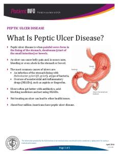
Overview • Peptic Ulcer – breach in the mucosa extending from muscularis mucosae into submucosa or deeper • Can occur anywhere in GIT but more commoner in duodenum & stomach • Acute gastric ulcer can occur from severe systemic stress e.g. Stress ulcers from severe infection
Peptic Ulcer • • • •
Chronic – long history Solitary – single. Site – along GIT exposed to action of acid-peptic juices Site of occurrence: – – – – – –
Duodenum – first portion Stomach – antrum Gastroesophageal junction – setting of reflux Within magins of a gastrojejunostomy Duodenum, stomach or jejunum in pts with ZE syndrome Within or adjacent to Meckel diverticulum – ectopic gastric mucosa
Epidermiology • 350,000 new cases diagnosed per year in US (2000) • PNG Data unknown but is a common clinical presentation • Lifetime risk in developing ulcer – 10% male & 4% females (US figures)
• Remitting & relapsing lesions – chronic history. • More common in middle-aged to older individuals. • Women more affected at or after menopause
Pathogenesis • Imbalance between mucosal defence mechanisms & damaging forces • Gastric acid & pepsin must be present • Hyperacidity is NOT a prerequisite • Only minority of pts with peptic ulcer have hyperacidity • H.pylori infection is known to cause peptic ulcer – All pts with duodenal ulcers have H.pylori infection – 70% of pts with gastric ulcers have H.pylori infection
Pathogenesis • Peptic ulcer can readily occur:
– Mucosal blood flow drops – hypovolumic shock – Gastric emptying is delayed – Epithelial restitution is impaired
H.Pylori & Peptic Ulcer Pathogenesis • Urease generates free ammonia & a protease breaks down glycoproteins in gastric mucus • Phospholipases which damage epithelial cells • Neutrophils release myeloperoxidase which produces hypochlorous & monochloramine that destroy cells • Mucosal epithelial cells & lamina propria endothelial cells are targets for destructions of H.pylori during colonisation.
H.Pylori & Peptic Ulcer Pathogenesis • Surface capillaries are also thrombosed. This is promoted by bacterial platelet-activating factor • Elaboration of other enzymes result in chronic inflammation. Chronically inflamed mucosa more susceptible to acid injury • Damage in mucosa allows leakage of nutrients into surface microenvironment sustaining the bacillus
Pathogenesis • Only 10-20% of individuals with H.pylori infection develop peptic ulcer. – Why some do & other do not?
• Actual infection limited to stomach but pts develop duodenal ulcer. Why?
– Some studies suggest ammonia stimulates gastrin release thereby increasing acid production
• Link between H.pylori & peptic ulcer is established but interactions leading to ulceration remains to be elucidated.
Hyperacidity & Peptic Ulcer Pathogenesis
• Gastric hyperacidity is strongly ulcerogenic • Causes of hyperacidity: – – – –
Increased parietal cell mass Increased sensitivity to secretory stimuli Increased basal secretory drive Impaired inhibition of stimulatory mechanisms such as gastrin release
• In ZE syndrome there is excess gastrin secretion from gastrin secreting tumor resulting in increased acid production
– ZE exhibits multiple ulcers in stomach, duodenum & jejunum
Peptic Ulcer Pathogenesis • Chronic NSAIDS use suppress mucosal prostaglandin synthesis • Cigarette smoking impairs mucosal blood flow & healing • Alcoholic cirrhosis is associated with increased incidence of peptic ulcers • Rapid gastric emptying exposes mucosa to excessive acid load
Peptic Ulcer Pathogenesis • Duodenal ulcers more frequent in pts with: – Alcoholic cirrhosis – COPD
• Chronic Renal Failure – hypercalcaemia stimulates gastrin production thereby increased acid production • Hyperparathyroidism – hypercalcaemia stimulates gastrin production thereby increased acid production • Genetic influences appear to play no role.
• Compelling arguments but no hard evidence to suggest these factors have role in peptic ulcer pathogenesis
Morphology • 98% occur in duodenum or stomach in a ratio of 4:1. • Gastric ulcers – occur along lesser curvature • 80-90% pts have single ulcer but – 10-20% have gastric & duodenal ulcer
• Small lesions (0.6cm are bona fide ulcers • 50% of ulcers are 4cm) do occur • Size does not differentiate a benign from a malignant ulcer
Morphology • Classic peptic ulcer – round to oval sharply punched out defect with straight walls and clean base. • Dept is variable depending on chronicity of ulcer presence and ongoing acute & chronic inflammation. • Involves mucosa or extending to muscularis propria • If perforation occur pancreas, omental fat or liver can form the base
Morphology • Base of ulcer is clean and smooth – peptic digestion of exudate & debris • Scarring may occur and involve entire thickness of stomach
– Puckering of surrounding mucosa creates mucosal folds radiating from the ulcer crater in a spokelike fashion
• Surrounding mucosa is oedematous & reddened due to gastritis
An acute duodenal ulcer is seen in two views on upper endoscopy in the panels below.
Here is a large 3 x 4 cm gastric ulcer that led to the resection of the stomach shown here. This ulcer is quite deep with irregular margins. Complications of gastric ulcers (either benign or malignant) include pain, bleeding, perforation, and obstruction
Peptic ulcer duodenum
On microscopic examination at high power, this gastric mucosa shows infiltration by neutrophils. This is acute gastritis
Histological Features • Varies from active necrosis to chronic inflammation & scaring to healing • In active ulcers 4 zones can be recognised
– Base & margins have thin layer of necrotic fibrinoid debris – Zone of non-specific inflammatory infiltrate (neutrophils predominate) – Active granulation tissue infiltrated with mononuclear leucocytes – Granulation tissue rests on fibrous/collagenous scar. Thickened vessel walls & thrombosed.
Morphology
Histological Features • Chronic gastritis is universal in pts with peptic ulcer • Occur in 85-100% of pts with duodenal or gastric ulcers • H.pylori is almost always demonstable in pts with gastritis • Gastritis can remain after ulcer has healed • Recurrence of ulcer is not related to progression of gastritis – Helps distinguish peptic ulcer from acute erosive gastritis or stress ulcers which have normal adjacent mucosa
Clinical Presentation Epigastric gnawing, burning or aching pain Anaemia, frank hemorrhage or perforation Pain Worse at night, 1-3 hrs after meals at daytime Pain relieved by alkalis or food Nausea, vomiting, bloating, belching or wt loss should raise suspicion occult malignancy • Pain referred to back in penetrating ulcers, left upper quadrant or back • Pain can be mistaken as being of cardiac origin • Malena stool • • • • • •
Treatment & Prognosis • • • •
Notoriously chronic recurring lesions Impair quality of life more than shorten it Left untreated may take up to 15 years to heal Principles of treatment: – Reduce acid production – Promote mucus secretion – Neutralize acidity – H.pylori treatment
Complications •
Bleeding – – – – –
15-20% of pts Most frequent Can be life threatening Accounts for 25% of ulcer deaths First indication of an ulcer
•
Perforation
•
Obstruction from oedema or scarring
•
Chronic intractable pain
– 5% of pts – Accounts for 66% of ulcer deaths – Late presentation
– 2% of pts – Occur in pyloric channel ulcers but can occur in duodenal ulcers – Chronic abdominal pain
Acute peptic ulcer
ACUTE GASTRIC ULCERATION
Acute Gastric Ulceration • Focal acutely developing gastric mucosal defects are well known complication of NSAIDs use • Can occur after severe physiological stress hence name of stress ulcers
– Pts with shock, extensive burns, sepsis or severe trauma or post intracranial surgery
• Generally multiple lesions mainly in stomach but can occur in duodenum • Range in depth from superficial erosions to entire mucosal involvement (bona fide ulcer) • Shallow erosions are extension of acute erosive gastritis • Well defined ulcers are not precursors of chronic peptic ulcers
Acute Gastric Ulceration • Curling ulcers – patients with severe burns – Proximal duodenum – Associated with severe burns or trauma
• Cushing ulcers – intracranial injury, surgery or tumors – Stomach – Duodenum – Esophagus – Have high incidence of perforations
Pathogenesis – Acute Gastric Ulcer • Poorly understood • Impaired oxygenation to mucosa may have role • Cranial lesions – direct vagal stimulation by increase intracranial – Vagal stimulation causes increased acid secretion
• Systemic acidosis may contribute to mucosal compromise by lowering intracellular pH of mucosal cells already hypoxic by splanchnic vasoconstriction