Bajopas Volume 2 Number 2 December, 2009 Bayero Journal of Pure and Applied Sciences, 2(2): 79 - 83 Received: August, 2009 Accepted: October, 2009
HELICOBACTER PYLORI: THE CAUSATIVE AGENT OF PEPTIC ULCER *Shamsuddeen U., Yusha’u M. and Adamu I. A.
Biological Sciences Department, Bayero University Kano, Nigeria. *Correspondence author:
[email protected]
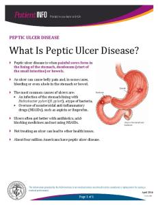
ABSTRACT This review examines Helicobacter pylori as an organism and as the causative agent of peptic ulcers. The review also examined the classification of ulcers, how the bacterium produces the ulcer, some of the virulence factors possessed by the organism, its metabolism and growth requirements. The incidence and prevalence of peptic ulcers were originally believed to have resulted from some factors such as stress, eating spicy food, long term use of nonsteroidal anti-inflammatory agents (NSAIDs), like aspirin and ibuprofen, smoking, and many alike. presently due to the development in the field of research, researchers from different geographical locations have reported a spiralshaped Gram-negative bacterium called Helicobacter pylori to be responsible for the pathogenesis of peptic ulcer. This confirmation has been supported by the strong and effective diagnostic procedures such as urea breath test, stool test, endoscopy, and blood test. Elimination of Helicobacter pylori by treatment with antibiotics in peptic ulcer patients resulted in the healing of the ulcer. Prevention of Helicobacter pylori infections is difficult because the mode of transmission is not well known. Key words: Helicobacter pylori, peptic ulcer, causative agent. INTRODUCTION A peptic ulcer also known as ulcus pepticum, PUD or peptic ulcer disease , is an ulcer (defined as mucosal erosions equal to or greater than 0.5 cm) of an area of the gastrointestinal tract that is usually acidic and thus extremely painful (GI consult, 2007). A peptic ulcer can also be defined as a sore on the lining of the stomach or duodenum, which is the beginning of the small intestine. One cause of peptic ulcer is a bacterial infection, but some ulcers are caused or worsen by long term use of non steroidal anti-inflammatory agents (NSAIDs), like aspirin and ibuprofen (NDDIC, 2004). As many as 80% of ulcers are associated with Helicobacter pylori, a spiral-shaped bacterium that live in acidic environment of the stomach (GI consult, 2007). Peptic ulcers are not caused by stress or eating spicy food, but these can make ulcers worse (NDDIC, 2004). Contrary to general belief, more peptic ulcers arise in the duodenum (first part of the small intestine, just after the stomach) than in the stomach (GI consult, 2007). Classification of Peptic Ulcer A peptic ulcer may arise at various locations: • • • o
Stomach (called gastric ulcer) Duodenum (called duodenal ulcer ) Esophagus (called esophageal ulcer) Meckel’s Diverticulum (called Meckel’s Diverticulum ulcer) Types of peptic ulcers: • Type I: ulcer along the lesser curve of the stomach • Type II: Two ulcers present –one gastric, one duodenal
79
• Type III: Prepyloric ulcer • Type IV: Proximal gastro esophageal ulcer • Type V: Anywhere along gastric body, NSAID induced (GI consult, 2007).
Helicobacter pylori Helicobacter pylori is a spiral-shaped Gram-negative rod (Brooks et al., 2004). Helicobacter pylori play a
major role in the pathogenesis of peptic ulceration and gastric cancers. The G+C content of their DNA is 35.2 moles % (Collier et al., 1998). The surface of the human stomach mucosa is the major habitat of Helicobacter pylori. Almost all isolations are from gastric biopsy specimens, but the organism has occasionally been detected in gastric juice, saliva, dental plaque, bile and feces (Owen, 1995). H. pylori cells take the form of curved, or s-shaped Gramnegative rods, 0.5-0.9µm wide by about 3µm long with a wave length of about 2.6µm. In agar cultures, spiral forms are less obvious and cells appear more as singly curved rods. H. pylori undergo coccal transformation on exposure to air within 1-2 hours at room temperature, and in this state it fails to grow on subculture. Such coccoid forms do not appear to be virulent (Collier et al., 1998). Cultural Characteristics, Metabolism and Growth Requirements of H. pylori H. pylori is strictly microaerophilic and CO2 (5-20%) and high humidity are required for growth. H. pylori requires media containing supplements similar to those used for Campylobcters; blood, haemin, serum, starch or charcoal. However, H. pylori is inhibited by the bisulphate in the FBP Campylobacter aero tolerance supplement.
Bajopas Volume 2 Number 2 December, 2009 Growth is best on media such as moist freshly prepared heated (chocolated) blood agar or brain – heart infusion agar with 5% horse blood and 1% isovitalex. Strains grow in various liquid media supplemented with fetal calf or horse serum (Shahamat and Mai, 1991), some strains grow in serum–free media, notably bisulphate-free brucella broth (Hawrylik and Wasilko, 1994). Sheep blood, either whole or laked, when used as a broth supplement, has been shown to inhibit the growth of H. pylori (Coudron and Stratton,1995). All strains grow at 37oC, some grow poorly at 30 and 42oC but none grows at 25oC. Colonies from primary cultures at 37oC usually take 3-5 days to appear and are circular, convex, and translucent. They seldom grow bigger than 2mm in diameter even if incubation is extended beyond 1 week. They are weakly hemolytic on 5%
horse blood agar. Motility is weak or absent when grown on agar (Collier et al., 1998.) H. pylori is inactive in most conventional biochemical tests. Notable exceptions are the strong production of urease, catalase and alkaline phosphatase. All strains produce DNAase, leucine amino peptidase, and γglutamylaminopeptidase. The urease is a nickel containing high molecular weight protein (c. 600 KDa) with maximum activity at 45oC and PH 8.2. Remarkably, it forms 6% of protein produced by the organism. In the presence of urease (5M) it enables H. pylori to tolerate pH values down to 2.6, because the production of urease raises the pH in the immediate vicinity of the bacterial cell. The importance of this action for colonization of the stomach is obvious (Collier et al., 1998).
Table 1: Cultural and biochemical characteristics of H. Pylori Feature Number of flagella Distribution of flagella Periplasmic fibers Growth at 42oC Catalase Nitrate reduction Urease Alkaline phosphatase γ –Glutamyl transpeptidase Indoxyl acetate hydrolysis Growth in the presence of 1% glycine Susceptibility to nalidicxic acid Susceptibility to cephalothin
Characteristic 4-8 MP _ V + _ + + + _ _ R S
KEY; MP = mono polar. + = positive reaction. - = negative reaction. V = some positive/sensitive, some negative/resistant; R, resistant; S, sensitive. Source; Collier et al., (1998). LABORATORY ISOLATION AND IDENTIFICATION Direct examination of specimens: Various methods have been used for the direct detection of H. pylori pathological samples, including Gram stain, immunofluorecence acridine orange staining, histochemical stains, and molecular methods such as PCR (VanZwet and Thijs, 1993). H. pylori can also be detected by testing biopsy tissue for urease activity. A non invasive method for detecting radiolabelled CO2 after oral administration of (13C) urea known as the urea breath test is also used for the diagnosis of Helicobacter infection (Collier et al., 1998). Isolation and identification Gastric biopsy specimens are the only ones likely to be used for the primary isolation of H. pylori. They should be transported in a moist state and cultured within 2hours of collection. Storage beyond this time should be at 4oC , or at -20oC if the period is more than 2days. Various transport media have been described for transporting biopsy samples, including cystein brucella broth, normal saline, glucose, milk,
80
Stuart’s medium, semi-solid agar, brain-heart infusion broth, Cary-Blair medium and more recently, Cystein Albini medium containing 20% glycerol (Han and Flamm, 1995). Various non selective and selective culture media have been used for the isolation of H. pylori and other Helicobacter species (Jerris, 1995). Chocolate agars, or more complex media such as brain-heart infusion or brucella-agar supplemented with 5-7% horse, blood, are suitable non selective media. Examples of selective media are Dent’s, Glupczynski’s Brussels Charcoal medium and skirrow’s Campylobacter medium. Samples should be prepared for direct examination and plating on non-selective and selective media and plates should be incubated in a micro-aerobic environment at 35-37oC. Colonies may be visible after 3-5days of incubation but may take longer to appear on primary isolation. Colonies are small, domed, translucent, and sometimes weakly hemolytic. Because of the unique characteristic of H. pylori, only a few simple tests are needed for identification, including Gram stain appearance, catalase, oxidase and urease tests (Jerris, 1995).
Bajopas Volume 2 Number 2 December, 2009 product of a cytotoxin associated gene (cagA), is produced by about 60% of H. pylori strains and is associated with the expression of vacA. CagA-positive strains are also associated with the presence of duodenal ulceration, although it is unclear whether cagA ha an independent pathogenic role (Cover and Glupczynski, 1995; Owen, 1995). Stress and Ulcers Despite the finding that a bacterial infection is the cause of ulcers in 80% of cases, bacterial infection does not appear to explain all ulcers and researchers continue to look at stress as a possible cause, or at least a complication in the development of ulcers. An expert panel convened by the Academy of Behavioral Medicine Research concluded that ulcers are not purely an infectious disease and that psychological factors do play a significant role. Researchers are examining how stress might promote H. pylori infection. For example, Helicobacter pylori thrive in an acidic environment, and stress has been demonstrated to cause the production of excess stomach acid (GI Consult, 2007). A study on mice showed that both long term water immersion restraint stress and H. pylori infection were independently associated with the development of peptic ulcers (Kim et al., 2002). Symptoms of Ulcer Abdominal discomfort is the most common symptom. This discomfort usually is a dull, gnawing ache which comes and goes for several days or weeks, occurs two to three hours after a meal, occurs in the middle of the night when the stomach is empty and is relieved by antacid medications. Other symptoms include weight loss, poor appetite, bloating, burping, nausea, vomiting, sharp sudden persistent stomach pain, bloody or black stools, bloody vomit or vomit that looks like coffee grounds. They could be signs of a serious problem, such as perforation when the ulcer burrows through the stomach or duodenal wall bleeding when acid or the ulcer breaks a blood vessel, obstruction when the ulcer blocks the path of food trying to leave the stomach Some people experience only very mild symptoms, or none at all (NDDIC, 2004).
Most typing success has been with molecular methods that exploit the considerable genetic diversity of H. pylori. Methods such as random amplified polymorphic DNA analysis or RFLP analysis of specific amplified genes have been used successfully for studying strains within families or answering other epidemiological questions (Taylor and Fox, 1995). How Does Helicobacter pylori Cause a Peptic Ulcer H. pylori weaken the protective mucous coating of the stomach and duodenum, which allows acid to get through to the sensitive lining beneath. Both the acid and the bacteria irritate the lining and cause a sore, or ulcer. H. pylori is able to survive in stomach acid because it secretes enzymes that neutralize the acid. This mechanism allows H. pylori to make its way to the "safe" area—the protective mucous lining. Once there, the bacterium's spiral shape helps it burrow through the lining (NDDIC, 2004). Pathogenicity and Virulence Factors H. pyloris cause chronic active gastritis in humans and is the cause of most cases of duodenal and gastric ulcers that are not associated with administration of non-steroidal anti-inflammatory agents (Rauws and Tytgat, 1995). In addition H. pylori is associated with primary mucosa-associated lymphoid tissue, gastric lymphoma and some types of gastric adenocarcinoma (Webb and Forman, 1995). Most H. pylori bacteria found in the stomach are in the layer of mucus overlying the epithelium. Some penetrate the mucous layer and adhere to the gastric epithelial cells with formation of a pedestal, as is seen with enteropathogenic E. coli strain, which require cytoskeletal rearrangements (Smoot and Resau, 1993). A few H. pylori are seen between adjacent cells in proximity to tight junctions. H. pylori urease appears to be necessary for the establishment of gastric mucosal colonization according to animal experimental studies with urease negative mutants and urease inhibitors. Urease may serve to protect the organism from gastric acid by converting urea to ammonia and carbon dioxide, thus surrounding the bacterium with an alkaline layer that neutralizes the acid. Urease may also be an important cause of the inflammation seen in the lamina propria that is the hallmark of chronic H. pylori infections, as it is a phlogistic and cytotoxic agent (Mai and Prez-Prez, 1992; Segal et al., 1992). Bacterial motility is also important for the colonization of mucous layer, as non-motile mutants do not colonize the stomachs of gnotobiotic piglets (Eaton et al., 1989). Several H. pylori adhesins have been described that bind to Lewis b antigens with terminal fucose residues (found in humans with blood group O), to sialic acid-lactose residues and to phosphatidylethanolamine, a glycolipid receptor on the gastric antral mucosa. The adhesin binding to blood group O-specific antigens may help explain the predisposition of people with blood group O to peptic ulcer disease and gastric adenocarcinoma (Lee et al., 1993). Apart from urease which may cause damage to host cells, CagA, a high molecular weight protein
Diagnosis of Peptic Ulcer Caused By H. pylori Diagnosing an Ulcer To see whether symptoms are caused by an ulcer, the doctor may do an upper gastrointestinal (GI) series or an endoscopy. An upper GI series is an x-ray of the esophagus, stomach, and duodenum. The patient drinks a chalky liquid called barium to make these organs and any ulcers show up more clearly on the xray. An endoscopy is an exam that uses an endoscope, a thin, lighted tube with a tiny camera on the end. The patient is lightly sedated, and the doctor carefully eases the endoscope into the mouth and down the throat to the stomach and duodenum. This allows the doctor to see the lining of the esophagus, stomach, and duodenum.
81
Bajopas Volume 2 Number 2 December, 2009 The doctor can use the endoscope to take photos of ulcers or remove a tiny piece of tissue to view under a microscope. This procedure is called a biopsy. If an ulcer is bleeding, the doctor can use the endoscope to inject drugs that promote clotting or to guide a heat probe that cauterizes the ulcer (NDDIC, 2004).
differ across regions of the world because different areas have begun to show resistance to particular antibiotics. The use of only one medication to treat H. pylori is not recommended. At this time, the most proven effective treatment is a 2-week course of treatment called triple therapy. It involves taking two antibiotics to kill the bacteria and either an acid suppressor or stomach-lining shield. Two-week triple therapy reduces ulcer symptoms, kills the bacteria, and prevents ulcer recurrence in more than 90 percent of patients. Unfortunately, patients may find triple therapy complicated because it involves taking as many as 20 pills a day. Also, the antibiotics used in triple therapy may cause mild side effects such as nausea, vomiting, diarrhea, dark stools, metallic taste in the mouth, dizziness, headache, and yeast infections in women. Most side effects can be treated with medication withdrawal. Nevertheless, recent studies show that two weeks of triple therapy is ideal. Early results of studies in other countries suggest that one week of triple therapy may be as effective as the two week therapy, with fewer side effects. Another option is two weeks of dual therapy. Dual therapy involves two drugs: an antibiotic and an acid suppressor. It is not as effective as triple therapy. Two weeks of quadruple therapy, which uses two antibiotics, an acid suppressor, and a stomach-lining shield, looks promising in research studies. It is also called bismuth triple therapy (NDDIC, 2004). Drugs Used to Treat H. pylori Peptic Ulcers Antibiotics: Metronidazole, tetracycline, clarithromycin, amoxicillin H2 blockers: Mimetidine, ranitidine, famotidine, nizatidine Proton pump inhibitors: Omeprazole, lansoprazole, rabeprazole, esomeprazole, pantoprozole Stomach-lining protector: Bismuth subsalicylate (NDDIC, 2004).
Diagnosing H. pylori If an ulcer is found, the doctor will test the patient for H. pylori. This test is important because treatment for an ulcer caused by H. pylori is different from that for an ulcer caused by NSAIDs. H. pylori is diagnosed through blood, breath, stool, and tissue tests. Blood tests are most common. They detect antibodies to H. pylori bacteria. Blood is taken at the doctor's office through a finger stick (NDDIC, 2004). Urea breath tests are an effective diagnostic method for H. pylori. They are also used after treatment to see whether it worked. In the doctor's office, the patient drinks a urea solution that contains a special carbon atom. If H. pylori is present, it breaks down the urea, releasing the carbon. The blood carries the carbon to the lungs, where the patient exhales it. The breath test is 96 – 98% percent accurate. Stool tests may be used to detect H. pylori infection in the patient's fecal matter. Studies have shown that this test, called the Helicobacter pylori stool antigen (HpSA) test, is accurate for diagnosing H. pylori. Tissue tests are usually done using the biopsy sample that is removed with the endoscope. There are three types: • The rapid urease test detects the enzyme urease, which is produced by H. pylori. • A histology test allows the doctor to find and examine the actual bacteria. • A culture test involves allowing H. pylori to grow in the tissue sample. In diagnosing H. pylori, blood, breath, and stool tests are often done before tissue tests because they are less invasive. However, blood tests are not used to detect H. pylori following treatment because a patient's blood can show positive results even after H. pylori has been eliminated (NDDIC, 2004). Treatment of Peptic Ulcer Caused By H. pylori H. pylori peptic ulcers are treated with drugs that kill the bacteria, reduce stomach acid, and protect the stomach lining. Antibiotics are used to kill the bacteria. Two types of acid-suppressing drugs might be used: H2 blockers and proton pump inhibitors. H2 blockers work by blocking histamine, which stimulates acid secretion. They help reduce ulcer pain after a few weeks. Proton pump inhibitors suppress acid production by halting the mechanism that pumps the acid into the stomach. H2 blockers and proton pump inhibitors have been prescribed alone for years as treatments for ulcers. But used alone, these drugs do not eradicate H. pylori and therefore do not cure H. pylori-related ulcers. Bismuth subsalicylate, a component of Pepto-Bismol, is used to protect the stomach lining from acid. It also kills H. pylori. Treatment usually involves a combination of antibiotics, acid suppressors, and stomach protectors. Antibiotic regimens recommended for patients may
Can H. pylori infection be prevented No one knows for sure how H. pylori spreads, so prevention is difficult. Researchers are trying to develop a vaccine to prevent infection (NDDIC, 2004). Epidemiology H. pylori is present on the gastric mucosa of less than 20% of persons under 30 years of age but increases in prevalence to 40-60% of persons aged 60years, including persons who are asymptomatic. In developing countries, the prevalence of infection may be 80% or higher in adults. Person-toperson transmission of H. pylori is likely because intrafamilial clustering of infection occurs (Brooks et al., 2004). Why don’t all doctors automatically check for H.
pylori
Changing medical belief and practice takes time. For nearly 100 years, scientists and doctors thought that ulcers were caused by stress, spicy food, and alcohol. Treatment involved bed rest and a bland diet. Later, researchers added stomach acid to the list of causes and began treating ulcers with antacids.
82
Bajopas Volume 2 Number 2 December, 2009 Since H. pylori was discovered in 1982, studies conducted around the world have shown that using antibiotics to destroy H. pylori cures peptic ulcers. The prevalence of H. pylori ulcers is changing. The infection is becoming less common in people born in developed countries. The medical community, however, continues to debate H. pylori’s role in peptic ulcers. If you have a peptic ulcer and have not been tested for H. pylori infection, talk to your doctor (NDDIC, 2004). Conclusion Despite all the general belief that peptic ulcers are caused by many factors including stress, malnutrition,
smoking, eating spicy food, long-term use of non steroidal anti-inflammatory agents (NSAIDs) and many alike, contrary to these, a peptic ulcer is caused by a spiral-shaped, Gram-negative, 0.3-1.0µm wide by 1.5-7.5µm long bacterium called Helicobacter pylori. Helicobacter pylori is believed to be responsible for more than 80% of cases of peptic ulcer and nowadays researchers often believed that all the aforementioned factors other than Helicobacter pylori contribute to the pathogenesis of peptic ulcer or make the ulcer worse, but do not cause it.
Scand. in C57BL/6 mice". Gastroenterology. 37 (11): 1259– 64.
REFERENCES Brooks, G.F., Butel, J.S., and Morse, S.A. (2004).
Jawetz, Melnick, and Adelberg’s Medical Microbiology, 23rd edition, Mc Graw
J.
Lee, A., Fox, J., and Hazell, S. (1993). Pathogenicity of Helicobacter pylori: a perspective, Infect.Immun., 61: 1601-10. Mai, U.E., and Perez-perez, G.I. (1992). Surface protein from Helicobacter pylori exhibit chemotactic activity for human leucocytes and are present in gastric mucosa, Journal of Experimental Medicine, 175: 517-25. National Digestive Disease Information Clearinghouse, (NDDIC), (2004). 2 Information Way Bethesda, NIH Publication. Owen, R.J. (1995). Bacteriology of Helicobacter pylori, Bailler’s Clinical Gastroenterology, 9: 415-46.
Hill International Edition, New York. Pp 2756. Collier, L., Balows, A., and Sussman, M. (1998).Topley
and Wilson’s Microbiology and Microbial Infections. Ninth edition, Arnold
publishers, 338 Euston Road, London: 12471251. Coudron, P.E., and Stratton, C.W. (1995). Factors affecting growth and susceptibility testing of Helicobacter pylori in liquid media, Journal of Clinical Microbiology, 33: 1028-30. Cover, T.L., and Glupczynski, Y. (1995). Serologic detection of infection with cagA+ Helicobacter pylori strains, Journal of Clinical Microbiology, 33: 1496-500. Eaton, K.A. Morgan, D.R., and Krakowka, S. (1989). Campylobacter pylori virulence factor in gnotobiotic piglets, Infect. Immun., 57: 1119-25. GI.Consult(2007):Perforated peptic Ulcer". http://www.emedmag.com/html/pre/gic/con sults/071503.asp. Han, S.W., and Flamm, R.(1995). Transport and storage of Helicobacter pylori from gastric mucosal biopsies and clinical isolates,
Rauws, E.A.J., and Tytgat, G.N.J. (1995). Helicobacter pylori in duodenal and gastric ulcer disease, Bailliere’s Clinical Gastroenterology, 9: 52947. Segal,E.D., Shon, J.and Tompkins, L.S. (1992). Characterization of Helicobacter pylori urease mutants , Infect. Immun., 60: 1883-9. Shahamat, M., and Mai, U.E.(1991). Evaluation of liquid media for growth of H. pylori, Journal of Clinical Microbiology, 29 : 2835-7. Smooth, D.T. and Rasau, J.H. (1993). Adherence of Helicobacter pylori to cultured human gastric epithelial cells, Infect. Immun. 61: 350-5. Taylor, N.S., and Fox, J.G. (1995). Long-term colonization with single and multiple strains of Helicobacter pylori assessed by DNA finger printing, Journal of clinical Microbiology, 33: 918-23. VanZwet, A.A., and Thijs, J. C. (1993). Sensitivity of culture compared with that of polymerase chain reaction for detection of Helicobacter pylori from antral biopsy samples, Journal of Clinical Microbiology, 31: 1918-52. Webb, P.M., and Forman, D. (1995). Helicobacter pylori as a risk factor for cancer, Bailliere’s Clinical Microbiology, 9: 563-82.
European Journal of Clinical Microbiology and Infectious Diseases, 14:
347-52. Hawrylik, S.J., and Wasilko, D.J. (1994). Bisulphite or sulphite inhibits growth of Helicobacter pylori, Journal of Clinical Microbiology, 32: 790-2. Jerris, R.C. (1995). Helicobcter, Manual of Clinical Microbiology, 6th edition, eds Murry, P.R.,and Baron, E.J., AM press, Washington, DC, 4928. Kim, Y.H., Lee, J.H., and Lee, S.S. (2002)."Long-term stress and Helicobacter pylori infection independently induce gastric mucosal lesions
83