SPECIAL SBE SPECIAL SECTION: SECTION: PROCESS TRANSLATIONAL INTENSIFICATION MEDICINE Reprinted with permission from Chemical Engineering Progress (CEP), May 2018. Copyright © 2018 American Institute of Chemical Engineers (AIChE).
Designing Drug Delivery Systems for Articular Joints Brett C. Geiger Alan J. Grodzinsky Paula T. Hammond Massachusetts Institute of Technology
O
No disease-modifying drug exists for osteoarthritis due to poor drug delivery within joints. Engineered biomaterials could address this challenge by improving the duration and targeting of therapies.
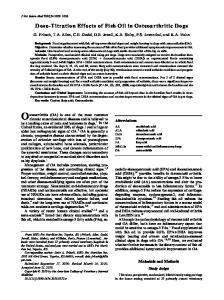
steoarthritis is a debilitating disease of individual joints, marked by progressive joint tissue degeneration, which causes pain and loss of mobility. It is a widespread disease that affects 30 million people in the U.S., including 19% of adults aged 45 and older (1). However, despite decades of research and development, no diseasemodifying drug for osteoarthritis has been approved for use in humans (2). Such a drug could slow disease progression by reducing the rate of cartilage degeneration or even regenerating new tissue. The current standard of care focuses on pain relief only after symptoms are present. Even approved drugs in this category, such as corticosteroids and hyaluronic acid suspensions, are subject to debate with respect to their safety and/or efficacy (3–5). Underlying the clinical failures of disease-modifying drugs and the shortcomings of approved drugs is inadequate drug delivery to target joint tissues (6, 7). Despite the use of intra-articular injection as a technique for local delivery to the joint, free drugs are unable to remain within the joint space for adequate time periods and thereby do not reach their biological targets at sufficient levels (8). The key obstacle for drug delivery in osteoarthritis is the hostile pharmacokinetics of the joint. Upon injection into the articular joint capsule (Figure 1), the drug enters synovial fluid, which is subject to rapid physiological turnover (8). The fluid and the drug contained within it are rapidly drained via the venules and lymphatic vessels located in the synovial membrane; hence, most drugs are lost to systemic circulation (9). Free drugs are cleared from articular joints in a matter of hours to days, with some dependence on the molecular weight of the drug molecule. In contrast to this short
46
www.aiche.org/cep May 2018 CEP
therapeutic time frame, most clinicians seek to minimize the frequency of repeat intra-articular injections. Time between injections varies based on the physician’s judgment and the drug being used, but an interval of 2–12 weeks is considered reasonable. It is therefore unsurprising that many treatments for osteoarthritis are ineffective. Moreover, articular cartilage, which is often the therapeutic target of disease-modifying drugs, presents a formidable biological barrier to drug delivery. Cartilage is avascular (i.e., it has no blood vessels), and thus penetration of drugs through the tissue to interact with the resident cell type, chondrocytes, occurs only by diffusion through the cartilage. Diffusive transport through cartilage is significantly hindered by its dense, highly anionic extracellular matrix and small
p Figure 1. Intra-articular injection is commonly used for local drug delivery into knee joints. However, upon injection into the fluid-filled joint capsule encased in the synovial membrane (or synovium), the drug is typically lost to systemic circulation within a matter of hours to days.
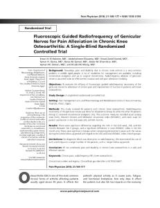
pore size of less than 15 nm (10). Diffusion through cartilage is slower than the clearance rate of the joint, so free drug in the joint space is typically cleared before it can penetrate the depth of cartilage at a therapeutic concentration. Fortunately, advanced formulation techniques for intra-articular injection using engineered biomaterials show promise in overcoming these delivery challenges. Even modest improvements in intra-articular penetration and halflife could have a considerable impact on therapeutic drug exposure time between injections (Figure 2). This article provides an overview of some of the design strategies used in drug delivery systems for joints, and discusses important considerations and challenges for clinical translation of these technologies.
In humans, TA released from FX-006 was measurable in synovial fluid in most patients through 12 weeks post- injection, whereas TA in crystalline suspension was below the lower limit of quantification by six weeks (13). The greatly improved pharmacokinetics of FX-006 produced statistically significant improvements in joint pain, function, and stiffness that warranted FDA approval of the therapy. Importantly, the possibility that clinical improvement can be achieved using already approved therapeutics for osteoarthritis pain suggests that further advanced delivery approaches could enable the success of true disease- modifying drugs. A cartilage drug delivery system could sufficiently improve the efficacy of a previously failed disease-modifying drug to show clinical benefit.
Strategies to avoid joint clearance: Microparticles One approach to prevent clearance of a drug from synovial fluid is to encapsulate the drug in a biomaterial package that is simply too large to enter the synovial micro vasculature. The biomaterial, with its longer joint residence time, can serve as a controlled release depot for the drug over a much longer timescale than an injection of a free drug (Figure 3). This tactic can also reduce the maximum concentration of the drug to which joint tissues are exposed. A reduced maximum concentration of drug is particularly important for corticosteroids, which have shown concerning side effects at repeated high doses (5, 11), as well as for potent biologic drugs. Flexion Therapeutics used this approach in developing FX-006, a therapy that was recently approved by the U.S. Food and Drug Administration (FDA). FX-006 is a poly(lactic-co-glycolic acid) (PLGA) microparticle that encapsulates triamcinolone acetonide (TA), a clinically used corticosteroid that targets synovial tissue to reduce inflammation and pain. The PLGA microparticles have a median size of 42 µm, which is large enough to prevent clearance through joint microvasculature (12).
Strategies to avoid joint clearance: Hydrogels Like microparticles, hydrogels serve as drug material reservoirs that are too large to clear from the joint, thereby extending the time of therapy (Figure 3). This technique has been used to extend time between injections in visco supplementation therapy — a medical procedure in which commercial hyaluronic acid formulations are injected into a joint with the goal of enhancing joint lubrication and alleviating pain. One advantage over microparticles is that hydrogels are capable of encapsulating disease-modifying biologic drugs, such as growth factors or cytokine receptor antagonists, without loss of bioactivity. Polymer microparticle formulations often involve degradable polymers that are not watersoluble and require solvent or heat for processing — harsh conditions that are likely to denature most biologics drugs.
30
Drug Alone With Drug Delivery Repeat Injection
Repeat Injection
Drug Concentration
Time at Therapeutic Dose
Time, days
60
Minimum Effective Dose 90
p Figure 2. Improved drug delivery would extend the residence time of intra-articular drug therapies in joints, which would markedly increase the total time at therapeutic dose over the course of treatment. Clinicians aim to reduce injection frequency as much as possible while still maintaining an effective drug concentration. Advanced drug delivery systems developed by researchers can make this goal possible.
p Figure 3. In articular joints, free drugs and unbound nanoparticles are quickly cleared into lymphatic vessels and venules in the synovium. Microparticles and hydrogels are retained within the synovial fluid and slowly release drugs until they degrade. Cartilage-penetrating nanocarriers enter the tissue and interact with cells, although those that do not bind to cartilage are cleared.
CEP May 2018 www.aiche.org/cep
47
SPECIAL SBE SPECIAL SECTION: SECTION: PROCESS TRANSLATIONAL INTENSIFICATION MEDICINE
Betre et al. designed a system using thermoresponsive elastin-like polypeptides (ELPs) that undergo a solutiongel transition upon injection at human body temperature to form micron-sized aggregates (14). These aggregated ELPs had a half-life of 3.7 days in rat joints, and a release span of 28 days without accumulation in non-target tissues such as the liver or lungs (15). ELP proteins can be expressed as fusions with biologic drugs, which would enable controlled drug release based on enzymatic degradation of the fusion linker.
Strategies to penetrate cartilage: Tissue binding The aforementioned techniques are effective at prolonging exposure of the drug to the joint space, but do not provide a means for encapsulated drugs to navigate the cartilage extracellular matrix to interact with chondrocytes. This is acceptable for therapies with molecular targets within synovial tissue or fluid, as is the case for many pain-alleviating therapies. However, a disease-modifying effect can often be most effectively achieved by targeting the chondrocytes directly (Figure 3). A challenge in designing biomaterials for cartilage penetration while avoiding rapid synovial clearance is the pore size of cartilage extracellular matrix. Research by our groups and others has shown that cartilage has an effective pore size of less than 15 nm, which precludes the use of microparticles, hydrogels, and many types of nanoparticles as cartilage-penetrating carriers (10). Smaller nanocarriers would be capable of transport through cartilage, but such carriers are susceptible to rapid clearance from the joint by the lymphatic vessels and venules in the synovium. To enable cartilage penetration while mitigating clearance, scientists have endeavored to design small nano materials that are capable of binding to cartilage at rates faster than the joint clearance rate. Early work by Rothenfluh et al. established this concept using a phage-panned peptide, WYRGRL (described in single-letter amino acid code), optimized for high-affinity binding with Type II collagen, a major constituent of cartilage extracellular matrix (16). When the researchers conjugated WYRGRL to fluorescent nano particles, the nanoparticles exhibited increased fluorescence intensity relative to untargeted nanoparticles within mouse cartilage four days after intra-articular injection. Nano particles with a volume-average size of 30 nm (measured by dynamic light scattering) were present throughout the depth of thin (~50 µm) mouse cartilage, but 90-nm (volume- average sized) nanoparticles were restricted to the surface. Hu and colleagues applied the same concept to the chelating small molecule DOTAM (17, 18). DOTAM conjugated with three WYRGRL peptides (DOTAM(WYRGRL)3) was retained in the mouse joint for seven days and penetrated at least 200 µm into ex vivo porcine 48
www.aiche.org/cep May 2018 CEP
cartilage. Interestingly, DOTAM-(WYRGRL)3 exhibited more cartilage binding and penetration than DOTAM(WYRGRL)1, suggesting that increasing the binding to cartilage can improve transport into the tissue (17).
Cartilage-penetrating nanocarriers: Cationic proteins In a manner analogous to the concept of using bio molecular peptide-protein interactions, electrostatic inter actions between cationic biomaterials and anionic cartilage can accelerate penetration into cartilage, and electrostatic binding interactions can augment retention within the tissue. This concept was thoroughly explored in our group by Bajpayee et al. with a cationic 7-nm protein, avidin, and its neutral counterpart, neutravidin (10). These two proteins are nearly identical in size and structure, yet avidin, with a net charge of +20, was able to penetrate 1,000-µm-thick ex vivo bovine cartilage within 24 hr, whereas neutravidin penetrated only 50–100 µm within the same time frame (10). In rat joints, avidin was detected up to seven days after intra-articular injection and had an intra-tissue half-life of 1.2 days (19). In rabbit joints, which contain thicker cartilage, the intra-tissue half-life of avidin ranged from 1.0 to 6.4 days, depending on the location of the cartilage within the joint and its thickness (20). These findings suggest that the electrostatic binding mechanism of cartilage retention and penetration is more effective in thicker cartilage (21). Cartilage-penetrating nanocarriers: Synthetic polyelectrolytes Our research groups are also developing polyelectrolyte complex systems to deliver biologic therapeutics, such as growth factors, throughout the depth of cartilage. To encapsulate insulin-like growth factor 1 (IGF-1) without loss of bioactivity, we created a nanoscale polyelectrolyte complex (nanoplex) by controlling the complexation of cationic IGF-1 with anionic poly (L-glutamic acid) and then introducing cationic poly (L-arginine) to modify the surface with excess positive charge (22). The nanoplex had a 16-nm mean diameter as measured by cryogenic transmission electron microscopy (cryo-TEM) and bioactivity equivalent to that of free IGF-1. The IGF-1 nanoplex achieved a joint residence time of 30 days, whereas IGF-1 alone was cleared within seven days. The nanoplex also penetrated through at least 500 µm of ex vivo bovine cartilage tissue, a degree of penetration on par with that of IGF-1 alone. To further engineer the surface charge of IGF-1 systems, we designed a unimolecular polyelectrolyte nanocarrier for IGF-1. By covalent modification of some fraction of cationic side groups of the nanocarrier with polyethylene glycol (PEG) oligomers, we created a small library of