Am J Transl Res 2017;9(1):136-145 www.ajtr.org /ISSN:1943-8141/AJTR0037604
Original Article MicroRNA-410 promotes chondrogenic differentiation of human bone marrow mesenchymal stem cells through down-regulating Wnt3a Yanjie Zhang, Xiaohan Huang, Yanhao Yuan Department of Knee Injury, Henan Luoyang Orthopedic Hospital, Luoyang 471002, Henan, China Received May 13, 2016; Accepted November 28, 2016; Epub January 15, 2017; Published January 30, 2017 Abstract: Background: Chondrogenic differentiation of mesenchymal stem cells (MSCs) is important for osteoarthritis (OA) treatment. However, the specific mechanisms involved are undefined. MicroRNAs (miRNAs) downregulate protein synthesis by binding to the 3’UTR of target mRNA. Methods: Bone marrow aspirates were obtained from OA patients undergoing total hip arthroplasty (n=8) to isolate MSCs. MiR-410 or miR-410 inhibitor were transfected into MSCs using lentivirus and the effects were assessed. Alcian blue staining detected differences in chondrogenic differentiation. An MTT assay and flow cytometry determined changes in cell proliferation and cell cycle, respectively. Real time PCR assessed differences in miRNA and mRNA expression levels and western blotting detected changes in protein levels. ChIP assessed differences in transcriptional activation. TOP/FOP determined changes in the activity of the Wnt signaling pathway. A dual-luciferase reporter assay was used to confirm the miR-410 target protein. Results: miR-410 was elevated during transforming growth factor β3 (TGF-β3)-induced chondrogenic differentiation of MSCs. miR-410 targeted a putative binding site in the 3’-UTR of the Wnt3a gene, thus regulating the Wnt signaling pathway. miR-410 transfection increased mRNA and protein levels of four chondrogenic markers, type II collagen (Col2a1), SRY-box 9 (Sox9), aggrecan (ACAN), and hyaluronan synthase 2 (Has2). miR-410 overexpression decreased Wnt3a protein expression. Wnt3a levels increased in OA patient cartilage concomitant with OA severity and significantly negatively correlated with miR-410 levels. Conclusion: miR-410 is a key regulator of MSC chondrogenic differentiation and directly targets Wnt3a triggering the Wnt signaling pathway. Keywords: BMSCs, chondrogenic differentiation, miR-410, Wnt3a
Introduction Osteoarthritis (OA) is a common musculoskeletal disease with a high socioeconomic impact [1]. It is characterized by alterations in bonecartilage homeostasis resulting in progressive degeneration of synovial joints [2]. Although the OA etiology is still unknown, it has been associated with susceptibility factors including age, prior joint injury, and other alterations linked to genetic and epigenetic factors [3, 4]. Currently, no effective treatment for OA exists beyond pain relief. However, cell therapy is being explored as an OA treatment [5]. Therapeutic approaches using bone marrow mesenchymal stem cells (MSCs) are considered one of the best options for treatment of injured cartilage and bone in OA. This approach is based on the potential of MSCs differentiating into chondrocytes or osteoblasts and their immunomodulatory characteristics.
MSCs chondrogenic differentiation is a critical step in MSC therapy for OA. Dysfunction in the canonical Wnt/β-catenin signaling is involved in OA pathogenesis [6]. Numerous components including Wnts, frizzled, secreted frizzled related protein (sFRP), Dickkopf, and LDL-receptor related proteins (LRPs) play crucial roles during cartilage and bone development and joint maintenance. Moreover, abnormal β-catenin expression is observed in degenerative cartilage, indicating that Wnt signaling might contribute to cartilage loss. sFRP3 polymorphisms have a reduced ability to limit β-catenin signaling leading to an increased susceptibility to OA [7]. MicroRNAs (miRNAs) are small non-coding RNA molecules 21-25 nucleotides long. miRNAs either suppress translation or degrade mRNAs by recognizing the specific and complementary sequences of the 3’UTR of target mRNAs [8, 9]. Various miRNAs play different roles in the
microRNA-410 promotes chondrogenic differentiation via targeting Wnt3a Table 1. Mankin Score of the eight samples
MSC culture
Samples OA-mild
MSCs were obtained from 10 ml of bone marrow aspirates. Briefly, the aspirate was diluted 1:1 with Dulbecco’s Modified Eagle’s Medium (DMEM) and layered over an equal volume of Ficoll. After centrifugation at 900 g for 30 min, the mononuclear cell layer was recovered, washed with DMEM, and suspended in DMEM (supplemented with 10% fetal bovine serum, 100 U/ml penicillin, 100 mg/ml streptomycin, and 2 ml L-glutamine). The cultures were washed to remove the non-adherent cells and further expanded until approximately 80% confluence. Cells at confluence were digested with 0.25% trypsin-EDTA for 5 min at 37°C and cultured for continued passaging. Cells at the third passage were used for experiments. The chondrogenic differentiation medium containing dexamethasone, ascorbate, ITS Supplement, sodium pyruvate, proline, and TGF-β3 was replaced every 2 days.
OA-moderate
OA-severe
Patient 1 Patient 2 Patient 3 Patient 4 Patient 5 Patient 6 Patient 7 Patient 8
Mankin Score 2.3 2.6 2.3 5.2 5.8 5.5 11.5 10.6
development of chondrogenic differentiation and may serve as potential biomarkers [10]. miR-495 inhibits chondrogenic differentiation in human MSCs by targeting Sox9 [11]. miR30a, miR-488, miR-221, and miR-410 regulate chondrogenic differentiation processes [12], and the target genes are important in OA [13]. However, to our best knowledge, there has been no investigation on the potential involvement of miRNAs during MSC chondrogenic differentiation.
Lentivirus vector construction
In this study, we report miR-410 upregulation during the MSC chondrogenic differentiation process. Next, miR-410 was transfected into MSCs using a lentivirus vector to assess its effects on chondrogenic differentiation. The transfected miR-410 greatly promoted chondrogenic differentiation through the Wnt signaling pathway. In the present study, we provide proof-of-concept and experimental methods combining miRNA techniques with MSC therapy in the treatment of OA.
The 293T cells were transfected with the psPAX, Pmd2.G lentivirus package system. In brief, the recombinant lentiviruses containing the entire coding sequence of miR-410 or miR410 inhibitor (miR-410-in) were digested, conjugated, and transformed. Then, the 293T cells were cultured in a 60-mm plate and transfected with the miR-410 plasmid at a confluency of 40%. The transfection reaction was carried out using the Lipofectamine 2000 reagent as described by the manufacturer (Promega). The viruses were harvested from the supernatant after 24 h using a 0.2 μm filter.
Materials and methods
Cell transfection
Clinical information
For transfection of MSCs with lentivirus, the cells were seeded in 6-well plates in growth medium containing DMEM and 10% FBS and incubated for 12 h. Virus stock was diluted with DMEM supplemented with 10% FBS and added to the cells. Following determination of the multiplicity of infection (MOI), MSCs were transfected with miR-410 or miR-410-in at the appropriate MOI. The miR-410 or miR-410-in transfected MSCs were analyzed by RT-PCR and western blotting for identification.
Bone marrow aspirates were obtained from eight OA patients undergoing total hip arthroplasty. The average age of the patients was 56.5 ± 4.6 years. OA diagnosis was determined using the American Rheumatism Association criteria [14]. The disease severity was evaluated by the Mankin Scoring System criteria for articular cartilage (Table 1). All samples were processed after obtaining written informed consent. The institutional ethics committee of Luoyang Orthopedic-Traumatological Hospital approved the study according to the principles presented in the Declaration of Helsinki.
137
Alcian blue staining Representative cultures were collected after induction and sulfated cartilage glycosamino-
Am J Transl Res 2017;9(1):136-145
microRNA-410 promotes chondrogenic differentiation via targeting Wnt3a
Figure 1. MicroRNA expression in MSCs after cartilage induction. MSCs were induced for chondrogenic differentiation by TGF-β3. The expression of miRNAs in MSCs and HC-α cell was detected by RT-PCR. A. miR-572, miR-130b, miR-193b, miR-410, miR-152, miR-560, and miR-28 expression in MSCs after cartilage induction. B. miR-410 expression in MSCs and HC-α cell. MSCs were induced for 15 or 25 days. *P < 0.05 vs. MSCs without induction.
glycans (GAGs) were measured by alcian blue staining to assess the deposition of cartilage matrix proteoglycans. The cells used for alcian blue staining were fixed using 4% paraformaldehyde, dehydrated, and paraffin imbedded. The 5 mm sections were stained with 1% alcian blue 8GX (Amresco) for 30 min. The cells were observed under the microscope.
Cambridge, UK), COL2A1 (ab34712, Abcam), Sox9 (ab26414, Abcam), ACAN (ab36861, Abcam), Has2 (ab131364, Abcam), p84 (ab487, Abcam), β-catenin (ab16051, Abcam), and GAPDH (ab9485, Abcam), and incubated with a peroxidase-conjugated secondary antibody. Proteins were visualized using a Millipore ECL Western Blotting Detection System.
Real time PCR
MTT assay
Total RNA was extracted from the cells with TRIzol reagent (Invitrogen) and cDNA was synthesized following the manufacturer’s protocol (Fermentas). Real-time PCR (qRT-PCR) analysis was conducted with SYBR Green Realtime PCR Master Mix (Toyobo) according to the manufacturer’s instructions. qRT-PCR reactions were performed in the PTC-220 Real-Time PCR Machine (Bio-rad). Results were normalized to the expression of U6 or GAPDH and were calculated with the 2-ΔΔCt method [15]. Western blot Proteins (30 μg) were separated by electrophoresis on 12% polyacrylamide gels containing 0.1% SDS and transferred to nitrocellulose membranes. The membranes were incubated overnight at 4°C in blocking buffer (5% nonfat dry milk), probed with antibodies against the following proteins: Wnt3a (ab28472, Abcam, 138
The MTT assay was used to detect MSC proliferation. The cells transfected with either miR410 or miR-410-in were induced to chondrocytes for 3-5 days. Subsequently, all cells were treated with 20 μl MTT at 5 mg/ml for 4 h. After adding 150 μl DMSO, the plate was read at 490 nm to obtain the cell growth curve. All experiments were repeated in triplicate. Flow cytometry Cell cycle was analyzed by flow cytometry after MSCs were treated with miR-410 and chondrogenic induction. After treatment, MSCs were fixed in ethanol at -20°C, washed in PBS, and stained with propidiumiodide (PI, 50 mg/mL) with RNaseA treatment (50 mg/mL) for 15 minutes at room temperature in the dark. Analysis was performed on the FACS Calibur (BD Biosciences). Flow Jo software was used to determine the percentage of MSCs in G0, G1, and S/G2/M. Am J Transl Res 2017;9(1):136-145
microRNA-410 promotes chondrogenic differentiation via targeting Wnt3a suspended in lysis buffer containing 0.1% SDS and sonicated to achieve fragment sizes of 200 to 600 bp. The IP was conducted with ChIP-grade protein G magnetic beads using an antibody against β-catenin. IgG protein was used as the negative control. To validate the enrichment, qPCR was performed with tiled primers. Dual-luciferase reporter gene assay
Figure 2. miR-410 promotes MSC proliferation. The empty vector (lentivirus) or recombinant lentivirus containing the entire coding sequence of miR-410 or miR-410 inhibitor were transfected into MSCs. The expression of miR-410 in MSCs with different treatments was measured by RT-PCR. Cell proliferation was assessed by the MTT assay. The cell cycle was detected by flow cytometry. A. miR-410 expression in MSCs after empty vector, miR-410, or miR-410-in transfection. B. Cell proliferation detected by the MTT assay after empty vector, miR-410, or miR-410-in transfection. C. Cell cycle tested by flow cytometry after empty vector, miR-410, or miR-410-in transfection. Vector group: MSCs transfected with lentiviruses; miR-410 group: MSCs transfected with recombinant lentiviruses containing the entire coding sequence of miR-410; miR-410-in group: MSCs transfected with recombinant lentiviruses containing the entire coding sequence of miR-410 inhibitor. *P < 0.05 vs. vector group.
TOP/FOP detection The activity of the Wnt signaling pathway was detected by a Wnt signal reporter assay using the TOPglow/FOPglow TCF reporter kit (Millipore). MSCs were seeded in 6-well plates and transfected with TOPglow and FOPglow according to the manual. All transfections were done in triplicate and repeated at least 3 times.
For luciferase activity analysis, MSCs (2×10⁵ cells per well) were co-transfected with the luciferase reporter construct phRL-TK Renilla luciferase plasmid (Promega) and Wnt3a-3’UTR using Lipofectamine 2000 according to the manufacturer’s instructions (Promega). After incubation for 24 h, the luciferase assay and subsequent luminescence calculations were performed using the DualGlo luciferase reporter assay system (Promega). Three independent experiments were performed in triplicate. Statistical analysis
Data were expressed as the mean ± SD and analyzed by SPSS 17.0 software. Statistical comparisons were made between two groups using the t-test and among multiple groups using one-way ANOVA. A value of P < 0.05 was considered to be statistically significant. Results
ChIP
miR-410 is upregulated during MSC chondrogenic differentiation
ChIP assays were performed as described previously [16]. Briefly, MSCs were cross-linked for 10 minutes with 1% formaldehyde and quenched with 125 mM glycine. After nuclei were isolated by centrifugation, the pellet was
To screen the miRNAs with potential effects on MSC chondrogenic differentiation, we selected overexpressed miRNAs reported in cartilage for detection. Real time PCR showed that eight miRNAs were significantly upregulated in MSCs
139
Am J Transl Res 2017;9(1):136-145
microRNA-410 promotes chondrogenic differentiation via targeting Wnt3a
Figure 3. miR-410 accelerated MSC chondrogenic differentiation. The empty vector (lentivirus) or recombinant lentivirus containing the entire coding sequence of miR-410 or miR-410 inhibitor were transfected into MSCs. The MSCs were stained with alcian blue. The mRNA and protein levels of cartilage marker proteins were detected by RT-PCR and western blot, respectively. A. Alcian blue staining of miR-410 or miR-410-in transfected MSCs for detection of chondrogenic differentiation. B. RT-PCR detection of mRNA expression of cartilage markers in miR-410 or miR-410-in transfected MSCs during chondrogenic differentiation. C. Western blot detection of protein expression of cartilage markers in miR-410 or miR-410-in transfected MSC during chondrogenic differentiation. Vector group: MSCs transfected with lentivirus; miR410 group: MSCs transfected with recombinant lentivirus containing the entire coding sequence of miR-410; miR-410-in group: MSCs transfected with recombinant lentivirus containing the entire coding sequence of miR-410 inhibitor. *P < 0.05 vs. vector group.
after cartilage induction with the largest increase found for miR-410 (Figure 1A). Next, to confirm the expression changes of miR-410 during MSC chondrogenic differentiation, MSCs were induced for 15 or 25 days for chondrogenic differentiation. The results show that miR410 was slightly increased after 15 days induction. It was significantly elevated after 25 days induction, but still lower than in the human chondrocyte cell line HC-a (Figure 1B). These results demonstrate that miR-410 is gradually upregulated during the process of MSC chondrogenic differentiation. miR-410 promotes MSC proliferation Because miR-410 was upregulated during MSC chondrogenic differentiation, we hypothesized that miR-410 may play a critical role in MSCs. Lentivirus was selected for cell transfection to increase or reduce miR-410 expression in 140
MSCs. Real time PCR confirmed that we successfully changed miR-410 levels in MSCs (Figure 2A). Furthermore, the effect of miR-410 on cell viability was tested. The results from the MTT assay revealed that miR-410 obviously suppressed MSC viability, whereas miR-410-in markedly enhanced MSC viability after four days incubation (Figure 2B). Flow cytometry also suggested that miR410 blocked MSCs in the G0/G1 phase, whereas miR410-in promoted cell proliferation by accelerating the cell cycle (Figure 2C). The data indicate that miR-410 may affect MSC proliferation through regulating the cell cycle. miR-410 accelerates MSC chondrogenic differentiation
The cell cluster became larger and the surface presented as a colloid during the process of chondrogenic differentiation. Morphological observation showed that miR410 transfection significantly promoted MSC chondrogenic differentiation compared with normal controls. However, miR-410-in markedly suppressed chondrogenic differentiation (Figure 3A). The mRNA and protein levels of cartilage related markers, such as COL2A1, SOX9, ACAN, and Has2, were tested by real time PCR and western blot after miR-410 transfection. Real time PCR results show that mRNA levels of the cartilage related markers were enhanced in the miR-410 group compared with the vector group, whereas they declined in the miR410-in group (Figure 3B). Western blot results also demonstrated that the changes in protein levels were similar to that of the mRNA levels (Figure 3C), suggesting that miR-410 promoted MSC chondrogenic differentiation. miR-410 triggers the Wnt signaling pathway The Wnt signaling pathway plays an important role in MSC chondrogenic differentiation. Thus, Am J Transl Res 2017;9(1):136-145
microRNA-410 promotes chondrogenic differentiation via targeting Wnt3a nucleus, the protein levels were detected by western blot. p84 levels in the nucleus were markedly increased in the miR-410-in group, revealing that miR-410 suppression facilitated β-catenin entering the nucleus during MSC chondrogenic differentiation (Figure 4B). miR-410 regulates the Wnt signaling pathway by targeting Wnt3a Because we found that miR410 affected the activity of the Wnt signaling pathway, we searched the miRNA database and found that miR410 pairs with Wnt3a mRNA (Figure 5A). Western blot results showed that Wnt3a expression was upregulated in the miR-410-in group (Figure 5B). Luciferase assay results Figure 4. miR-410 triggered the Wnt signaling pathway. The activity of the revealed that the level of Wnt signaling pathway was detected by a Wnt signal reporter assay. MSCs Wnt3a 3’UTR was reduced in were transfected with TOPglow and FOPglow expression plasmids. The valmiR-410 transfected MSCs ues of TOPglow and FOPglow were detected and the TOP/FOP ratio reflected (Figure 5C), suggesting that the activity of the Wnt signaling pathway. A. TOP/FOP ratio in miR-410 or miR-410-in transfected MSCs during chondrogenic differentiation. B. ChIP miR-410 regulates Wnt3a exdetection of b-FGF, IGF-1, and TGF-β1 in miR-410 or miR-410-in transfected pression by directly binding MSCs during chondrogenic differentiation. C. The protein levels of β-catenin to the 3’UTR of Wnt3a. Corand p84 in both the cytoplasm (C) and nucleus (N) were detected by western relation analysis demonstrblot. Vector/NC group: control group, MSCs transfected with lentivirus; miRated that Wnt3a express410 group: MSCs transfected with recombinant lentivirus containing the entire coding sequence of miR-410; miR-410-in group: MSCs transfected with ion was negatively correlated recombinant lentivirus containing the entire coding sequence of miR-410 with miR-410 expression in inhibitor. *P < 0.05 vs. vector/NC group. the cartilage from OA patients (Figure 5D), suggesting a rewe explored whether miR-410 regulates the lationship in OA. Moreover, Wnt3a mRNA exactivity of the Wnt signaling pathway using pression in clinical samples from OA patients a reporter assay. As shown in Figure 4A, the was tested. Wnt3a mRNA was markedly eleTOP/FOP ratio was significantly decreased in vated in OA patients in a severity dependent MSCs after miR-410 transfection, whereas it manner (Figure 5E). was upregulated after miR-410-in transfection. Discussion Furthermore, a ChIP assay was used to evaluate the activation of the Wnt signaling pathAs endogenous small inhibitory molecules of way. miR-410 overexpression facilitated nugene expression, miRNAs play critical roles in clear β-catenin binding to the promoters of stem cell function and differentiation [17, 18]. b-FGF, IGF-1, and TGF-β1, and promoted gene In this study, we identified that miR-410 protranscription and MSC chondrogenic differenmoted TGF-β3-induced chondrogenic differentiation (Figure 4C). Next, we examined the eftiation of MSCs by directly targeting Wnt3a at fect of miR-410 on β-catenin nuclear translocation. After extraction from the cytoplasm and the post-transcriptional level. TGF-β3 treatment
141
Am J Transl Res 2017;9(1):136-145
microRNA-410 promotes chondrogenic differentiation via targeting Wnt3a
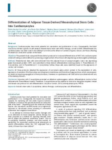
Figure 5. miR-410 regulates the Wnt signaling pathway by targeting Wnt3a. To confirm whether miR-410 targeted the 3’-UTR of Wnt3a, a luciferase activity assay was performed. MSCs were co-transfected with wild type Wnt3a 3’-UTR (Wnt3a-3’-UTR) or mutant Wnt3a 3’-UTR (Wnt3a-3’-UTR-mut) and miR-410 or miR-410-in. The relative luciferase activity was detected. A. Predicted miR-410 target sequence in the 3’ UTR of Wnt3a. B. Western blot detection of Wnt3a protein expression in miR-410 or miR-410-in transfected MSCs during chondrogenic differentiation. C. Dual-luciferase reporter assay of MSCs transfected with the Wnt3a-3’ UTR reporter and miR-410 or miR-410-in. D. Correlation analysis of miR-410 and Wnt3a expression in OA patients. E. RT-PCR detection of Wnt3a mRNA expression in OA patients with different degrees of severity. Vector group: MSCs transfected with lentivirus; miR-410 group: MSCs transfected with recombinant lentivirus containing the entire coding sequence of miR-410; miR-410-in group: MSCs transfected with recombinant lentivirus containing the entire coding sequence of miR-410 inhibitor. Normal control group: people without OA; OA-mild group: patients with mild OA; OA-moderate group: patients with moderate OA; OA-severe group: patients with severe OA. *P < 0.05 vs. vector group; #P < 0.05 vs. normal control group.
caused an elevation in miR-410 expression and affected the Wnt signaling pathway, facilitating chondrogenic differentiation. Recent research shows that miR-410 regulates MET, which is involved in influencing glioma cell proliferation and invasion [19]. In a pituitary gonadotroph tumor, miR-410 blocked cell cycle progression by targeting cyclin B1 [20]. In our data, miR-410 markedly accelerated the cell
142
proliferation rate in MSCs. These results indicate that miR-410 can regulate chondrogenic differentiation of MSCs by affecting cell proliferation. In conclusion, our results demonstrate that miR-410 is a positive regulator of MSC chondrogenic differentiation accelerating the cell cycle and enhancing cell viability. Our real time PCR data demonstrate that miR410 exhibited the largest enrichment in MSCs
Am J Transl Res 2017;9(1):136-145
microRNA-410 promotes chondrogenic differentiation via targeting Wnt3a after cartilage induction among numerous reported overexpressed miRNAs. The expression of mature miR-410 gradually increased during TGF-β3-induced chondrogenic differentiation of MSCs. In addition, miR-410 was also increased in the cartilage cell line HC-a, as validated by real time PCR analysis. Therefore, miR-410 is a candidate with significant potential to participate in the regulation of chondrogenic differentiation. Although miR-410 has been known to be involved in the carcinogenesis of various types of cancer and regulation of renal fibrosis in the pathogenesis of lupus nephritis [21, 22], we have identified the complementary function of inhibiting TGF-β3-mediated chondrogenesis. Wnts can either suppress or trigger chondrocyte differentiation in different in vivo and in vitro models [23-28]. The different effects of Wnt signaling appear to be due to the different stages of development or cell type [26, 29]. For example, activation of Wnt signaling using Wnt3a inhibits chondrogenesis of the prechondrocyte cell line ATDC540, however, it promotes chondrogenic differentiation of undifferentiated mesenchymal cells [27]. Thus, the demonstration that Wnt signaling promotes the chondrogenic differentiation of MSCs in this study is consistent with a previous report that miR410 may regulate the Wnt signaling pathway [21]. These results suggest that miR-410 may affect MSC chondrogenesis through the Wnt signaling pathway. Consistent with the important role of the Wnt signaling pathway in regulating chondrogenesis, our results indicate that over-expression of miR-410 promotes MSC chondrogenic differentiation, as reflected by increased mRNA and protein expression of chondrogenic markers including COL2A1, SOX9, ACAN, and Has2. In contrast, inhibition of miR-410 weakened chondrogenic differentiation, as shown by down-regulated mRNA and protein levels of COL2A1, SOX9, ACAN, and Has2. Furthermore, miR-410 has an obvious effect on accelerating β-catenin nuclear translocation and binding with the promoters of b-FGF, IGF-1, and TGF-β1. The results suggest that miR-410 has a specific influence on genes associated with chondrogenesis in response to TGF-β3. Taken together, miR-410 represents a key regulator for promoting the chondrogenic phenotype in MSCs by regulating 143
the Wnt signaling pathway, which may be responsive to TGF-β3. The luciferase reporter analysis shows that exogenous miR-410 and miR-410-in regulated the activity of luciferase. These results indicate that miR-410 potentially regulates Wnt3a expression by binding to the miRNAs regulatory element in the Wnt3a 3’UTR. The results indicate that miR-410 affects chondrogenesis by targeting Wnt3a. Consistent with the prominent role of the Wnt signaling pathway in the regulation of chondrogenesis, our results show that over-expression of miR-410 resulted in enhancement of MSC chondrogenic differentiation, whereas inhibition of miR-410 inhibited chondrogenic differentiation. miRNAs inhibit target gene expression through two distinct pathways, which are dependent on the complementary sequences between miRNAs and the target mRNA. miRNAs inhibit mRNA translation with imperfect complementary target sequences and degrade mRNA via perfect complementary target sequences [30, 31]. Computational algorithms predicted that miR-410 binds to Wnt3a 3’-UTR with perfect complementation sequences, indicating that it might degrade Wnt3a mRNA. Consistent with the mechanisms of miRNAs regulation, we found Wnt3a was differentially expressed at both the protein and mRNA levels in gain- or loss-of function miR-410 experiments. Wnt3a mRNA was also increased in OA patients. Correlation analysis indicated that Wnt3a mRNA was significantly negatively correlated with miR-410. This study revealed that miR-410 suppressed Wnt3a expression by binding to the Wnt3a mRNA 3’-UTR causing translational inhibition. In conclusion, our studies demonstrate that miR-410 was increased during TGF-β3-induced chondrogenic differentiation of MSCs. During the process of chondrogenic differentiation of MSCs, the attenuation of miR-410 expression negatively regulated its target gene, Wnt3a, and resulted in inhibiting the Wnt signaling pathway. Our findings indicate that miR-410 plays a key role in chondrogenesis and provides a novel mechanism in miRNA mediated regulation of MSC chondrogenic differentiation. However, an in vivo study is needed for further confirmation and clarification. Am J Transl Res 2017;9(1):136-145
microRNA-410 promotes chondrogenic differentiation via targeting Wnt3a Acknowledgements The authors gratefully thank Dr. Xiaohan Huang for his assistance with the experiments and his useful suggestions on experimental design. Disclosure of conflict of interest
[11]
[12]
None. Address correspondence to: Yanhao Yuan, Department of Knee Injury, Henan Luoyang Orthopedic Hospital, 82 South Qi’ming Road, Luoyang 471002, Henan, China. Tel: +86-0379-63546480; E-mail:
[email protected]
References [1]
Bitton R. The economic burden of osteoarthritis. Am J Manag Care 2009; 15: S230-235. [2] Goldring MB, Goldring SR. Articular cartilage and subchondral bone in the pathogenesis of osteoarthritis. Ann N Y Acad Sci 2010; 1192: 230-237. [3] Blagojevic M, Jinks C, Jeffery A, Jordan KP. Risk factors for onset of osteoarthritis of the knee in older adults: a systematic review and metaanalysis. Osteoarthritis Cartilage 2010; 18: 24-33. [4] Reynard LN, Loughlin J. Insights from human genetic studies into the pathways involved in osteoarthritis. Nat Rev Rheumatol 2013; 9: 573-583. [5] Diekman BO, Guilak F. Stem cell-based therapies for osteoarthritis: challenges and opportunities. Curr Opin Rheumatol 2013; 25: 119126. [6] Corr M. Wnt-beta-catenin signaling in the pathogenesis of osteoarthritis. Nat Clin Pract Rheumatol 2008; 4: 550-556. [7] Loughlin J, Dowling B, Chapman K, Marcelline L, Mustafa Z, Southam L, Ferreira A, Ciesielski C, Carson DA, Corr M. Functional variants within the secreted frizzled-related protein 3 gene are associated with hip osteoarthritis in females. Proc Natl Acad Sci U S A 2004; 101: 9757-9762. [8] Shukla GC, Singh J, Barik S. MicroRNAs: processing, maturation, target recognition and regulatory functions. Mol Cell Pharmacol 2011; 3: 83-92. [9] Bartel DP. MicroRNAs: target recognition and regulatory functions. Cell 2009; 136: 215233. [10] Tornero-Esteban P, Hoyas JA, Villafuertes E, Garcia-Bullon I, Moro E, Fernandez-Gutierrez B, Marco F. [Study of the role of miRNA in mesenchymal stem cells isolated from osteoarthri-
144
[13]
[14]
[15]
[16]
[17] [18]
[19]
[20]
[21]
[22]
tis patients]. Rev Esp Cir Ortop Traumatol 2014; 58: 138-143. Lee S, Yoon DS, Paik S, Lee KM, Jang Y, Lee JW. microRNA-495 inhibits chondrogenic differentiation in human mesenchymal stem cells by targeting Sox9. Stem Cells Dev 2014; 23: 1798-1808. Karlsen TA, Jakobsen RB, Mikkelsen TS, Brinchmann JE. microRNA-140 targets RALA and regulates chondrogenic differentiation of human mesenchymal stem cells by translational enhancement of SOX9 and ACAN. Stem Cells Dev 2014; 23: 290-304. Tian Y, Guo R, Shi B, Chen L, Yang L, Fu Q. MicroRNA-30a promotes chondrogenic differentiation of mesenchymal stem cells through inhibiting Delta-like 4 expression. Life Sci 2016; 148: 220-228. Arnett FC, Edworthy SM, Bloch DA, McShane DJ, Fries JF, Cooper NS, Healey LA, Kaplan SR, Liang MH, Luthra HS, et al. The American rheumatism association 1987 revised criteria for the classification of rheumatoid arthritis. Arthritis Rheum 1988; 31: 315-324. Livak KJ, Schmittgen TD. Analysis of relative gene expression data using real-time quantitative PCR and the 2(-Delta Delta C(T)) method. Methods 2001; 25: 402-408. Lee TI, Johnstone SE, Young RA. Chromatin immunoprecipitation and microarray-based analysis of protein location. Nat Protoc 2006; 1: 729-748. Gangaraju VK, Lin H. MicroRNAs: key regulators of stem cells. Nat Rev Mol Cell Biol 2009; 10: 116-125. Lakshmipathy U, Hart RP. Concise review: MicroRNA expression in multipotent mesenchymal stromal cells. Stem Cells 2008; 26: 356-363. Chen L, Zhang J, Feng Y, Li R, Sun X, Du W, Piao X, Wang H, Yang D, Sun Y, Li X, Jiang T, Kang C, Li Y, Jiang C. MiR-410 regulates MET to influence the proliferation and invasion of glioma. Int J Biochem Cell Biol 2012; 44: 1711-1717. Mussnich P, Raverot G, Jaffrain-Rea ML, Fraggetta F, Wierinckx A, Trouillas J, Fusco A, D’Angelo D. Downregulation of miR-410 targeting the cyclin B1 gene plays a role in pituitary gonadotroph tumors. Cell Cycle 2015; 14: 2590-2597. Zhang X, Ke X, Pu Q, Yuan Y, Yang W, Luo X, Jiang Q, Hu X, Gong Y, Tang K, Su X, Liu L, Zhu W, Wei Y. MicroRNA-410 acts as oncogene in NSCLC through downregulating SLC34A2 via activating Wnt/beta-catenin pathway. Oncotarget 2016; 7: 14569-85. Liu D, Zhang N, Zhang J, Zhao H, Wang X. miR410 suppresses the expression of interleukin-6 as well as renal fibrosis in the pathogen-
Am J Transl Res 2017;9(1):136-145
microRNA-410 promotes chondrogenic differentiation via targeting Wnt3a
[23]
[24]
[25]
[26]
145
esis of lupus nephritis. Clin Exp Pharmacol Physiol 2016; 43: 616-25. Hill TP, Spater D, Taketo MM, Birchmeier W, Hartmann C. Canonical Wnt/beta-catenin signaling prevents osteoblasts from differentiating into chondrocytes. Dev Cell 2005; 8: 727738. Hu H, Hilton MJ, Tu X, Yu K, Ornitz DM, Long F. Sequential roles of Hedgehog and Wnt signaling in osteoblast development. Development 2005; 132: 49-60. Day TF, Guo X, Garrett-Beal L, Yang Y. Wnt/beta-catenin signaling in mesenchymal progenitors controls osteoblast and chondrocyte differentiation during vertebrate skeletogenesis. Dev Cell 2005; 8: 739-750. Hartmann C, Tabin CJ. Dual roles of Wnt signaling during chondrogenesis in the chicken limb. Development 2000; 127: 3141-3159.
[27] Fischer L, Boland G, Tuan RS. Wnt-3A enhances bone morphogenetic protein-2-mediated chondrogenesis of murine C3H10T1/2 mesenchymal cells. J Biol Chem 2002; 277: 30870-30878. [28] Rudnicki JA, Brown AM. Inhibition of chondrogenesis by Wnt gene expression in vivo and in vitro. Dev Biol 1997; 185: 104-118. [29] Hartmann C. A Wnt canon orchestrating osteoblastogenesis. Trends Cell Biol 2006; 16: 151158. [30] Carrington JC, Ambros V. Role of microRNAs in plant and animal development. Science 2003; 301: 336-338. [31] Zeng Y, Yi R, Cullen BR. MicroRNAs and small interfering RNAs can inhibit mRNA expression by similar mechanisms. Proc Natl Acad Sci U S A 2003; 100: 9779-9784.
Am J Transl Res 2017;9(1):136-145