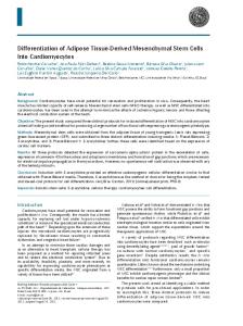
MOLECULAR MEDICINE REPORTS 14: 5065-5071, 2016
Notch1 is associated with the differentiation of human bone marrow‑derived mesenchymal stem cells to cardiomyocytes ZIPU YU*, YU ZOU*, JINGYA FAN, CHENGCHEN LI and LIANG MA Department of Cardiac Surgery, First Affiliated Hospital, Zhejiang University, Hangzhou, Zhejiang 310000, P.R. China Received October 8, 2015; Accepted August 16, 2016 DOI: 10.3892/mmr.2016.5862 Abstract. Notch signaling is involved in the early process of differentiation to determine the fate of stem cells. However, the precise role of Notch in human bone marrow‑derived mesenchymal stem cells (hBMSCs) remains unclear. The present study aimed to investigate the involvement of Notch signalling during the course of hBMSC differentiation into cardiomyocytes using hBMSCs, with multilineage differentiation ability, isolated and purified from human bone marrow. Flow cytometric analysis revealed that CD29, CD44 and CD90 were highly expressed on the surface of cells in their fifth passage, whereas detection of CD34, CD45, CD54 and HLA‑DR was negative. Visualization of morphological changes, western blotting, immunocytochemistry and reverse transcription‑quantitative polymerase chain reaction (RT‑qPCR) demonstrated that hBMSCs differentiate into cardiomyocytes through treatment with 5‑azacytidine (5‑aza). Transmission electron microscopy revealed ultramicroscopic details of differentiated hBMSCs. Western blotting and immunocytochemistry demonstrated increased protein expression levels of α‑actin and cardiac troponin T expression, and RT‑qPCR revealed increased mRNA expression of Notch1 early in the process of differentiation (days 1, 4 and 7), and increased mRNA expression levels of the transcription factors GATA binding protein‑4 and NK2 homeobox 5 at day 28 day. In conclusion, differentiation of hBMSCs into cardiomyocytes was induced in vitro by 5‑aza, and was associated with upregulation of Notch1, GATA binding protein‑4 and Nkx2.5 expression. Overexpression of the Notch1 signaling pathway may represent a potential mechanism underlying the differentiation of hBMSCs.
Introduction Stem cells are a major focus of modern scientific research into the treatment of heart diseases, as their plasticity may help overcome the limited self‑repair capacity of cardiomyocytes. Human bone marrow‑derived mesenchymal stem cells (hBMSCs) have been widely used in numerous studies and clinical trials due to their unique properties, including ease of isolation, expansion in vitro, multipotency and immunological tolerance (1‑4). Previous studies have revealed that 5‑azacytidine (5‑aza) induces MSC differentiation into myocardial cells (5‑7). It has also been demonstrated that hBMSCs can differentiate into cardiac troponin T (cTnT)‑expressing cardiomyocytes (2,8‑11); however, the precise mechanisms controlling this process currently remain unclear. The mechanism of differentiation is complex and requires various signaling pathways. Notch signaling, which is important in cell fate specification during embryogenesis, has previously been implicated in regulating differentiation of stem cells in adults (12). Numerous studies have demonstrated that Notch signaling regulates a wide variety of processes during embryonic and post‑natal development, including proliferation, apoptosis and cell fate decisions (13‑15). However, the association between Notch signalling and the differentiation of hBMSCs to cardiomyocytes remains unclear. The aim of the present study was to investigate the function of Jagged1‑Notch1 signaling during hBMSCs differentiation into cardiomyocytes. As a result of its widespread expression, it was hypothesized that Notch signaling may be involved in the differentiation of hBMSCs. Materials and methods
Correspondence to: Dr Liang Ma, Department of Cardiac Surgery, First Affiliated Hospital, Zhejiang University, 79 Qinchun Road, Hangzhou, Zhejiang 310000, P.R. China E‑mail:
[email protected] *
Contributed equally
Key words: human bone marrow‑derived mesenchymal stem cells, differentiation, Notch1, 5‑azacytidine
Isolation and culture of hBMSCs. hBMSCs were isolated by bone marrow aspiration from the sternums of 20 male patients (age, 23‑48 years) with heart valve diseases awaiting cardiac surgery, but were healthy with regard to the circulatory system. Informed consent was obtained from all patients prior to inclusion in the study. The study used a protocol approved by the Research Ethics Committee of the First Affiliated Hospital, Zhejiang University (Hangzhou, China). Bone marrow mononuclear cells were purified by Ficoll‑Paque density‑gradient centrifugation as previously described (16). The purified mononuclear cells were allowed to adhere to culture flasks in Dulbecco's modified Eagle's medium (Gibco; Thermo Fisher
5066
YU et al: Notch1 IS ASSOCIATED WITH MYOCARDIAL DIFFERENTIATION OF MSCs
Scientific, Inc., Waltham, MA, USA) supplemented with 10% fetal bovine serum (Invitrogen; Thermo Fisher Scientific, Inc.) overnight at 37˚C in 5% CO2. Cells from passages 3‑6 were used for subsequent experiments.
Table I. Primers used for quantitative polymerase chain reaction.
Cell viability assay. hBMSCs (~5,000 cells/well) were seeded into 96‑well plates. Following overnight incubation, the medium was removed and Cell Counting Kit 8 (CCK8; Dojindo Molecular Technologies, Inc., Kumamoto, Japan) solution was added to each well and incubated at 37˚C for 1 h. The absorbance of the solution was measured spectrophotometrically at 450 nm with a MRX II absorbance reader (Dynex Technologies, Inc., Chantilly, VA, USA). The growth assay was performed each day for 5 days consecutively.
GAPDH Jagged1 Notch‑1 GATA‑4 NKx2.5
Analysis of hBMSCs by flow cytometry. Culture‑expanded cells (passages 3‑6) were washed with phosphate‑buffered saline (PBS) containing 0.5% (w/v) bovine serum albumin (BSA), their concentration adjusted to 1x10 6 cells/100 µl, and phenotypic analyses performed via flow cytometry. The hBMSCs were blocked with 1% BSA and incubated with phycoerythrin‑ or fluorescein isothiocyanate‑conjugated mouse monoclonal antibodies against human CD34 (catalog no. sc‑19587; Santa Cruz Biotechnology, Inc., Dallas, TX, USA), CD54 (catalog no. ab27582; Abcam, Cambridge, UK), CD45 (catalog no. 560976; BD Biosciences, San Jose, CA, USA), CD44 (catalog no. 560977; BD Biosciences), CD29 (catalog no. sc‑59829; Santa Cruz Biotechnology, Inc.), human leukocyte antigen‑antigen D related (HLA‑DR; catalog no. 560944; BD Biosciences) and CD90 (catalog no. 561969; BD Biosciences) for 60 min in the dark at 4˚C, at dilutions recommended by the manufacturers. Subsequently, cells were washed with PBS and fixed with 2% paraformaldehyde. Immunoglobulin isotype incubation was performed as a negative control. Flow cytometry was performed with a FACSCalibur system (FC500; Beckman Coulter, Inc., Brea, CA, USA) and analysed using FlowJo software version 7.6.5 (FlowJo, LLC, Ashland, OR, USA). Multilineage differentiation assays. To induce osteogenic differentiation, hBMSCs were cultured in a commercially available osteogenic differentiation medium (catalog no. HUXMA‑90021; Cyagen Biotechnology Co. Ltd., Taicang, China). On day 21, cells were stained with Alizarin Red in accordance with the manufacturer's protocol. To induce adipogenic differentiation, hBMSCs were cultured in a commercially available adipogenic differentiation medium (catalog no. HUXMA‑90031; Cyagen Biotechnology Co. Ltd.). On day 21, cells were stained for 30 min with Oil Red O, diluted 3:2 with distilled water and filtered. To induce chondrogenic differentiation, hBMSCs were cultured in a commercially available chondrogenic differentiation medium purchased from Cyagen (catalog no. HUXMA‑90041; Cyagen Biotechnology Co. Ltd.). On day 28, cells were stained with 1 mg/ml Alcian blue for 30 min. Cells were observed under a Nikon Eclipse E200 light microscope (Nikon Corporation, Tokyo, Japan). hBMSC5‑aza‑induced differentiation to cardiomyocytes. Following the third passage, the cells were washed twice with PBS, adjusted to a density of 2x10⁴ cells/ml, and seeded in a 6‑well plate. Following 24 h incubation, 10 µmol/l 5‑aza was
Gene
Primer sequence (5'‑3') F AAGGTGAAGGTCGGAGTCA R GGAAGATGGTGATGGGATTT F AGCTATTTGCCGACAAGGCT R CACTGCCAGGGCTCATTACA F AACGCCTACCTCTGCTTCTG R CTCACAGGCACACTCGTAGC F GGAAGCCCAAGAACCTGAAT R TGCCCGTAGTGAGATGACAG F CTACCAGGCTCGGATACCAT R GCCAACAACAACTTCGTGAAC
GAPDH, glyceraldehyde 3‑phosphate dehydrogenase; F, forward; R, reverse; GATA‑4, GATA binding protein 4; Nkx2.5, NK2 homeobox 5.
added (for control group, 0 µmol/l 5‑aza was added), and cells were incubated for a further 24 h. The culture medium containing 5‑aza was then removed and complete culture medium was added. The cells went through the same treatment process course following each cell passage. The cells were maintained in culture for 4 weeks following treatment. Samples were taken for detection of morphological changes by transmission electron microscopy (TEM) on day 28, for α‑actin and cTnT expression by immunocytochemistry and western blot analysis on day 28, and for analysis of mRNA expression by reverse transcription‑quantitative polymerase chain reaction (RT‑qPCR) for GATA binding protein‑4 (GATA‑4), and NK2 homeobox 5 (Nkx2.5) on day 28, and Jagged1 and Notch1 on days 1, 4 and 7. TEM analysis. Cultured cells were rinsed with PBS and immersed in 2.5% glutaraldehyde for 4 h, rinsed in 0.1 mol/l sodium cacodylate buffer (pH 7.3), and postfixed for 1 h in 1% OsO4. The cells were embedded in Epon resin and cut into 60‑nm thick sections with a Sorvall MTB2 ultramicrotome (Thermo Fisher Scientific, Inc.). Sections were stained for 20 min at room temperature with uranyl acetate and lead citrate and examined under a Hitachi H‑600 electron microscope (Hitachi, Ltd., Tokyo, Japan). RNA isolation and RT‑qPCR. Total RNA was extracted using TRIzol (Invitrogen; Thermo Fisher Scientific, Inc.) following the manufacturer's protocol and reverse transcribed into cDNA using the PrimeScript RT reagent kit (Takara Biotechnology Co., Ltd., Dalian, China). The resulting cDNA was quantified by qPCR using SYBR Green (Takara Biotechnology Co., Ltd.) and an ABI 7500 fast real‑time PCR System (Applied Biosystems; Thermo Fisher Scientific, Inc.). An initial denaturation step was performed at 95˚C for 30 sec, followed by 40 cycles of denaturation at 95˚C for 30 sec and annealing at 60˚C for 30 sec. Glyceraldehyde 3‑phosphate dehydrogenase (GAPDH) mRNA was used as an internal control. The mRNA and miRNA expression levels were normalized to GAPDH mRNA. The quantification cycle (Cq) value of mRNA was calculated using the 2‑ΔΔCq method (17). The qPCR primers
MOLECULAR MEDICINE REPORTS 14: 5065-5071, 2016
5067
Figure 1. Characterization of hBMSCs. (A) Representative images demonstrating stromal cell‑like morphology in hBMSCs. (B) Normalized growth curve demonstrating in vitro expansion ability of hBMSCs. (C) Phenotypic profile of hBMSCs determined by flow cytometry using labeled antibodies specific for the indicated human surface antigens (blue line) and isotype antibodies (red line). hBMSCs, human bone marrow‑derived mesenchymal stem cells; FITC, fluorescein isothiocyanate; HLA, human leukocyte antigen.
were provided by Sangon Biotech Co., Ltd. (Shanghai, China) and the sequences are presented in Table I.
RapidStep™ Enhanced Chemiluminescence reagent (EMD Millipore, Billerica, MA, USA).
Protein extraction and western blotting. The cells were lysed in cell lysis buffer (Sangon Biotech Co., Ltd.). A bicinchoninic acid protein assay kit (Pierce; Thermo Fisher Scientific, Inc.) was used to calculate the total protein concentration in every lysate. Equivalent amounts of protein samples (100 µg) were separated on 10% gels by sodium dodecyl sulphate‑polyacrylamide gel electrophoresis and transferred to polyvinylidene difluoride membranes. Membranes were blocked in 10% skimmed milk for 1 h at room temperature and then incubated overnight at 4˚C with the following primary antibodies: Rabbit anti‑α‑actin (catalog no. ab137346; Abcam) and rabbit anti‑cTnT (catalog no. ab45932; Abcam), used at dilutions recommended by the manufacturer. Following washing, membranes were incubated with the corresponding goat anti‑rabbit secondary antibody (catalog no. D111018; Sangon Biotech Co., Ltd.) for 1 h at a dilution recommended by the manufacturer. Protein bands were visualized using
Immunocytochemistry analysis. For immunocytochemical analyses, 1‑5x10⁶ cells/ml were seeded onto glass slides and allowed to adhere overnight. The following day the cells were washed in PBS and fixed for 10‑15 min with 3% H2O2. Non‑specific binding was prevented by blocking with 5% normal goat serum (Sangon Biotech Co., Ltd.). The cells were then incubated for 2‑3 h at 37˚C with the α‑actin and cTnT primary antibodies described previously, washed with PBS, and incubated with the secondary antibody, biotinylated anti‑rabbit IgG (catalog no. D111053; Sangon Biotech Co., Ltd.), at a dilution recommended by the manufacturer. Horseradish peroxidase was used as detection reagent and finally diaminobenzidine was used to visualize antibody binding using a Nikon Eclipse E200 light microscope (Nikon Corporation). Statistical analysis. All data are expressed as the mean ± standard deviation of three independent experiments.
5068
YU et al: Notch1 IS ASSOCIATED WITH MYOCARDIAL DIFFERENTIATION OF MSCs
Differences between samples were analyzed by t‑tests using SPSS version 17.0 software (SPSS, Inc., Chicago, IL, USA). P