Indian Journal of Basic and Applied Medical Research; June 2014: Vol.-3, Issue- 3, P. 338-343
Case Report
Multiple Giant Cervical Polyps: A Case Report with Literature Review Dr Bhagwan Singh Yadav, Dr Shirish S. Nandedkar, Dr Kamal Malukani, Dr Pallavi Agrawal 1,2,3 Dept of
Pathology, 4 Dept of Obstetrics & Gynaecology
Name of Institute: Sri Aurobindo Institute of Medical Sciences, Indore , India Corresponding author – Dr Bhagwan Singh Yadav
Abstract Cervical polyps are a common pathology in the female adult population. Giant cervical polyp is described as a polyp greater than 4cm in size and is rarely seen in clinical practice. The size and clinical presentation mimic neoplasia. So far only 14 cases of giant cervical polyps have been reported in medical literature, which are mostly single. We report a case of multiple giant cervical polyps in a 51 yrs old female, presented with white discharge and a mass protruding through vagina. Clinically it was thought to be a neoplasia and panhysterectomy was resorted to. The diagnosis, management and pathologic findings of this entity along with review of literature is presented. Key words : Polyp, Giant, Cervix.
INTRODUCTION:
cervix was reported. Routine haematological and
Cervical polyp is a quite common pathology in the
biochemical investigations were within normal
female adult population. Most polyps are less than
limits. Patient was subjected to abdominal pan-
1cm in diameter. Giant cervical polyp with a size
hysterectomy.
greater than 4cm is rare and till now only 14 cases
The
pan-hysterectomy
specimen
showed
have been reported [1]. They occur in adult women,
normal sized uterus with an attached cervical polyp
more rarely in children and are frequently
measuring 3.0 x 2.5 x 2.0 cm. and two separated
interpreted as malignant neoplasm at the time of the
polyps 5.0 x 4.0 x 3.0 cm and 4.0 x 4.0 x 3.0 cm.
presentation.
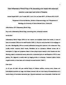
The polyps were pearly white in colour and soft to
CASE REPORT:
firm in consistency. Cut section of polyps showed
A 51year old multiparous female with menopause
mucin filled cystic spaces (Fig 1). Both adenexae
for 2 years, came with the complaints of white
were unremarkable.
discharge and something coming out from the
The microscopic examination of the polypoidal
vagina since one year. There was no history of
masses showed dilated glandular spaces lined by
menstrual irregularity. On examination a mass was
flattened to columnar mucin secreting epithelium
protruding through the vagina, which was mobile.
showing squamous metaplasia at places with fibro-
Cervical examination was not possible due to
muscular stroma (fig 2) and surface stratified
presence of the mass. There was no inguinal
squamous epithelial lining (Fig.3). There was no
lymphadenopathy. On abdominal ultrasonography
evidence of dysplasia or malignancy in the multiple
(USG) an ovarian mass just adjacent to bulky
sections taken from the polyps.
338 www.ijbamr.com P ISSN: 2250-284X , E ISSN : 2250-2858
Indian Journal of Basic and Applied Medical Research; June 2014: Vol.-3, Issue- 3, P. 338-343
DISCUSSION:
was done while in one case each radical th
Cervical polyps are most commonly seen in 4 &
hysterectomy and pan-hysterectomy was done
5 decade of life [2] and their incidence is 4–10%
because of clinical suspicion of malignancy and
of all cervical lesions [3]. Although they are mostly
endometrial hyperplasia respectively. In one case,
benign, carcinomatous change occurs in 1.7% of
the youngest one, exploratory laparotomy was done
cervical polyps [3]. Only fourteen cases (Table 1)
and polyp was resected through an incision in the
of giant cervical polyps have been reported so far
lower uterine segment because the polyp extended
and there is no case reported from India [1]. All the
into the endometrial cavity and the clinical impre-
previous cases are of single polyp except one case
ssion was rhabomyosarcoma. In the present case
in which there were two polyps but one of small
pan-hysterectomy was done because of suspicious
size (1cm.). The present case had three polyps and
ovarian tumour on USG, but ovaries were
is the first case of multiple giant cervical polyps. In
unremarkable in the specimen received.
th
previous cases age of presentation ranged from 5
The site of origin in ten out of fifteen giant
years to 61 years with mean age of 32 years [4].
cervical polyps (including present case) was
Most of the patients presented with leukorrhea,
ectocervix (Table 2), while in four cases it was
introital mass and variable per-vaginal bleeding. In
endocervix. In one case site of origin was not
one pregnant patient, the polyp protruded from the
reported.
external OS mimicking inevitable abortion [5]. The
CONCLUSION :
present case presented with leukorrhea and introital
To conclude giant cervical polyps are rare. They
mass. Out of thirteen previously reported cases,
can be multiple and polypectomy is sufficient to
two were post-menopausal and above 50 yrs in age
treat them. All the reported cases of giant cervical
and only 4 were parous. The present case is
polyps are benign and thought to be the result of
multiparous, postmenopausal and of above 50 yrs
reactive changes from long standing chronic
age. The possible diagnosis of mass protruding
inflammation. They are misdiagnosed as malig-
through
prolapsing
nancy and then pan-hysterectomy is resorted to. So
polyps,
proper knowledge of this entity and its clinical &
endocervical carcinoma, carcinosarcoma, products
USG presentation can save the patient from major
of conception and rhabdomyosarcoma [4].
surgery.
vaginal
submucous
introitus
fibroids,
include
endometrial
In nine out of thirteen previous cases polypectomy
Medworld-asia Dedicated for quality Research www.medworldasia.com
339 www.ijbamr.com P ISSN: 2250-284X , E ISSN : 2250-2858
Indian Journal of Basic and Applied Medical Research; June 2014: Vol.-3, Issue- 3, P. 338-343
Table 1. Clinical features of patients with giant cervical polyps [1, 4-15] Author
Age
Parity
Introital mass (duration)
Additional symptoms
Treatment
Saier et al [6]
61
0
+ (2 yrs)
Leukorrhea, PMB
Polypectomy
Lippert et al [7]
26
0
+
Leukorrhea, urinary retention
ARH+BSO+PLND
Duckman et al [8]
56
3
+
PMB
Polypectomy, D&C, TAH+BSO
Aridogan et al [9] Adinma [5]
17
0
+ (3 yrs)
Polypectomy
30
1
–
Bleeding, discharge 1st trimester bleeding for 2 wk
Branger et al [10] Gogus et al [11]
22
0
–
None
Polypectomy
5
0
–
Leukorrhea for 1 yr, bloody discharge for 1wk
Exploratory laparotomy, polyp resection
Khalil et al [12]
27
0
+ (2 days)
Mal-odorous discharge for 2 yr
Polypectomy, D&C
Amesse et al [13] Wu WY et al [4] Wu WY et al [4] Yi KW et al [14]
12
0
+ (1 mo)
None
Polypectomy
47
2
+(6 mo)
None
Polypectomy
45
0
+ (10 d)
Bleeding
Polypectomy
35
0
+
Vaginal bleeding, discharge
Polypectomy
Bucella D et al [15]
47
1
+
Bleeding
Polypectomy
Simavli S et al [1] Present case
46
Multipara
+
None
Electrosurgery
51
4
+(1 year )
White discharge
TAH
Polypectomy
PMB = postmenopausal bleeding; ARH = abdominal radical hysterectomy; BSO = bilateral salpingo-oophorectomy; PLND = pelvic lymph node dissection;D&C = dilatation and curettage; TAH = total abdominal hysterectomy.
340 339 www.ijbamr.com P ISSN: 2250-284X , E ISSN : 2250-2858
Indian Journal of Basic and Applied Medical Research; June 2014: Vol.-3, Issue- 3, P. 338-343
Table 2. Histopathologic features of giant cervical polyps [1, 4-15] Author
Polyp location
Size (cm)
Microscopic description Endocervical mucosa with squamous metaplasia Endocervical mucosa with squamous metaplasia
Diagnosis
Saier et al [6]
Ectocervix, right anterior lip
13 x 6
Lippert et al [7]
Ectocervix
17 x 12 x 4
Duckman et al [8]
Ectocervix, right posterior lip
10 x 3.5 x 1.8
Squamous mucosa with ulceration
Cervical polyp
Aridogan et al [9]
Ectocervix
14 x 4 x 3.5
Squamous mucosa with ulceration
Cervical polyp
Adinma [5] Branger et al [10]
External cervical os
5 x 1x 0.5
Not reported
Cervical Polyp
Not reported
15
Squamous mucosa with pseudo-papillary proliferations; chronic inflammation
Cervical polyp
5 x 4 x 1.5
Endocervical mucosa with squamous metaplasia Fibrovascular tissue with endocervical glands Squamous mucosa admixed with endocervical mucosa
Multiloculated endocervical polyp Giant cervical polyp
Gogus et al Endocervix, posterior midline [11]
Cervical polyp
Endocervical polyp
Khalil et al [12]
Endocervix, anterior lip
17 x 10 x 5
Amesse et al [13]
Ectocervix, right anterior lip
5.2 x 2.2 x 1.4
Wu WY et al [4]
Endocervix, posterior midline
7 x 2.5 x 1.5
Endocervical mucosa with squamous metaplasia
Endocervical polyp
Wu WY et al [4]
Ectocervix, posterior lip
5 x 2 x 0.7
Cervical polyp
Yi KW et al [14] Bucella D et al [15] Simavli S et al [1] Present case
Endocervical
12.6x8cm
Endocervical mucosa with focal squamous metaplasia Not reported
Ectocervix, posterior lip Ectocervix
5.5 cm in diameter 6x1,5cm
Focal squamous metaplasia Not known
Giant Cervical polyp Polypectomy
Ectocervix
Three polyps � 5x4x4 � 4x3x2.5 � 3x2.5 x 2
Endocervical glands with squamous metaplasia, lining epithelium squamous
Multiple giant cervical polyps
Cervical polyp
Cervical polyp
341 340 www.ijbamr.com P ISSN: 2250-284X , E ISSN : 2250-2858
Indian Journal of Basic and Applied Medical Research; June 2014: Vol.-3, Issue- 3, P. 338-343
Fig 1. Gross photograph of cut open uterus with multiple giant cervical polyps.
Fig 2. Microphotograph showing endocervical gland with focal squamous metaplasia and fibrovascular stroma in a giant cervical polyp ( H & E, 40X).
Fig 3. Microphotograph showing giant cervical polyp lined with stratified squamous epithelium ( H & E, 40X).
REFERENCES: 1.
Simavli S, Kinay T. Giant cervical polyp ; A case report and review of the literature. Turkiye Klinikleri J Gynecol Obst 2013;23(2):119-22.
2.
Danakas GT. Cervical polyps. In : Ferri FF. Ferri’s Clinical Advisor. Instant Diagnosis and Treatment. St. Louis : Mosby Inc, 2003:195.
3.
Israel SL. A study of cervical polyps. Am J Obstet Gynecol 1940;39:45-50.
4.
Wu WH, Sheu BC, Lin HH. Giant cervical polyps : Report of two cases and literature review. Taiwanese J Obstet Gynecol 2005;44(1):65-8.
5.
Adinma JI. Cervical polyp presenting as inevitable abortion. Trop Doct 1989;19:181.
6.
Saier FL, Hovadhanakul P, Ostapowicz F. Giant cervical polyp. Obstet Gynecol 1973;41:94-7.
7.
Lippert LJ, Richart RM, Ferenczy A. Giant benign endocervical polyp : report of a case. Am J Obstet Gynaecol 1974;118:1140-1.
8.
Duckman S, Suarez JR, Sese LO. Giant cervical polyp. Am J Obstet Gynaecol 1988;159:852-4.
9.
Aridogan N, Cetin MT, Kadayifci O, Atay Y, Bisak U. Giant cervical polyp due to a foreign body in a “virgin”. Aust NZ J Obstet Gynaecol 1988;28:146-7.
10. Branger C, Dreher E, Burkhardt A, Schmuckle U. Giant polyp of the cervix. Geburtshilfe Frauenheilkd
342 338 www.ijbamr.com P ISSN: 2250-284X , E ISSN : 2250-2858
Indian Journal of Basic and Applied Medical Research; June 2014: Vol.-3, Issue- 3, P. 338-343
1991;51:148-9. [In German] 11. Gogus S, Senocak ME, Arda IS, Buyukpamukcu N, Akcoren Z. Multilocular endocervical polyp in a five-year-old girl. Pediatr Pathol 1993;13:415-9. 12. Khalil AM, Azar GB, Kaspar HG, Abu Musa AA,Chararah IR, Seoud MA. Giant cervical polyp. A case report. J Reprod Med 1996;41:619-21. 13. Amesse LS, Taneja A, Broxson E, Pfaff-Amesse T. Protruding giant cervical polyp in a young adolescent with a previous rhabdomyosarcoma. J Pediatr Adolesc Gynecol 2002;15:271-7. 14. Yi KW, Song SH, Kim KA, Jung WY, Lee JK, Hur JY. Giant endocervical polyp mimicking cervical malignancy : Primary excision and hysterectoscopic resection. The Journal of Minimally Invasive Gynaecology. 2009;16(4):498-500. 15. Bucella D, Frederic B, Noel JC. Giant cervical polyp : A case report and review of a rare entity. Arch Gynaecol Obstet 2008;278:295-298.
Date of submission: 4 March 2014 Date of Publication: 22 June 2014
339 343 www.ijbamr.com P ISSN: 2250-284X , E ISSN : 2250-2858