Journal of Orthopaedic Surgery 2001, 9(1): 75–81
High cervical lipoma: A case report and review with classification Tomojiro Yamane, Ichiro Miyagawa, Akira Dezawa, Hiroto Mikami and Yukihiro Arai Department of Orthopaedic Surgery, Teikyo University School of Medicine, Tokyo, Japan
CASE REPORT ABSTRACT We experienced a 51 year-old female with high cervical lipoma who complained of neck pain and hypoaesthesia on her right lower extremity. She underwent surgery for removal of the tumour, but she was found unconscious after the surgery and then died after 19 days. Keywords: high cervical lipoma, spinal dysraphism, spina bifida occulta, spinal tumor, cervical spine
INTRODUCTION Lipomas of the spine are associated with or without spinal dysraphism. Lipoma unassociated with dysraphism is very rare, especially in high cervical spine,8 which is common at thoracic level4 while lipoma with spinal dysraphism in the lower lumbar spine is not infrequent.2 In the past lipoma unassociated with dysraphism was often an incidental finding at necropsy. With the advent of MRI there is increased opportunity to find such lipoma. A neurological deficit may have developed at birth or may develop later in aging.6 Some patients spend their lives unaware of having such as an anomaly in their spinal cord until symptoms appear such as foot deformity, dislocation of the hips, or bladder and bowel disturbance.
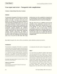
A 51 year-old housewife presented herself to our clinic with complaints of neck pain and hypoaesthesia of the right lower extremity. On neurological examination she showed slight hyperreflexia on the left upper and lower extremities, while the right side showed normal reflexes. Muscle testing demonstrated slight weakness of the right extremities as follows: Grip power was 16.5 kg in the right hand (right-handed) and 24.5 kg in the left hand. Deltoid, biceps, triceps, FCU, FCR, FDS and FDP in the right upper extremity were almost normal in motor power. However, quadriceps femoris, tibialic anterior and gastrocnemius on the right side were grade 4 in motor power. Muscles of the left upper and lower extremities revealed normal motor power. No clear segmental muscle atrophy was recognized. Subclinical radiculopathy was not confirmed, because needle EMG was not performed. Sensation to pain, temperature and touch was reduced on the right side of her extremities (Fig. 1). No bladder or bowel disturbance was recognized. Her clinical picture was characterized by slight myelopathy. The plain cervical radiographs showed no special findings at the high cervical level (Fig. 2). However, MRI of the cervical spine in the lateral view revealed a mass having the same intensity as subcutaneous fatty tissue continuing from the subcutaneous tissue to within the intraspinal canal (Fig. 3). The CTM also demonstrated the mass having isodensity to subcutaneous fatty tissue with laminar dysplasia of the atlas (Fig 4). Both MRI and CTM revealed compression on the posterior spinal cord to almost half
Address correspondence and reprint requests to: Dr Tomojiro Yamane, Department of Orthopaedic Surgery, Teikyo University School of Medicine, 2–11–1 Kaga, Itabashi, Tokyo 173–8605, Japan. E-mail:
[email protected]
76
Journal of Orthopaedic Surgery
Tomojiro Yamane et al.
its anteroposterior diameter by the tumor. But it did not seem that the spinal cord was markedly shifted to the anterior part of the canal, where the anterior subdural space still remained. The cerebrospinal fluid
showed no finding suggesting complete spinal canal occlusion. The patient was diagnosed as having lipoma associated with spina bifida occulta of Cl from the MRI and CTM findings.
Figure 1. The patient showed Brown-Squard type myelopathy.
Figure 2. Plain radiograms of the cervical spine showed no specific finding.
Vol. 9 No. 1, June 2001
High cervical lipoma: A case report and review with classification
77
Figure 3. MRI (T2 weighed) revealed the mass as high intensity subcutaneous fatty tissue which suggested lipoma.
Figure 4. CTM demonstrated low density tumor at C1-C2 levels with laminar dysplasia. The tumor compressed the cord and developed in the posterior space.
78
Tomojiro Yamane et al.
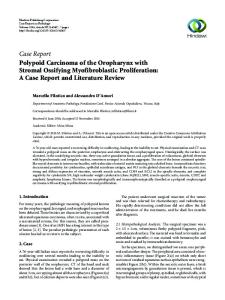
She underwent surgery for removing the tumour. The tumour was fatty tissue that invaded the dura mater of the spinal cord through the laminar defect of the atlas. After the dura was cut, we tried to separate the tumour from the spinal cord under microscopic magnification, but we were unable to remove the tumour completely because of adhesions. She never became conscious, showing anisocoria and absence of light reflex after the surgery. The brain stem infarct was suggested with brain CT, brain MRI and angiogram (Fig. 5). The CT, just after the accident, demonstrated edematous change in the brain stem, which was thought to be caused by embolism. She died 19 days after the surgery. Histopathological examination confirmed lipoma with mature cells. Postmortem examination was refused by the relatives.
DISCUSSION Frequent benign spinal tumours are neurinoma, meningioma and neurofibroma. Neurinoma is the most frequent intradural extraspinal tumor. Neurofibroma is sometimes seen as a dumbbell type tumour, while neurinoma mostly remains in the spinal
Journal of Orthopaedic Surgery
canal. Intradural lipoma accounts for about 1% of the primary intradural tumor. MRI is a very useful tool for differential diagnosis of the tumour. Generally neurinoma shows low intensity in T1 weighted image and relatively high intensity in T2 weighted image. Lipoma shows high intensity in T1 weighed image characteristically and also high intensity in T2 weighted image. Both the tumors develop their symptoms very slowly, sometimes after middle age. Giuffre’s review of 100 intraspinal lipoma cases revealed that the second and the third decade cases were 54.9% and the fifth decade cases were 16%.6 High cervical lipomas are rare. In lipoma of the spinal cord, it should be borne in mind that there are two conditions, i.e. associated with or without spinal dysraphism. They must be distinguished from each other. The theories of genesis are still controversial.2,6 Spinal lipoma is thought to be a primary tumour of the spine, that is, direct change into fat from the pericapillary mesenchymal cells. Lipoma with spinal dysraphism is subcutaneous adipose tissue, which is often found in the lower lumbar spine. However, those two conditions are sometimes difficult to differentiate from each other.
Figure 5. CT, just after the accident, demonstrated edematous change in the brain stem, suggesting embolism.
Vol. 9 No. 1, June 2001
High cervical lipoma: A case report and review with classification
Chapmann3 classifies lumbar spinal lipoma with tethered cord. However, there is no appropriate classification for high cervical lipoma. We reviewed the reports available for analyzing the details of lipoma. We classified the high cervical lipomas into three types according to the radiographs, MRI, CTM and the surgical findings that could be obtained from 18 cases in the world literature including our case (Fig. 6 and Table 1).1–13 Type I-a is lipoma without spinal dysraphism which is localized in the spinal canal. Type I-b is a kind of Type I that extends to the posterior cranial fossa. Type II-a is lipoma with spinal dysraphism which is connected with subcutaneous lipoma. Type II-b is a kind of Type II that extends to the posterior fossa. Type III is a type with or without spinal dysraphism where the lipoma is connected with a fibrous string to subcutaneous lipoma. In the high cervical lipomas that we reviewed, there were 9 cases with dysraphism and 9 cases without dysraphism. The numbers of cases in the classification were as follows: Type I-a — 5, Type I-b — 4, Type II-a — 4, which included our case, Type II-b — 3 and Type III — 3 (Fig. 6).
79
The operation was performed very carefully with a microscope and proceeded very smoothly. However, a sudden blood pressure drop and bradycardia was recognized once temporarily during the middle of the surgery. At that time the embolism might have occurred in the brain stem. We could not remove the lipoma completely because of tight adhesion of the lipoma to the cord. Many authors4,6,8 advise not to try to remove the tumor completely to prevent damage to the cord because of tight adhesion. On this point, the lipoma has the character of an intramedullary tumour , while neurinoma can be completely removed from the spinal cord. Fat and air embolism is not infrequent during anesthesia. Fat embolism occurs most frequently following surgery of long bone fractures. The following theories are believed: first, direct entry of marrow fat into the bloodstream, secondly, the loss of intravascular fat emulsion stability is involved and then coalescence of blood-borne lipids may occur, and, thirdly, venous air embolism may occur whenever venous pressure at the site of surgery is below atmospheric pressure. The third theory may be most applicable to our case.
Figure 6. Classification of high cervical lipoma in reviewing the literature.
80
Journal of Orthopaedic Surgery
Tomojiro Yamane et al. Table 1 References related to high cervical lipomas
Year
Author
Age
Level
Type
Duration
Op. method
Prognosis
Reference
1978
Watanabe
33M
C1-C3
II-a
12y
Part. Removal
No change
10,11
1983
Kusunoki
23M
C1-C6
III
19y
Subtotal rem.
No information
7,10
1991
Tauchi
52F
C1-C6
III
2y
Dura plasty
Improved
10
1918
Oppenheim
44M
C1-C4
I-a
13y
Subtotal rem.
Improved
2,4,6
1974
Drapkin
24M
C1-C7
I-a
24y
Subtotal rem.
Improved
4,5
1939
Jobotinshi
13M
C1-C4
I-a
13y
Necropsy
–
4,6
1972
Hubert
16M
C1-C4
I-a
16y
Subtotal excision
Dead
4
1964
Slooft
61F
C1-C6
I-a
14y
Subtotal removal
Alive-3yrs
4,6
1966
Beaudoing
6F
C1-C2
II-a
1/2y
–
Good
4
1953
Novotny
30M
C1-C6
II-a
14y
Decompression
Improved
2,4,6
1953
Wycis
48M
C1-C7
II—b
48y
Part. Removal
Improved
4,6
1938
Bucy
18M
C1-C7
II-a
18y
Subtotal removal
Improved
2,4,6
1989
Fan
18?
F.M.-C7
I-b
–
Subtotal removal
Improved
5
1987
McGillicuddy
24M
F.M.-C3
I-b
1.5y
Good
8
1985
Bertolino
62F
F.M.-C2
I-b
1986
Mori
7M
P.F.-C7
I-b
1983
White
11F
P.F.-C7
II-b
1920
Ritter
43M
C1-T1
II-b
–
1,5 At birth
Air embolism occurs at craniectomy and cervical laminectomy during neurosurgical procedures, particularly in the sitting position, although it is detected in orthopaedic hip replacement and abdominal surgical procedures in the prone, lateral or supine position. Michenfelder and his colleagues reported an incidence of 2.5% of 418 cases during occipital cranioectomy in the sitting position.9 Our case, who had no recognized heart problem before the surgery, had the surgery in the prone position with a
Part. Removal
Necropsy
Improved
10
Unchanged
13 13
slight head up position. But we could not clearly identify the reason why the brain stem embolism had occurred and we were unable to obtain post-mortem findings about the brain stem by a necropsy, but CT after the accident suggested oedema. We should always consider the possibility of air or fat embolism during cervical laminectomy and pay attention to a patient’s position during surgery. Surgical trauma certainly has to be considered in this case, despite the greatest of care during surgery.
REFERENCES 1. Bertolino GC, Cusmano F, Pichezzi P, Contini C, DeDonatis M, Pazza P, Bruschi G, Biassi P. Computed tomography of intradural cervical lipoma. Neuroradiology 1985; 27:184. 2. Bucy PC, Gustafson WA. Intradural lipoma of the spinal cord. Zentral blatt fur Neurochirurgie 1938; 6:341–349. 3. Chapmann PH Congenital Intraspinal Lipomas; Anatomic Consideration and Surgical Treatment. Child’s Brain 1982; 9:37– 47. 4. Drapkin, AJ. High cervical intradural lipoma. J Neurosurg 1974; 41:699–704. 5. Fan CJ, Veerapen RJ, Tan CT. Case report: Subdural spinal lipoma with posterior fossa extension. Clinical Radiology 1989; 40:91–4. 6. Giuffre R. Inraspinal spinal lipomas. Review of the literature (99 cases) and report of an additional case. Acta Neurochi 1996;14, 69–95. 7. Kusunoki S, Inoue K, Asano T. A case of cervical lipoma. J Neurological Medicine, Shinkei Naika 1983; 18:406–8.
Vol. 9 No. 1, June 2001
High cervical lipoma: A case report and review with classification
81
8. McGillicuddy,GT, Shucart W, Kwan ESK. Intradural spinal lipomas. Neurosurgery 1987; 21:343–6. 9. Michenfelder JD, Terry HR, Daw EF, Miller RH. Air embolism during neurosurgery: A new method of treatment. Anesthesia and Analgesia 1966; 45:390–95. 10. Mori K, Kamimura Y,Uchida Y, Kurisaka M, Eguchi S. Large intramedullary lipoma of the cervical cord and posterior fossa. J. Neurosurg 1986;64:974–6. 11. Tauchi T, Mochizuki M, Murakami M, Sodeyama T, Kobayashi Y, Minami S, Goto S, Kitahara H, Moriya H, Watanabe T. Cervical spinal lipoma: A case report. J Orthopedic Trauma Kanto Seikeisaigaigeka 1993; 24:580–4. 12. Watanabe S. Iwata K, Ando Y, Okabe Y. A case of the upper spinal cord and medulla oblongata lipoma. J Clinical Neurology 1978; 18:436 13. White WR, Fraser RAR. Cervical spinal cord lipoma with extension into the posterior fossa. Neurosurgery 1983; 12:460–2.