Controversies and Considerations in the Diagnosis of Primary Cutaneous CD4þ Small/Medium T-Cell Lymphoma Thanh T. Ha Lan, MD; Noah A. Brown, MD; Alexandra C. Hristov, MD
� Context.—Primary cutaneous CD4þ small/medium T-cell lymphoma is a provisional and controversial entity with a broad differential diagnosis. Despite being an uncommon lymphoma, it is a frequent diagnostic consideration in cutaneous biopsies with a dense lymphoid infiltrate because it shows overlapping features with reactive lymphoid hyperplasia (pseudolymphoma) and a variety of other primary cutaneous and systemic lymphomas. However, proper classification of this process is important for determining patient prognosis and treatment options. Objectives.—To review the clinical, morphologic, immunophenotypic, and genetic features of primary cutaneous CD4þ small/medium T-cell lymphoma and contrast those features with entities in the differential diagnosis. Data Sources.—Applicable literature will be reviewed
T
he concept of primary cutaneous CD4þ small/medium Tcell lymphoma (PC-SMTCL) began in the early 1990s when the updated Kiel classification system1,2 was applied to primary cutaneous T-cell lymphomas that did not meet criteria for mycosis fungoides (MF) or Sezary ´ syndrome.3,4 These early studies established that lymphoid infiltrates with predominantly small, pleomorphic T-cells had a more favorable prognosis than those with predominantly large, pleomorphic T-cells.1,3–5 Although cases with mediumsized, pleomorphic T-cells were not initially classified consistently,1,3,4 eventually infiltrates with small- to medium-sized, pleomorphic T cells were grouped together as a single clinicopathologic entity. Because of the association of Accepted for publication June 6, 2014. From the Departments of Pathology (Drs Lan, Brown, and Hristov) and Dermatology (Drs Lan and Hristov), University of Michigan Health System, Ann Arbor. The authors have no relevant financial interest in the products or companies described in this article. This material was presented as part of the ‘‘Dermatopathology Greatest Hits’’ short course at the annual meeting of the United States and Canadian Academy of Pathology; March 7, 2013; Baltimore, Maryland. Also presented in part at the New Frontiers in Pathology: An Update for Practicing Pathologists meeting; University of Michigan; September 26–28, 2013. Reprints: Alexandra C. Hristov, MD. Department of Pathology, University of Michigan Health System, Medical Science I, 1301 Catherine St, M-3261, Ann Arbor, MI 48109 (e-mail: ahristov@med. umich.edu). Arch Pathol Lab Med—Vol 138, October 2014
with emphasis on current controversies and distinguishing characteristics. Conclusions.—Although many consider primary cutaneous CD4þ small/medium T-cell lymphoma to be indistinguishable from reactive lymphoid hyperplasia/ pseudolymphoma, it can be differentiated from other primary cutaneous and systemic lymphomas. Patients with solitary lesions of primary cutaneous CD4þ small/medium T-cell lymphoma generally have an excellent prognosis. Nevertheless, a subset of patients who have been reported to meet criteria for this lymphoma have followed a moreaggressive course; however, those patients show some differing clinical, morphologic, and immunophenotypic features. (Arch Pathol Lab Med. 2014;138:1307–1318; doi: 10.5858/arpa.2014-0299-CC) some CD8þ cases with a significantly worse prognosis,6,7 CD8þ cases were excluded, and PC-SMTCL became a provisional lymphoma in the current World Health Organization (WHO)–European Organization for Research and Treatment of Cancer classification system.7,8 Notably, many experts now allow for CD8þ variants of PC-SMTCL.9,10 At approximately the same time, there were immunophenotypic and genetic investigations into a group of cutaneous lesions with a dense lymphoid infiltrate that were classified as reactive lymphoid hyperplasia or cutaneous pseudolymphoma. These pseudolymphomas had a concerning histopathologic appearance but a benign clinical course; no loss of CD2, CD3, or CD5; and no clonal T-cell population, features that were thought to distinguish them from true lymphomas.11–13 However, as cases of pseudolymphoma were further examined, examples were identified that showed loss of pan–T-cell markers and a clonal T-cell population (as reviewed in Ploysangam et al14). Currently, the distinction between PC-SMTCL and pseudolymphomas, if any, is uncertain, and the precise classification and clinicopathologic features of PC-SMTCL remain topics of ongoing research and debate. Here, we review the current clinicopathologic features described for PC-SMTCL, and we compare and contrast those features with pseudolymphoma and a variety of other primary cutaneous and systemic lymphomas, many with significantly more-aggressive clinical behavior (Table). Despite its broad differential diagnosis, accurate recognition of Cutaneous Small/Medium T-Cell Lymphoma—Lan et al 1307
Summary of Features Supporting a Diagnosis of Primary Cutaneous CD4þ Small/Medium T-Cell Lymphoma (PC-SMTCL) or Entities in Its Differential Diagnosis Features
PC-SMTCL
Pseudolymphoma
PCMZL
Skin lesions Cell size T cell or B cell predominant
Usually solitary Small/medium Variable
Usually solitary Small/medium Variable
Solitary to multiple Small/medium Variable
Epidermotropism Secondary follicles
� �
� þ
Loss of pan–T-cell markers T-cell clone B-cell clone PD1þ cells
þ/ � þ/ � � þ Scattered cells and pseudorosettes
Other differentiating features
� �
� /þ � /þ � þ Scattered cells and pseudorosettes12 or scattered, small cells22 � Spontaneous resolution or resolution without aggressive/destructive therapy
� þ Colonization � � þ � Positive cells within reactive follicles
No clinical resolution TFH phenotype, less commonly CD10þ
� � � � �
Monocytoid B cells Sheets of plasma cells Dutcher bodies CD43þ mature B cells Light-chain restriction
Abbreviations: AITL, angioimmunoblastic T-cell lymphoma; ATLL, adult T-cell leukemia lymphoma; MF, mycosis fungoides; PCFHTCL, primary cutaneous follicular helper T-cell lymphoma; PCMZL, primary cutaneous marginal zone lymphoma; PTCL-NOS, peripheral T-cell lymphoma, not otherwise specified; TFH, follicular helper T-cells.
PC-SMTCL is important for patient prognosis and treatment options. CLINICAL, MORPHOLOGIC, AND IMMUNOPHENOTYPIC FEATURES Primary cutaneous CD4þ small/medium T-cell lymphoma represents approximately 2% to 3% of all cutaneous T-cell lymphomas.7,8 Although it affects individuals with a wide age range, including children,10,12,15�18 most patients are older adults with an average age in the 50s to 60s and without a clear sex predilection.3,4,7,10,12,15–21 Patients typically present with a solitary, asymptomatic lesion on the face, neck, upper trunk, or upper extremities, which may appear as a violaceous papule, plaque, nodule, or tumor.* Some early series, before PC-SMTCL was a well-defined entity, reported a relatively high frequency (50%–74%) of multiple lesions in a localized or generalized distribution.3,4,6,19,24 However, recent studies describe a single lesion for most patients.† Pain or pruritus has been occasionally noted.16,17,19,21,23 The clinical course for these lesions is variable. They may appear suddenly, enlarge rapidly over a few months, slowly grow over years, persist with minimal change, wax and wane over a course of weeks to years, or spontaneously resolve.3,4,6,16,17,19–21,23 Ulceration has been described.7,10,18�21 Unusual clinical presentations include alopecia,16 poikilodermic plaques,22 pigmented purpura,21 and an association with annular elastolytic giant cell granuloma.25 By definition, patients should be free of a background of patches and plaques typical of MF.7,8 Lesions of PC-SMTCL are composed of a mixed hematolymphoid infiltrate with a bandlike, nodular, or sheetlike distribution (Figure 1, A through C). Frequently, the infiltrate extends into the subcutaneous fibroadipose tissue, and it may also involve skeletal muscle, vessels, and adnexal structures, sometimes with invasion and destruction * References 7, 8, 10, 15, 17, 18, 20–23. † References 7, 8, 10, 12, 15, 17, 20�23. 1308 Arch Pathol Lab Med—Vol 138, October 2014
of vessels and adnexae.4,6,7,10,15–22,26 Lymphoid cells are most often separated from the epidermis by a grenz zone, but focal epidermotropism may be seen.3,4,7,8,19,20,22 Notably, prominent epidermotropism should prompt consideration of a diagnosis of MF. As the name implies, most cells in PCSMTCL are small- to medium-sized, pleomorphic lymphocytes. They are usually found in association with numerous, admixed inflammatory cells, including small lymphocytes, plasma cells, eosinophils, and histiocytes.‡ Granulomatous inflammation may be present.10,12,15,18,19,22 Notably, secondary lymphoid follicles have not been described in association with this lymphoma.17,21,22 A subset of large, atypical, pleomorphic cells may be present, but they make up less than 30% of the infiltrate by definition.4,7,8 Importantly, similar lymphoid proliferations with more than 30% large Tcells are better classified as primary cutaneous peripheral Tcell lymphoma, unspecified/not otherwise specified and have a significantly worse prognosis with a 5-year survival of less than 20%.4,6,7 Primary cutaneous CD4þ small/medium T-cell lymphoma is a neoplasm of CD3þ/CD4þ T cells that do not express CD30 or cytotoxic markers (Figure 2, A through G).7,8 Loss of one or more pan–T-cell markers, most commonly CD7, is noted in 13% to 90% of cases.4,10,17,19–21,23 Intermixed CD20þ B cells are often numerous (10%–60% of the infiltrate),10,17,20–23 and intermixed plasma cells are typically polytypic.8,17,20 CD8þ T cells generally make up a substantial minority of cells (5%–47%).10,15,17,18,20–23 Scattered CD30þ immunoblasts are usually identified.10,12,15,18,21,22 Thus, immunophenotypic studies confirm the mixed hematolymphoid infiltrate seen morphologically in this lymphoma. Although PC-SMTCL generally has a low Ki-67 proliferation index, usually ranging from 5% to 30%,10,17,18,20,21,23,26 in some cases it may be as high as 50% to 70%.21,23 Consistently, PC-SMTCL is negative for Epstein-Barr virus.6,16,20�22 A clonal T-cell receptor gene rearrangement ‡ References 6, 10, 12, 15, 17, 18, 20, 22. Cutaneous Small/Medium T-Cell Lymphoma—Lan et al
Extended PTCL-NOS
AITL
Multiple Variable T cell predominant
MF
Multiple Medium/large T cell predominant
Multiple Variable T cell predominant
Multiple Medium/large Variable
þ �
� �
Multiple Variable T cell predominant, numerous B cells � �
þ �
� �
þ þ � þ/ � Extensive/diffuse expression when positive � Long-standing patches and plaques characteristic of MF
þ þ � � /þ
þ þ � þ
þ þ � þ
Not reported þ � þ
�
�
�
.30% large cells; and/or Evidence of systemic disease
� � � �
TFH phenotype, commonly CD10þ Vascular proliferation Perivascular infiltrate Vasculitis EBVþ
is detected in 54% to 100% of cases,10,15,17–23,26 but immunoglobulin heavy-chain (IgH) rearrangements are polyclonal.8,17,21,22 The recent demonstration that the neoplastic CD4þ Tcells in PC-SMTCL coexpress PD1, BCL6, CXCL13, and ICOS has provided valuable insights into the potential derivation of PC-SMTCL and its microenvironment.12,22,23 PD1, or programmed death 1, is expressed on cell membranes of follicular helper T-cells (TFH) found in reactive germinal centers, as well as activated T cells. PD1 appears to negatively regulate T-cell function and immune response, creating an atmosphere of immune tolerance and helping neoplastic cells to evade the host’s immune surveillance.12,27,28 BCL6, CXCL13, and ICOS are important mediators of germinal center development.28 Although CXCL13 (chemokine [C-X-C motif] ligand 13) has an important role in the homing and entry of B cells into germinal centers, BCL6 is required for the differentiation of TFH and germinal-center B cells, and it is involved in B-cell proliferation.28–30 ICOS, or inducible Tcell costimulator, is required for germinal-center formation and TFH differentiation.30 The expression of these markers indicates that the tumor cells in PC-SMTCL show a TFH phenotype. Because TFH cells are required for germinal-center formation and stimulate B-cell proliferation and differentiation (as reviewed in Crotty30), this phenotype may help to explain the numerous background B cells in this T-cell lymphoma.22 In support of this hypothesis, enlarged CD4þ T cells expressing PD1, BCL6, and CXCL13 form pseudorosettes around CD20þ and CD30þ immunoblasts in PC-SMTCL.22 These markers highlight a few lymphocytes in PC-SMTCL (usually ,30%) as scattered cells, small aggregates, and pseudorosettes.22,23 They also provide additional immunophenotypic support for the diagnosis of PC-SMTCL as they are expressed in PC-SMTCL, but are less common in MF and peripheral T-cell lymphoma, not otherwise specified.12,21,22,31,32 In addition, PD1þ cells are infrequent in dermatitis, including drug reactions.23 Interestingly, CD10, another TFH marker, is uncommonly positive in PCSMTCL.12,22,23 Arch Pathol Lab Med—Vol 138, October 2014
ATLL
� �
HTLV-1 Extracutaneous disease
PCFHTCL
� � �
TFH phenotype Rapidly developing lesions Extracutaneous clonal T cells, yet indolent clinical course
The prognosis for PC-SMTCL is favorable, with 5-year survival rate of 60% to 100%.7,8,10,20,24,26 In particular, patients with one to few localized lesions have an excellent prognosis,§ prompting some to consider PC-SMTCL to be a form of reactive lymphoid hyperplasia (T-cell pseudolymphoma) rather than a true lymphoma. Indeed, patients with limited disease are often ‘‘cured’’ with surgical excision or radiation therapy, although recurrences may occur.7,8,10,17,20–23 Importantly, however, some studies have reported a subset of patients who meet diagnostic criteria for PC-SMTCL but who follow a significantly more-aggressive course.4,6,20 In one such study, Garcia-Herrera et al20 reported 5 patients with a fatal outcome and a median survival of 23 months, despite multiagent chemotherapy in 4 patients. However, many of those patients with a worse prognosis have had distinguishing features. In particular, rapidly developing lesions,20,21 multifocality,3,6,21 larger lesion size (.5 cm),20,21 higher proliferation indices,20,21 diminished expression of CD4,20 and more-monotonous infiltrates with significantly decreased numbers of background inflammatory cells, including infiltrating B cells and CD8þ T cells,20,21 have been associated with a poorer outcome.8,20 Further studies are needed to more fully understand aggressive cases. Their differing clinical and pathologic features suggest that they may best be categorized separately in the future. DIFFERENTIAL DIAGNOSIS Reactive Lymphoid Hyperplasia/T-Cell Pseudolymphoma Reactive lymphoid proliferations in the skin are heterogeneous and, unfortunately, lack precise diagnostic criteria. The term pseudolymphoma is not a diagnosis suis generis, but rather refers to a reactive process that clinically and/or pathologically simulates lymphoma.14,33,34 It has been defined as a ‘‘reactive, polyclonal, benign lymphoproliferative process’’ and may be localized or disseminated.35 It includes a heterogeneous group of bandlike, nodular, or diffuse proliferations of B cells and/or T cells that are § References 7, 8, 10, 12, 17, 18, 20, 22, 23, 26. Cutaneous Small/Medium T-Cell Lymphoma—Lan et al 1309
morphologically worrisome for lymphoma.11 Despite a concerning histopathologic appearance, pseudolymphomas ultimately behave in a clinically benign fashion: They are not associated with systemic involvement, and they eventually resolve, either spontaneously, after removal of an identified, initiating agent or with nonaggressive therapy (such as topical or intralesional steroids).13,14,20,33–35 The underlying cause of most cases of T-cell–rich pseudolymphomas is idiopathic.11,14,34 Common known causes include lymphomatoid drug reaction,14,34,36–41 lymphomatoid contact dermatitis,14,33,42–45 and persistent, nodular, arthropod-bite reaction,14,33 among others. Morphologically, reactive lymphoid infiltrates in the skin (pseudolymphomas) may display a marked similarity to PCSMTCL.8,10,12,15,17,20–22 Indeed, most consider PC-SMTCL to be indistinguishable clinically and pathologically from pseudolymphoma and have advocated that these proliferations be descriptively termed cutaneous nodular proliferation of pleomorphic T lymphocytes of undetermined significance,10 solitary small- to medium-sized pleomorphic T-cell nodules of undetermined significance,18 spectrum of pseudo–T-cell lymphoma/PC-SMTCL,12 or lymphoid infiltrate of uncertain nature9 until we more fully understand the nature of these lesions. Pseudolymphomas may be composed of a predominant population of reactive, small to medium CD4þ T cells, often with admixed polyclonal B-cells, plasma cells, eosinophils, and histiocytes (Figure 3, A through E).8,11,13,14,33–35 Unfortunately, there are no precise morphologic criteria to differentiate pseudolymphoma from PC-SMTCL. However, pseudolymphomas may include reactive, secondary follicles, 13,35 which have not been described for PCSMTCL.20,22,35 Given the morphologic similarities, immunohistochemistry and molecular techniques are often used to help support a diagnosis of PC-SMTCL.9,11,17,20,35 Loss of pan–Tcell markers, including CD2, CD5, and CD7, and a clonal T-cell receptor gene rearrangement help to favor a diagnosis of PC-SMTCL, although they are not present in every case.** Conversely, pseudolymphomas may show loss of CD7 and, less-commonly, CD5,13,14,34 as well as clonal T-cell receptor gene rearrangements.14 Interestingly, PD1þ and CXCL13þ cells are also present in pseudolymphomas and in inflammatory infiltrates, including dermatitis.12,22,23 However, in many of these inflammatory processes, the TFH cells tend to be small, fewer in number, and do not form pseudorosettes.22,23 In addition, in cases with reactive follicles, these TFH cells tend to be confined to germinal centers.22 In summary, PC-SMTCL and pseudolymphoma share overlapping features, and the current distinction between the 2 requires the analysis of clinical, morphologic, immunophenotypic, and genetic data.7,8,10,17,20,21 Primary Cutaneous Marginal Zone Lymphoma þ
Figure 1. Primary cutaneous CD4 small/medium T-cell lymphoma. A 57-year-old man presented with a posterior neck mass that enlarged for several months. A, There is a patchy, dermal hematolymphoid infiltrate that is separated from the epidermis by a grenz zone. B, Lymphoid cells are predominantly small to medium and display irregular nuclear contours. C, There are intermixed histiocytes and plasma cells (hematoxylin-eosin, original magnifications 340 [A], 3400 [B], and 3200 [C]).
The presence of numerous B cells and plasma cells in PC-SMTCL may prompt consideration of primary cutaneous marginal zone lymphoma (PCMZL) (Figure 4, A through F).10,17,21 Primary cutaneous marginal zone lymphoma, a variant of extranodal marginal zone lymphoma that is limited to the skin at diagnosis, is a low-grade B-cell lymphoma with a favorable prognosis (5-year survival of 90%–100%).7 Clinically, it presents similar to PC-SMTCL ** References 6–9, 11, 14, 17, 20, 21, 35.
1310 Arch Pathol Lab Med—Vol 138, October 2014
Cutaneous Small/Medium T-Cell Lymphoma—Lan et al
Figure 2. Primary cutaneous CD4þ small/medium T-cell lymphoma. The lymphoid cells are predominantly CD3þ (A) and CD4þ (B) T cells and show some loss of CD7 (C). There are few CD8þ (D) and CD20þ (E) cells. F and G, PD1 marks scattered cells, including rosettes. A T-cell receptor gene rearrangement study revealed a clonal T-cell population (data not shown) (original magnification 340 [A through F]; original magnification 3400 [G]).
Arch Pathol Lab Med—Vol 138, October 2014
Cutaneous Small/Medium T-Cell Lymphoma—Lan et al 1311
Figure 3. T-cell–rich reactive lymphoid hyperplasia/pseudolymphoma. Sections show a dense, dermal lymphoid infiltrate with a patchy distribution that is separated from the epidermis by a grenz zone (A) and includes scattered reactive lymphoid follicles (B). Higher magnification reveals mostly small- to medium-sized lymphocytes with irregular nuclear contours and admixed histiocytes, eosinophils, and plasma cells (C). Lymphoid cells include a mixture of CD3þ T-cells (D) and CD20þ B-cells (E). Gene rearrangement studies for both B-cell and T-cell receptors were polyclonal (data not shown) (hematoxylin-eosin, original magnifications 340 [A], 3100 [B], and 3600 [C]; original magnification 3100 [D and E]).
as one to multiple, red to violaceous papules, plaques, or nodules on the trunk or extremities in an older patient.7,46 Primary cutaneous marginal zone lymphoma may also show an architecture similar to that of PC-SMTCL, with a 1312 Arch Pathol Lab Med—Vol 138, October 2014
dermal to subcutaneous collection of atypical lymphoid cells that are separated from the epidermis by a grenz zone. Cytologically, the neoplastic lymphocytes in PCMZL include variable combinations of centrocyte-like (marginal Cutaneous Small/Medium T-Cell Lymphoma—Lan et al
Figure 4. Primary cutaneous marginal zone lymphoma. A patchy, dense lymphoid infiltrate replaces much of the dermis (A) and is composed of small- to medium-sized lymphocytes with irregular nuclear contours (B). Plasma cells are found in sheets (C) and include Dutcher bodies (D). Reactive follicles (E) and monocytoid B cells (F) are typical (hematoxylin-eosin, original magnifications 340 [A and E], 3400 [B, C, and F], and 3600 [D]).
zone) B cells; lymphocytes with relatively abundant, pale cytoplasm, imparting a monocytoid appearance (monocytoid B cells); and cells with plasmacytic differentiation.7 In many cases, the marginal zone cells predominate and are Arch Pathol Lab Med—Vol 138, October 2014
morphologically similar to the neoplastic lymphocytes in PC-SMTCL, showing a small to medium size and irregular nuclear contour.46,47 Additionally, there may be background histiocytes and eosinophils.46 Finally, both lymCutaneous Small/Medium T-Cell Lymphoma—Lan et al 1313
Figure 5. Primary cutaneous marginal zone lymphoma. The lymphoid cells include a near equal mixture of CD3þ T cells (A) and CD79aþ B cells/ plasma cells (B). Plasma cells are j restricted (C) with few k-positive cells present (D) (original magnification 340 [A through D]).
phomas may show near-equal mixture of B cells and T cells. Primary cutaneous marginal zone lymphomas typically have numerous, intermixed T cells (50%–75% of cells),48 whereas PC-SMTCL often includes numerous background B cells (10%–60% of cells; Figure 5, A and B).10,17,20–23 Despite the similarities, several features help to distinguish PC-SMTCL from PCMZL. The presence of sheets of plasma cells, often located at the periphery of the infiltrate, and Dutcher bodies favors PCMZL.7,46 Similarly, reactive lymphoid follicles are often seen in PCMZL and may be colonized by neoplastic lymphocytes,46 but follicles have not been reported in PC-SMTCL, and follicular colonization is not identified.17,21 In addition, PCMZL frequently shows light-chain restriction (Figure 5, C and D)9 and may show CD43 coexpression by mature B cells.47 Finally, a clonal IgH gene rearrangement also supports a diagnosis of PCMZL.7,9,21,46,47 In contrast, B cells and plasma cells within the reactive background of PC-SMTCL are typically polyclonal by immunohistochemical or in situ hybridization assessment of j and k and by B-cell receptor gene rearrangement studies.13,21,22 In addition, PC-SMTCL includes a clonal T-cell population that expresses TFH markers 1314 Arch Pathol Lab Med—Vol 138, October 2014
(PD1, BCL6, ICOS, CXCL13, and sometimes CD10). Although PD1- and CXCL13-expressing T cells are present in PCMZL B-cell lymphoma, the cells are largely limited to reactive follicles.22,48 Mycosis Fungoides Although the patch and many-plaque stages of MF can be easily distinguished from PC-SMTCL because of the presence of significant epidermotropism, some plaque and many-tumor stage MF lesions may histopathologically be confused with PC-SMTCL when clinical information is lacking because these more-advanced stages often do not include epidermotropism.7,17,21,49 Tumor-stage MF appears clinically as solitary to multiple, discrete lesions of 1 cm or greater (Figure 6, A).50 These lesions are characterized by a dense, dermal infiltrate of small- to medium-sized CD4þ T cells with variable loss of pan–T-cell markers (Figure 6, B and C). Despite the histopathologic similarity, clinical features help to distinguish these 2 processes. Patients with plaque- and/or tumor-stage MF have a long history of patches and plaques characteristic of MF, whereas PCSMTCL arises de novo.7,21,50 In addition, the neoplastic cells in MF are less likely to express PD1 and tend to show more Cutaneous Small/Medium T-Cell Lymphoma—Lan et al
Peripheral T-Cell Lymphoma, Not Otherwise Specified Peripheral T-cell lymphoma, not otherwise specified (PTCL-NOS) includes a heterogeneous group of lymphomas that, by definition, do not meet criteria for other categories of T-cell lymphoma.7,52 Thus, these lymphomas lack clearly defined clinical, histopathologic, and immunophenotypic characteristics. Peripheral T-cell lymphomaNOS may be primary to the skin (primary cutaneous PTCL-NOS) or may secondarily involve the skin. Importantly, the prognosis for primary or secondary cutaneous PTCL-NOS is dismal, with a 5-year survival rate less than 20%.4,6,7,12 Like PC-SMTCL, primary cutaneous and secondary cutaneous PTCL-NOS usually present in older patients as nodules or tumors. Histopathologically, they are characterized by a nodular or diffuse, dermal to subcutaneous infiltrate of CD4þ T cells, with little or no epidermotropism and a variable, mixed, inflammatory background (Figure 7, A through E).6 However, primary and secondary cutaneous PTCL-NOS more commonly presents with multiple lesions.4,6,7 In addition, primary cutaneous PTCL-NOS includes more than 30% large T cells4,7,8 and generally does not express PD1,12 features that distinguish it from PC-SMTCL. Patients with secondary cutaneous involvement by PTCL-NOS have evidence of extracutaneous disease, more commonly have a predominance of large neoplastic cells6 and less commonly show a TFH immunophenotype.12,32 Angioimmunoblastic T-Cell Lymphoma Angioimmunoblastic T-cell lymphoma is a peripheral Tcell lymphoma of follicular helper T cells that is associated with Epstein-Barr virus and frequently includes cutaneous manifestations. Patients are typically older and may present with skin papules or nodules composed of a diffuse dermal infiltrate of small- to medium-sized cells.53 Like PC-SMTCL, angioimmunoblastic T-cell lymphoma shows a CD4þ TFH immunophenotype, often with loss of pan–T-cell markers, and includes numerous background B cells, CD8þ T cells, and perivascular eosinophils.54,55 However, angioimmunoblastic T-cell lymphoma often presents as a diffuse, pruritic, maculopapular eruption and shows a perivascular infiltrate, vascular proliferation, and/or vasculitis on biopsy,53,55 features that are not typical of PC-SMTCL. In addition, angioimmunoblastic Tcell lymphoma may include Epstein-Barr virus–positive cells53,55 and commonly expresses CD10, including in extranodal sites.53 Finally, patients with angioimmunoblastic T-cell lymphoma have evidence of systemic disease and typically follow an aggressive course with a 5-year survival less than 30%.53–55 Figure 6. Tumor stage mycosis fungoides (MF). A 36-year-old man with a 15-year history of MF. A, Multiple tumors are found in a background of patches and plaques. B, The dermis has been virtually entirely replaced by a lymphoid infiltrate that does not enter the epidermis. C, Lymphocytes are small to medium, and there are background histiocytes (hematoxylin-eosin, original magnifications 340 [B] and 3400 [C]).
extensive to diffuse PD1 expression when positive.12,31,51 Moreover, other TFH markers, including BCL6, CXCL13, and ICOS are uncommonly positive in MF.51 Importantly, PD1 expression alone does not define TFH and may be seen with T-cell activation.31,51 Arch Pathol Lab Med—Vol 138, October 2014
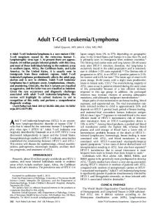
Adult T-Cell Leukemia/Lymphoma Adult T-cell leukemia lymphoma (ATLL), a T-cell lymphoma caused by the human T-cell leukemia virus type I, has a broad morphologic, immunophenotypic, and clinical spectrum. Although most patients present with disseminated disease by highly pleomorphic T-cells, ATLL may present similarly to PC-SMTCL as a primary cutaneous, dermal or subcutaneous infiltrate of small- to medium-sized CD4þ T-cells that form a clinical papule, plaque, or tumor.7,56–58 Epidermotropism may be limited or absent,56,58 and histiocytes and admixed granulomas may be prominent.57 In addition, the T cells in ATLL Cutaneous Small/Medium T-Cell Lymphoma—Lan et al 1315
Figure 7. Peripheral T-cell lymphoma, not otherwise specified (PTCL-NOS). An 81-year-old man with a history of nodal PTCL-NOS presented with a nodule on his left arm. Biopsy revealed a patchy dermal infiltrate (A) that included numerous small- to medium-sized lymphocytes with irregular nuclear contours, histiocytes forming granulomas, eosinophils, and plasma cells (B and C). D and E, A lymph node biopsy performed a year earlier revealed diffuse effacement by a morphologically similar T-cell infiltrate. T-cell receptor gene rearrangement analysis showed an identical clone in the skin and lymph node (data not shown) (hematoxylin-eosin, original magnifications 320 [A and D], 3200 [B], and 3400 [C and E]).
often lack CD7,56,58 and they commonly express PD1.57,59 Although these features overlap with PC-SMTCL, the presence of multiple lesions,58 systemic disease, and human T-cell leukemia virus type I infection help to 1316 Arch Pathol Lab Med—Vol 138, October 2014
favor ATLL. Although prognosis depends on clinical subtype/disease extent, patients with ATLL and cutaneous disease generally have a significantly worse prognosis than do those with PC-SMTCL.57 Cutaneous Small/Medium T-Cell Lymphoma—Lan et al
Primary Cutaneous Follicular Helper T-Cell Lymphoma Primary cutaneous follicular helper T-cell lymphoma (PCFHTCL) is a recently proposed lymphoma with few reported cases.60 This category was suggested by Battistella et al,60 who described 5 cases of primary cutaneous T-cell lymphoma with follicular helper phenotype. These cases were considered morphologically and immunophenotypically similar to the recently described nodal PTCL showing follicular growth and TFH phenotype (follicular PTCL).52,61 Similar to PC-SMTCL, most of the patients with PCFHTCL were older than 50 years (4 of 5; 80%) and presented with erythematous to violaceous papules, plaques, and nodules.9 However, unlike PC-SMTCL these lesions were multiple in most cases (4 of 5, 80%) and rapidly evolved during a few months (5 of 5; 100%). Morphologically both PC-SMCTL and PCFHTCL include a dermal, nonepidermotropic, lymphoid infiltrate with TFH immunophenotype and numerous associated B cells, and both lack Epstein-Barr virus and CD30. However, the neoplastic cells in PCFHTCL are described as medium to large, although presence of numerous background, small lymphocytes could cause morphologic overlap with PC-SMTCL. PC-SMTCL also tends to have a higher Ki-67 proliferation index than is typical for PC-SMTCL (30%–50%). PCFHTCL commonly has evidence of extracutaneous disease; many of these patients had a clonal T-cell population in the peripheral blood (4 of 5; 80%), lymph node (1 of 5; 20%), and bone marrow (2 of 5; 40%), in addition to the skin (5 of 5; 100%). In addition, patients with PCFHTCL showed resistance to multiagent chemotherapy, despite an overall indolent course, with no deaths after 5 years.60 Interestingly, PCFHTCL and follicular PTCL may mimic follicular B-cell lymphomas.60,61 Some have suggested that at least a subset of cases of PCFHTCL share overlapping features with angioimmunoblastic T-cell lymphoma.23 Given the few reported cases, it seems reasonable that further studies will be necessary to fully establish this proposed primary cutaneous lymphoma and to delineate it from others with a TFH phenotype.9,23 Other Considerations Although subcutaneous panniculitis-like T-cell lymphoma could also be considered in the differential diagnosis, its preferential subcutaneous localization, normally without dermal involvement, and its CD8þ cytotoxic T-cell phenotype help to distinguish this lymphoma.7,62 Similarly, lymphomatoid papulosis and other CD30þ lymphoproliferative disorders usually have more-numerous CD30þ cells that are also generally larger and more atypical than those seen in PC-SMTCL.7,62,63 Moreover, the neoplastic cells in CD30þ lymphoproliferative disorders do not have a TFH immunophenotype.12,22 CONCLUSION Our understanding of PC-SMTCL remains incomplete. Although it shares many overlapping features with cutaneous reactive lymphoid hyperplasia/pseudolymphoma, the extent of the infiltrate, cytologic atypia, and the frequent presence of immunophenotypic abnormalities, T-cell clonality and clinical persistence have led to the provisional classification of this process as a lymphoma. The differential diagnosis for PC-SMTCL includes a variety of other primary cutaneous and systemic lymphomas, some with a significantly worse prognosis. Given its indolent nature, accurate Arch Pathol Lab Med—Vol 138, October 2014
recognition of PC-SMTCL is important for prognostication and to prevent overtreatment. References 1. Suchi T, Lennert K, Tu LY, et al. Histopathology and immunohistochemistry of peripheral T cell lymphomas: a proposal for their classification. J Clin Pathol. 1987;40(9):995–1015. 2. Wright DH. Updated Kiel classification for lymphomas. J Pathol. 1989; 157(4):283–284. 3. Sterry W, Siebel A, Mielke V. HTLV-1-negative pleomorphic T-cell lymphoma of the skin: the clinicopathological correlations and natural history of 15 patients. Br J Dermatol. 1992;126(5):456–462. 4. Beljaards RC, Meijer CJ, Van der Putte SC, et al. Primary cutaneous T-cell lymphoma: clinicopathological features and prognostic parameters of 35 cases other than mycosis fungoides and CD30-positive large cell lymphoma. J Pathol. 1994;172(1):53–60. 5. Willemze R, Beljaards RC, Meijer CJ. Classification of primary cutaneous T-cell lymphomas. Histopathology. 1994;24(5):405–415. 6. Bekkenk MW, Vermeer MH, Jansen PM, et al. Peripheral T-cell lymphomas unspecified presenting in the skin: analysis of prognostic factors in a group of 82 patients. Blood. 2003;102(6):2213–2219. 7. Willemze R, Jaffe ES, Burg G, et al. WHO-EORTC classification for cutaneous lymphomas. Blood. 2005;105(10):3768–3785. 8. Gaulard P, Berti E, Willemzi R, ES J. Primary cutaneous CD4 positive small/ medium T-cell lymphoma. In: Swerdlow S, Campo E, Harris N, et al, eds. WHO Classification of Tumours of Haematopoietic and Lymphoid Tissues. 4th ed. Lyon, France: IARC Press; 2008:304–305. World Health Organization Classification of Tumours; vol 2. 9. Ally MS, Robson A. A review of the solitary cutaneous T-cell lymphomas [published online ahead of print March 25, 2014]. J Cutan Pathol. doi:10.111/ cup.12353. 10. Beltraminelli H, Leinweber B, Kerl H, Cerroni L. Primary cutaneous CD4þ small-/medium-sized pleomorphic T-cell lymphoma: a cutaneous nodular proliferation of pleomorphic T lymphocytes of undetermined significance?: a study of 136 cases. Am J Dermatopathol. 2009;31(4):317–322. 11. Bakels V, van Oostveen JW, van der Putte SC, Meijer CJ, Willemze R. Immunophenotyping and gene rearrangement analysis provide additional criteria to differentiate between cutaneous T-cell lymphomas and pseudo-T-cell lymphomas. Am J Pathol. 1997;150(6):1941–1949. 12. Cetinozman F, Jansen PM, Willemze R. Expression of programmed death-1 in primary cutaneous CD4-positive small/medium-sized pleomorphic T-cell lymphoma, cutaneous pseudo-T-cell lymphoma, and other types of cutaneous Tcell lymphoma. Am J Surg Pathol. 2012;36(1):109–116. 13. Medeiros LJ, Picker LJ, Abel EA, et al. Cutaneous lymphoid hyperplasia: immunologic characteristics and assessment of criteria recently proposed as diagnostic of malignant lymphoma. J Am Acad Dermatol. 1989;21(5, pt 1):929– 942. 14. Ploysangam T, Breneman DL, Mutasim DF. Cutaneous pseudolymphomas. J Am Acad Dermatol. 1998;38(6, pt 1):877–95; quiz 896–897. 15. Baum CL, Link BK, Neppalli VT, Swick BL, Liu V. Reappraisal of the provisional entity primary cutaneous CD4þ small/medium pleomorphic T-cell lymphoma: a series of 10 adult and pediatric patients and review of the literature. J Am Acad Dermatol. 2011;65(4):739–748. 16. Volks N, Oschlies I, Cario G, Weichenthal M, Folster-Holst R. Primary cutaneous CD4þ small to medium-size pleomorphic T-cell lymphoma in a 12year-old girl. Pediatr Dermatol. 2013;30(5):595–599. 17. Grogg KL, Jung S, Erickson LA, McClure RF, Dogan A. Primary cutaneous CD4-positive small/medium-sized pleomorphic T-cell lymphoma: a clonal T-cell lymphoproliferative disorder with indolent behavior. Mod Pathol. 2008;21(6): 708–715. 18. Leinweber B, Beltraminelli H, Kerl H, Cerroni L. Solitary small- to mediumsized pleomorphic T-cell nodules of undetermined significance: clinical, histopathological, immunohistochemical and molecular analysis of 26 cases. Dermatology. 2009;219(1):42–47. 19. Friedmann D, Wechsler J, Delfau MH, et al. Primary cutaneous pleomorphic small T-cell lymphoma: a review of 11 cases—the French Study Group on Cutaneous Lymphomas. Arch Dermatol. 1995;131(9):1009–1015. 20. Garcia-Herrera A, Colomo L, Camos M, et al. Primary cutaneous small/ medium CD4þ T-cell lymphomas: a heterogeneous group of tumors with different clinicopathologic features and outcome. J Clin Oncol. 2008;26(20):3364–3371. 21. Williams VL, Torres-Cabala CA, Duvic M. Primary cutaneous small- to medium-sized CD4þ pleomorphic T-cell lymphoma: a retrospective case series and review of the provisional cutaneous lymphoma category. Am J Clin Dermatol. 2011;12(6):389–401. 22. Rodr´ıguez Pinilla SM, Roncador G, Rodr´ıguez-Peralto JL, et al. Primary cutaneous CD4þ small/medium-sized pleomorphic T-cell lymphoma expresses follicular T-cell markers. Am J Surg Pathol. 2009;33(1):81–90. 23. Ally MS, Prasad Hunasehally RY, Rodriguez-Justo M, et al. Evaluation of follicular T-helper cells in primary cutaneous CD4þ small/medium pleomorphic T-cell lymphoma and dermatitis. J Cutan Pathol. 2013;40(12):1006–1013. 24. Grange F, Hedelin G, Joly P, et al. Prognostic factors in primary cutaneous lymphomas other than mycosis fungoides and the Sezary syndrome—the French Study Group on Cutaneous Lymphomas. Blood. 1999;93(11):3637–3642.
Cutaneous Small/Medium T-Cell Lymphoma—Lan et al 1317
25. Boussault P, Tucker ML, Weschler J, et al. Primary cutaneous CD4þ small/ medium-sized pleomorphic T-cell lymphoma associated with an annular elastolytic giant cell granuloma. Br J Dermatol. 2009;160(5):1126–1128. 26. von den Driesch P, Coors EA. Localized cutaneous small to medium-sized pleomorphic T-cell lymphoma: a report of 3 cases stable for years. J Am Acad Dermatol. 2002;46(4):531–535. 27. Fife BT, Bluestone JA. Control of peripheral T-cell tolerance and autoimmunity via the CTLA-4 and PD-1 pathways. Immunol Rev. 2008;224(1): 166–182. 28. Ferenczi K. Could follicular helper T-cells play a role in primary cutaneous CD4þ small/medium-sized pleomorphic T-cell lymphomas? J Cutan Pathol. 2009; 36(6):717–718. 29. Ansel KM, Ngo VN, Hyman PL, et al. A chemokine-driven positive feedback loop organizes lymphoid follicles. Nature. 2000;406(6793):309–314. 30. Crotty S. Follicular helper CD4 T cells (TFH). Annu Rev Immunol. 2011;29: 621–663. 31. Wada DA, Wilcox RA, Harrington SM, Kwon ED, Ansell SM, Comfere NI. Programmed death 1 is expressed in cutaneous infiltrates of mycosis fungoides and Sezary syndrome. Am J Hematol. 2011;86(3):325–327. 32. Krishnan C, Warnke RA, Arber DA, Natkunam Y. PD-1 expression in T-cell lymphomas and reactive lymphoid entities: potential overlap in staining patterns between lymphoma and viral lymphadenitis. Am J Surg Pathol. 2010;34(2):178– 189. doi:10.1146/annurev-immunol-031210-101400. 33. Smolle J, Torne R, Soyer HP, Kerl H. Immunohistochemical classification of cutaneous pseudolymphomas: delineation of distinct patterns. J Cutan Pathol. 1990;17(3):149–159. 34. Rijlaarsdam JU, Scheffer E, Meijer CJ, Willemze R. Cutaneous pseudo-Tcell lymphomas: a clinicopathologic study of 20 patients. Cancer. 1992;69(3): 717–724. 35. Burg G, Kempf W, Sander C, Wood G, Schmid U, Cogliatti S. Lymphoid infiltrates of the skin mimicking lymphoma (cutaneous pseudolymphoma). In: LeBoit P, Burg G, Weedon D, Sarasin A, eds. Pathology and Genetics, Skin Tumours. 3rd ed. Lyon, France: IARC Press; 2006:212–214. World Health Organization Classification of Tumours; vol 6. 36. Rijlaarsdam U, Scheffer E, Meijer CJ, Kruyswijk MR, Willemze R. Mycosis fungoides-like lesions associated with phenytoin and carbamazepine therapy. J Am Acad Dermatol. 1991;24(2, pt 1):216–220. 37. Souteyrand P, d’Incan M. Drug-induced mycosis fungoides-like lesions. Curr Probl Dermatol. 1990;19:176–182. 38. Kardaun SH, Scheffer E, Vermeer BJ. Drug-induced pseudolymphomatous skin reactions. Br J Dermatol. 1988;118(4):545–552. 39. Furness PN, Goodfield MJ, MacLennan KA, Stevens A, Millard LG. Severe cutaneous reactions to captopril and enalapril; histological study and comparison with early mycosis fungoides. J Clin Pathol. 1986;39(8):902–907. 40. Henderson CA, Shamy HK. Atenolol-induced pseudolymphoma. Clin Exp Dermatol. 1990;15(2):119–120. 41. Welykyj S, Gradini R, Nakao J, Massa M. Carbamazepine-induced eruption histologically mimicking mycosis fungoides. J Cutan Pathol. 1990; 17(2):111–116. 42. Orbaneja JG, Diez LI, Lozano JL, Salazar LC. Lymphomatoid contact dermatitis: a syndrome produced by epicutaneous hypersensitivity with clinical features and a histopathologic picture similar to that of mycosis fungoides. Contact Dermatitis. 1976;2(3):139–143. 43. Ackerman AB, Breza TS, Capland L. Spongiotic simulants of mycosis fungoides. Arch Dermatol. 1974;109(2):218–220. 44. Wall LM. Lymphomatoid contact dermatitis due to ethylenediamine dihydrochloride. Contact Dermatitis. 1982;8(1):51–54. 45. Fisher AA. Allergic contact dermatitis mimicking mycosis fungoides. Cutis. 1987;40(1):19–21. 46. Cerroni L, Signoretti S, Hofler G, et al. Primary cutaneous marginal zone Bcell lymphoma: a recently described entity of low-grade malignant cutaneous Bcell lymphoma. Am J Surg Pathol. 1997;21(11):1307–1315. 47. Isaacson PG, Chott A, Nakamura S, Muller-Hermelink HK, Harris NL, ¨ Swerdlow SH. Extranodal marginal zone lymphoma of mucosa-associated
1318 Arch Pathol Lab Med—Vol 138, October 2014
lymphoid tissue (MALT lymphoma). In: Swerdlow SH, Campo E, Harris NL, et al, eds. WHO Classification of Tumours of Haematopoietic and Lymphoid Tissue. Lyon, France: IARC Press; 2008:214–217. World Health Organization Classification of Tumours; vol 2. 48. Cetinozman F, Koens L, Jansen PM, Willemze R. Programmed death-1 ¨ expression in cutaneous B-cell lymphoma. J Cutan Pathol. 2014;41(1):14–21. 49. Stansfeld AG, Diebold J, Noel H, et al. Updated Kiel classification for lymphomas. Lancet. 1988;1(8580):292–293. 50. Olsen E, Vonderheid E, Pimpinelli N, et al. Revisions to the staging and classification of mycosis fungoides and Sezary syndrome: a proposal of the International Society for Cutaneous Lymphomas (ISCL) and the Cutaneous Lymphoma Task Force of the European Organization of Research and Treatment of Cancer (EORTC). Blood. 2007;110(6):1713–1722. 51. Park JH, Han JH, Kang HY, Lee ES, Kim YC. Expression of follicular helper T-cell markers in primary cutaneous T-cell lymphoma. Am J Dermatopathol. 2014;36(6):465–470. 52. Pileri S, Weisenberger D. Peripheral T-cell lymphoma, not otherwise specified. In: Swerdlow SH, Campo E, Harris NL, et al, eds. WHO Classification of Tumours of Haematopoietic and Lymphoid Tissues. Lyon, France: IARC Press; 2008:306–308. World Health Organization Classification of Tumours; vol 2. 53. Balaraman B, Conley JA, Sheinbein DM. Evaluation of cutaneous angioimmunoblastic T-cell lymphoma. J Am Acad Dermatol. 2011;65(4):855– 862. 54. Dogan A, Gaulard P, Jaffe E, Ralfkiaer E, Muller-Hermelink H. Angioim¨ munoblastic T-cell lymphoma. In: Swerdlow SH, Campo E, Harris NL, et al, eds. WHO Classification of Tumours of Haematopoietic and Lymphoid Tissue. Lyon, France: IARC Press; 2008:309–311. World Health Organization Classification of Tumours; vol 2. 55. Martel P, Laroche L, Courville P, et al. Cutaneous involvement in patients with angioimmunoblastic lymphadenopathy with dysproteinemia: a clinical, immunohistological, and molecular analysis. Arch Dermatol. 2000;136(7):881– 886. 56. Ohshima K, Jaffe E, Kikuchi M. Adult T-cell leukaemia/lymphoma. In: Swerdlow SH, Campo E, Harris NL, et al, eds. WHO Classification of Tumours of Haematopoietic and Lymphoid Tissues. Lyon, France: IARC Press; 2008:281–284. World Health Organization Classification of Tumours; vol 2. 57. Tokura Y, Sawada Y, Shimauchi T. Skin manifestations of adult T-cell leukemia/lymphoma: clinical, cytological and immunological features. J Dermatol. 2014;41(1):19–25. 58. Bittencourt AL, Barbosa HS, Vieira MD, Farr´e L. Adult T-cell leukemia/ lymphoma (ATL) presenting in the skin: clinical, histological and immunohistochemical features of 52 cases. Acta Oncol. 2009;48(4):598–604. 59. Shimauchi T, Kabashima K, Nakashima D, et al. Augmented expression of programmed death-1 in both neoplastic and non-neoplastic CD4þ T-cells in adult T-cell leukemia/lymphoma. Int J Cancer. 2007;121(12):2585–2590. 60. Battistella M, Beylot-Barry M, Bachelez H, Rivet J, Vergier B, Bagot M. Primary cutaneous follicular helper T-cell lymphoma: a new subtype of cutaneous T-cell lymphoma reported in a series of 5 cases. Arch Dermatol. 2012;148(7):832–839. 61. de Leval L, Savilo E, Longtine J, Ferry JA, Harris NL. Peripheral T-cell lymphoma with follicular involvement and a CD4þ/bcl-6þ phenotype. Am J Surg Pathol. 2001;25(3):395–400. 62. Jaffe E, Gaulard P, Ralfkiaer E, Cerroni L, Meijer C. Subcutaneous Panniculitis-like T-cell lymphoma. In: Swerdlow SH, Campo E, Harris NL, et al, eds. WHO Classification of Tumours of Haematopoietic and Lymphoid Tissues. Lyon, France: IARC Press; 2008:294–295. World Health Organization Classification of Tumours; vol 2. 63. Ralfkiaer E, Willemze R, Paulli M, Kadin M. Primary cutaneous CD30positive lymphoproliferative disorders. In: Swerdlow SH, Campo E, Harris NL, et al, eds. WHO Classification of Tumours of Haematopoietic and Lymphoid Tissues. Lyon, France: IARC Press; 2008:300–301. World Health Organization Classification of Tumours; vol 2.
Cutaneous Small/Medium T-Cell Lymphoma—Lan et al