Lumbar Facet Joint Tropism Does Not Accelerate Degeneration of the Facet Joints John Grogan, Bruce H. Nowicki, Timothy A. Schmidt, and Victor M. Haughton PURPOSE: To study the relationship of lumbar facet joint tropism to degeneration of the cartilage and subcortical bone in the facet joints and the effect of tropism in intervertebral disk degeneration. METHODS: The orientation of 104 cadaveric lumbar facet joints with respect to sagittal plane was measured on CT scans, and the joints were classified as having no tropism, mild tropism, or severe tropism. On MR images, the severity of cartilage degeneration and bony sclerosis was measured. The correlation between tropism and degeneration was calculated, as was the relationship among age, spinal level, and degeneration. RESULTS: We identified four spinal levels with severe tropism, six with moderate tropism, and 94 without tropism. Cartilage degeneration was not significantly more severe in the joints with tropism than in the joints without. Sclerosis was slightly greater in the joints with tropism than in the joints without it. Sclerosis and cartilage degeneration were significantly related to age and spinal level. CONCLUSION: Age, spinal level, and overall facet joint angle are more important factors in facet joint degeneration than is tropism. Index terms: Spine, facet joints; Spine, intervertebral disks, degeneration AJNR Am J Neuroradiol 18:1325–1329, August 1997
In the lumbar spine, the majority of facet joints vary by less than 7° in orientation between the two sides (L. J. Grobler, P. A. Robertson, J. E. Novotny, M. H. Pope, “Facet Joint Morphology in Spinal Stenosis and Degenerative Spondylolisthesis at L4–5,” In: Proceedings of the Orthopaedic Research Society 39th Annual Meeting, February 1993:64), whereas they may vary by as much as 70° among levels. When the orientation differs from side to side by more than 5° or 7°, the facet joints at that level are defined as having tropism (Grobler et al, ”Facet Joint...“; and L. J. Grobler, P. A. Robertson, J. E. Novotny, J. Ahern, “Decompression for Degenerative Spondylolisthesis and Spinal Stenosis at L4–5: The Effects of Facet Joint Morphology,” In: Proceedings of the Orthopaedic Research Society 39th Annual Meeting, February 1993:400). One investigator defined moderate tropism as a difference of Received July 17, 1996; accepted after revision February 24, 1997. Supported by National Institutes of Health grant AR33667– 08. From the Department of Radiology, Medical College of Wisconsin, Froedtert Hospital, 9200 W Wisconsin Ave, Milwaukee, WI 53226. Address reprint requests to Victor M. Haughton, MD. AJNR 18:1325–1329, Aug 1997 0195-6108/97/1807–1325
© American Society of Neuroradiology
7° to 15° between the orientation of the joints (1 standard deviation of the mean difference) and severe tropism as more than 15° (2 SD of the mean) between the two sides (Grobler et al, “Facet Joint...”). Despite extensive studies on tropism, the clinical significance of it is not yet well established. Hypothetically, tropism could have a significant relationship to degenerative changes in the spine, either as the cause of degenerative changes or as the result of abnormal forces produced by degeneration. Most studies have focused on the relationship between tropism and disk degeneration (1–4)(Grobler et al, “Facet Joint...”; Grobler et al, “Decompression...”; and S. D. Boden, K. D. Riew, K. Yamaguchi, T. P. Branch, D. Schellinger, S. W. Wiesel, “Lumbar Facet Joint Orientation: Definitive Evidence for Clinical Relevance,” In: Proceedings of the International Society for the Study of the Lumbar Spine, June 1994:20). The purpose of this study was to investigate the role of facet joint tropism in degenerative changes of the facet joint itself. Materials and Methods Various methods have been used to measure the orientation of the facet joint. The plane that most nearly defines
1325
1326
GROGAN
AJNR: 18, August 1997
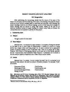
Fig 1. Axial CT scans show the measurement of the facet joint angles in this study at a disk level without (A) and with (B) tropism. In A, the left angle is 32° to the midsagittal plane and the right angle is 29° to the midsagittal plane. In B, the left angle is 34° to the midsagittal plane and the right angle is 56° to the midsagittal plane, which is a difference of 22° between the angulation of each facet joint. The images were obtained with parameters of 120 kV, 200 mA, 2.0-second scan duration, 12-cm2 field of view, 1.0-mm section thickness, and 512 3 256 matrix.
the facet joint surfaces has been determined in threedimensional space (5). A more practical and clinically useful method is to measure the angle of the articular surfaces as seen on computed tomographic (CT) scans or magnetic resonance (MR) images. The angle may be measured with reference to the sagittal plane or to the coronal plane (see Fig 1). The measurement by either technique can be converted to the other by geometric formulas. The precision of the method approaches 1% (Boden et al, “Lumbar...”). One hundred four lumbar facet joints were harvested from 21 cadavers, ranging in age from 49 to 85 years, that were selected from our institution’s body donation program within 48 hours postmortem. Subjects with a history of metastatic disease, spinal trauma, or spinal surgery were excluded. Cause of death was provided but not general medical records. Lumbar spinal columns were harvested by means of blunt and sharp dissection. The columns were transected into motion segments consisting of an intervertebral disk, two vertebral bodies, and the posterior spinal elements. The surrounding paraspinal and supraspinal muscles, connective tissue, and fat were removed while the ligaments and nerve roots in the neural foramen were preserved. The motion segments were imaged with a 1.5-T MR unit with a 5-inch-diameter send and receive solenoid coil. T1and T2-weighted axial images were obtained with a spinecho sequence. T1-weighted images were obtained with parameters of 650/40/2 (repetition time/echo time/excitations), 1.0-mm section thickness, 8-cm2 field of view, and 512 3 256 matrix. Long-repetition-time images were obtained with parameters of 2000/33/2 and 2000/80/2, 1.0-mm section thickness, 8-cm2 field of view, and 512 3 256 matrix. The specimens were also imaged with a high-speed helical CT scanner. A lateral localizer scan was obtained and axial scans were prescribed through each vertebral level with the plane of section chosen to bisect the intervertebral disk at that level. Axial scans were obtained with parameters of 120 kV, 200 mA, 2.0-second scan duration, medium body calibration, 12-cm field of view, 1.0-mm
section thickness, and 512 3 256 matrix. The imaging series was acquired in a plane parallel to the superior endplate of the inferior vertebral body of the motion segment to assure consistency in the anteroposterior and lateral inclinations of the scans. The orientation of the facet joints was measured on the axial CT scans using the method described by Noren et al (4). On an axial scan that bisected the intervertebral disk, one line was drawn in the midsagittal plane of the vertebra and one through each facet joint tangential to the superior articular process. Resident software was used to calculate the left and right facet joint angles subtended by each of the oblique lines and the sagittal plane. The difference between the two angles was determined. According to the method described by Vanharanta et al (2), moderate tropism was defined as a difference of 7° to 15°, and severe tropism as more than 15°. The sum of the two angles was calculated as a measure of the overall orientation of the joints. The facet joint orientation measurements were made by one investigator, who was blinded to the measurements of degeneration. The degree of articular cartilage degeneration and articular process sclerosis was scored by one observer using conventional criteria (6). The cartilage within the facet joint was classified into four grades based on the shape and amount of cartilage covering the articular joint surfaces: grade 1 was characterized by uniformly thick cartilage covering both articular surfaces completely; grade 2 by cartilage covering the entire surface with eroded or irregular regions; grade 3 by cartilage incompletely covering the articular surface, with the underlying bone exposed to the joint space; and grade 4 by complete absence of cartilage except for traces evident on the articular surface (Fig 2). The degree of articulating process sclerosis was classified into four grades: grade 1 was defined as a uniform thin band of cortical bone; grade 2 represented a thin band of cortical bone that extended into the space from the articular surface; grade 3 was defined as dense bone that extended into the joint space but covered less than half the facet; and grade 4 represented the presence of osteophytes or dense cortical bone that covered greater than half the facet joint (Fig 3).
AJNR: 18, August 1997
FACET JOINT TROPISM
1327
Fig 2. Axial MR images show the four grades of articular cartilage degeneration. Long repetition time images were obtained with parameters of 2000/33/2, 1.0-mm section thickness, 8-cm2 field of view, and 512 3 256 matrix. A, Grade 1: uniformly thick cartilage covers the articular surfaces completely. The interspace between the cartilage layers covering each articular process is well defined by a uniform dark band (arrows) of low MR signal intensity. B, Grade 2: cartilage covers the entire surface of the articular processes but with eroded or irregular regions evident. The interspace is irregular in pattern in the posterior aspects (arrowheads) and not crescentic, as that shown in (A). C, Grade 3: cartilage incompletely covers the articular surfaces (arrows), with regions of the underlying bone exposed to the joint space. D, Grade 4: cartilage is absent except for traces on the articular surface. Voids are (arrows) present, and are characterized by low MR signal intensity within the facet joint interspace.
The differences and the sums of the facet joint angles were compared with the grade of degeneration. The left and right facet joint angle measurements were compared with the cartilage and sclerosis grades in the ipsilateral joint. The total or transverse interfacet joint angle was compared with the sum of the degenerative scores of both facet joints. Regression analysis with a significance level set at P 5 .05 was used. The angles and angle differences and the degeneration scores were correlated with age and with spinal level.
Results The left and right individual facet joint angles ranged from 10° to 77°. The sums of the two angles for one level ranged from 28° to 154°. The differences between angles ranged from 0° to 39° (mean [absolute] value, 8.4°; SD, 7.4°). Of the 104 facet joints studied, four specimens had differences exceeding 15° (severe tropism),
six had differences between 7° and 15° (moderate tropism), and 94 had differences of less than 7° (no tropism). Facet joint articular cartilage degeneration scores ranged from 1 to 4 (mean, 2.2; SD, 0.8). The average sclerosis grade was 2.0 (range, 1 to 4). The lumbar segments with facet joint tropism greater than 15° had combined (left plus right) degenerative cartilage scores of 6 and combined sclerosis scores of 4 to 8, with an average of 6 (Table). The average for moderate tropism was 4, and for no tropism, 5. The average sclerosis score was 6 for severe tropism, 4 for moderate tropism, and 4 for no tropism. The correlation between the angle differences and the combined cartilage degeneration score was not significant (r 5 .16, P 5 .24); however, correlation of differences with the combined sclerosis score was significant (r 5 .33, P 5 .015). The 10
1328
GROGAN
AJNR: 18, August 1997
Fig 3. Axial MR images showing the four grades of sclerosis. Long-repetitiontime images were obtained with parameters of 2000/33/2, 1.0-mm section thickness, 8-cm2 field of view, and 512 3 256 matrix. A, Grade 1: articular processes have a thin layer of cortical bone (arrows). B, Grade 2: cortical bone of the articular processes is focally thickened (arrows) as compared with the thin band of low MR signal intensity shown in A. C, Grade 3: thickened cortical bone (arrows) covers less than half the articular processes, and areas of high MR signal intensity can be seen within the joint space. D, Grade 4: dense cortical bone (short arrows) covers greater than half the facet joint, with an osteophyte (long arrow) present.
facet joints with tropism had a mean cartilage degeneration score of 3.0; the other 94 joints had a mean score of 2.1. The difference between means was not significant (t test, P . .10). Comparing just the side with the larger or the smaller angle, no significant differences were found for the degenerative cartilage scores (r 5 .11, P 5 .4) or for the sclerosis scores (r 5 .04, P 5 .8). The lumbar facet joints with the larger individual angles tended to have higher degenerative change scores. The joints with angles of 35° or less had cartilage degeneration scores ranging from 1 to 3, whereas joints with angles of 65° or more had grades of 2 to 3. The correlation was significant (r 5 .26, P 5 .009). The sum of the angles correlated significantly with the grade of cartilage degeneration (r 5 .3, P 5 .03) but not with the grade of sclerosis (r 5 .01, P 5 .935).
Age and spinal level were significant factors. Age correlated with cartilage degeneration (r 5 .40, P , .001). The mean cartilage degeneration scores and facet joint angulation increased at each level from the T12-L1 to the L4 –5 disk levels and decreased from the L4 –5 to the L5-S1 levels. Age and facet angles were not correlated. Discussion Our findings do not support the hypothesis that facet joint tropism significantly affects the risk of articular cartilage degeneration or sclerosis in facet joints. Specifically, we found no correlation with degeneration of the cartilage and a very small effect on sclerosis of the facet joint. Our results showed a larger and significant effect of age, spinal level, and total facet joint angle on facet joint degeneration, which is what
AJNR: 18, August 1997
FACET JOINT TROPISM
1329
Average facet joint angle, cartilage, and sclerosis grade according to degree of tropism
Tropism, degree
None 0 to 6 Moderate 7 to 15 Severe 15 to 39
n
Average Facet Joint Angle
Age, y
Average Cartilage Classification
Average Sclerosis Grade
L
R
Sum
Difference
L
R
Sum
Difference
L
R
Sum
Difference
94
71
38
36
73
3
2
2
5
0
2
2
4
0
6
70
32
33
65
10
2
2
4
0
2
2
4
0
4
73
38
54
92
26
3
3
6
0
3
3
6
0
other studies have shown (6). The greater risk of degenerative changes in spinal levels with more coronally orientated facet joints is explained as the result of greater strain within the joint (Grobler et al, “Facet Joint...”). Side-toside differences might therefore not result in such significant alterations in the strains to which the joints are subjected. Disk degeneration, weight, scoliosis, and lordosis could also be factors in facet joint degeneration, although they were not evaluated in this study. The facet joint angles and the degenerative changes in the lumbar spine in this study were similar to those in previous studies. Facet joint angles were similar level by level to those reported by others (1– 4) (Boden et al, “Lumbar...”). The differences in angles were similar to those reported by Vanharanta (2) et al and Noren et al (4), and the severity of facet joint degeneration in our sample was similar to that reported in another cadaveric study (6). The small number of specimens in our study was a limitation; however, the low regression coefficients that were found suggest that a larger sample size would not have demonstrated a greater effect of tropism. The change in facet joint angulation relative to disk level could be a confounding factor in the correlation of facet joint angle and facet degeneration; however, this correlation was not a major focus of our investigation. The angle of the facet joints can be measured easily on MR and CT studies. The orientation of the facet joints is thought to affect the risk of degenerative changes in the spine. For example, in one study (Grobler et al, “Facet Joint...”)
the facet joint orientations were 17° greater in patients with degenerative spondylolisthesis than in matched control subjects. Asymmetric orientation of the facet joints (that is, tropism) is also thought to have a role. Tropism does not, however, consistently correlate significantly with disk degeneration (2, 4) (Boden et al, “Lumbar...”), disk protrusion (3), disk herniation (7), or segmental instability (8). The data reported here suggest that tropism is not a significant factor in the pathogenesis of facet joint degeneration. The role of tropism in degenerative changes in the spine may be smaller than has been assumed.
References 1. Van Schiak JPJ, Verbiest H, Van Schiak FDJ: The orientation of laminae and facet joints in the lower lumbar spine. Spine 1985; 10:59 – 63 2. Vanharanta H, Floyd T, Ohnmeiss DD, Hochschuler SH, Guyer RD. The relationship of facet tropism to degenerative disc disease. Spine 1993;18:1000 –1005 3. Hagg O, Wallner A. Facet joint asymmetry and protrusion of the intervertebral disc. Spine 1990;15:356 –359 4. Noren R, Trafimow J, Andersson GBJ, Huckman MS. The role of facet joint tropism and facet angle in disc degeneration. Spine 1991;16:530 –532 5. Panjabi MM, Oxland T, Takata K, Goel V, Duranceau J, Krag M. Articular facets of the human spine: quantitative threedimensional anatomy. Spine 1993;18:1298 –1310 6. Wang Z, Yu S, Haughton VM. Age-related changes in the lumbar facet joints. Clin Anat 1989;2:55– 62 7. Cassidy JD, Loback D, Yong-Hing K, Tchang S. Lumbar facet joint asymmetry: intervertebral disc herniation. Spine 1992;17: 570 –574 8. Cyron BM, Hutton WC. Articular tropism and stability of the lumbar spine. Spine 1980;5:168 –172