papr_346
113..123
EVIDENCE-BASED MEDICINE Evidence-based Interventional Pain Medicine according to Clinical Diagnoses
5. Cervical Facet Pain Maarten van Eerd, MD, FIPP*†; Jacob Patijn, MD, PhD*; Arno Lataster, MSc‡; Richard W. Rosenquist, MD§; Maarten van Kleef, MD, PhD, FIPP*; Nagy Mekhail, MD, PhD, FIPP¶; Jan Van Zundert, MD, PhD, FIPP*,** *Department of Anesthesiology and Pain Management, University Medical Centre Maastricht, Maastricht, The Netherlands; †Department of Anesthesiology and Pain Management, Amphia Ziekenhuis, Breda, The Netherlands; ‡Department of Anatomy and Embryology, Maastricht University, Maastricht, The Netherlands; §Department of Anesthesia, Pain Medicine Division, University of Iowa, Iowa City, Iowa, USA; ¶Department of Pain Management, Cleveland Clinic, Cleveland, Ohio, U.S.A.; **Department of Anesthesiology and Multidisciplinary Pain Centre, Ziekenhuis Oost-Limburg, Genk, Belgium 䊏 Abstract: More than 50% of patients presenting to a pain clinic with neck pain may suffer from facet-related pain. The most common symptom is unilateral pain without radiation to the arm. Rotation and retroflexion are frequently painful or limited. The history should exclude risk factors for serious underlying pathology (red flags). Radiculopathy may be excluded with neurologic testing. Direct correlation between degenerative changes observed with plain radiography, computerized tomography, and magnetic resonance imaging and pain has not been proven. Conservative treatment options for cervical facet pain such as physiotherapy, manipulation, and mobilization, although supported by little evidence, are frequently applied before considering interventional treatments. Interventional pain management techniques, including intra-articular steroid injections, medial branch blocks, and radiofrequency treatment, may be considered (0). At present, there is no evidence to support cervical intra-articular corticosteroid injection. When Address correspondence and reprint requests to: M. van Eerd, MD, Maastricht University Medical Centre, Department of Anesthesiology and Pain Management, PO Box 5800, 6202 AZ Maastricht, The Netherlands. E-mail:
[email protected]. DOI. 10.1111/j.1533-2500.2009.00346.x
© 2010 World Institute of Pain, 1530-7085/10/$15.00 Pain Practice, Volume 10, Issue 2, 2010 113–123
applied, this should be done in the context of a study. Therapeutic repetitive medial branch blocks, with or without corticosteroid added to the local anesthetic, result in a comparable short-term pain relief (2 B+). Radiofrequency treatment of the ramus medialis of the cervical ramus dorsalis (facet) may be considered. The evidence to support its use in the management of degenerative cervical facet joint pain is derived from observational studies (2 C+). 䊏 Key Words: evidence-based medicine, cervical pain, cervical facet joint, injection therapy, radiofrequency
INTRODUCTION This review on cervical facet joint syndrome is part of the series “Interventional practice guidelines based on clinical diagnosis.” Recommendations formulated in this chapter are based on “Grading strength of recommendations and quality of evidence in clinical guidelines” described by Guyatt et al.1 and adapted by van Kleef et al. in the editorial accompanying the first article of this series2. (Table 1) The latest literature update was performed in August 2009.
114 • van eerd et al.
Table 1. Summary of Evidence Scores and Implications for Recommendation Score
Description
1 A+ 1 B+
Effectiveness demonstrated in various RCTs of good quality. The benefits clearly outweigh risk and burdens One RCT or more RCTs with methodologic weaknesses, demonstrate effectiveness. The benefits clearly outweigh risk and burdens One or more RCTs with methodologic weaknesses, demonstrate effectiveness. Benefits closely balanced with risk and burdens
Positive recommendation
Multiple RCTs, with methodologic weaknesses, yield contradictory results better or worse than the control treatment. Benefits closely balanced with risk and burdens, or uncertainty in the estimates of benefits, risk and burdens. Effectiveness only demonstrated in observational studies. Given that there is no conclusive evidence of the effect, benefits closely balanced with risk and burdens
Considered, preferably study-related
0
There is no literature or there are case reports available, but these are insufficient to suggest effectiveness and/or safety. These treatments should only be applied in relation to studies.
Only study-related
2 C-
Observational studies indicate no or too short-lived effectiveness. Given that there is no positive clinical effect, risk and burdens outweigh the benefit One or more RCTs with methodologic weaknesses, or large observational studies that do not indicate any superiority to the control treatment. Given that there is no positive clinical effect, risk and burdens outweigh the benefit RCT of a good quality which does not exhibit any clinical effect. Given that there is no positive clinical effect, risk and burdens outweigh the benefit
Negative recommendation
2 B+ 2 B⫾
2 C+
2 B-
2 A-
Implication
RCT, randomized controlled trial.
Neck pain is defined as pain in the area between the base of the skull and the first thoracic vertebra. Pain extending into adjacent regions is defined as radiating neck pain. Pain may radiate into the head (cervicogenic headache), shoulder, or upper arm (radicular or nonradicular pain).3 Neck pain is common in the general population with a 12-month prevalence that varies between 30% and 50%. Neck pain results in incapacity to perform daily activities in 2% to 11% of the cases. It occurs more often in women, with peak prevalence in middle age. Risk factors include genetic disposition and smoking.4 Although a correlation between type of work and neck pain has not been demonstrated, high quantitative job demands (eg, sedentary jobs at a computer or repetitive precision work with a high level of muscular tension) and lack of social support in the work environment appear to have an effect.5,6 Psychological factors such as avoidance behavior and catastrophizing are not related to neck symptoms, in contrast to patients with low back problems.5 Although trauma-related neck pain (whiplash-associated disorders; WADs) and degenerative neck problems both may be caused by chronic degeneration of the facet joints, the distinction is made on etiologic basis, because WADs may involve other painful structures, certainly in the subacute phase.5 The causes of neck pain often are unclear, but the following innervated structures in the neck may be sources of pain: vertebrae, intervertebral disks, uncovertebral (Luschka)
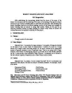
joints, ligaments, muscles, and facet (zygapophyseal) joints.5 Osseous and fibrocartilaginous degenerative disorders, identified by plain radiography, are frequently seen. The relationship between degenerative signs and pain, however, is unclear. There is a great deal of research into degenerative signs of the cervical vertebral column. In the intervertebral disk, (1) annular tears, (2) disk prolapse, (3) endplate damage and internal disk disruption have been identified as potential structural disk pathologies.7 Other structures in the neck, such as facet joints and uncovertebral joints, also show degenerative signs. The hypothesis that disk degeneration and disk narrowing increase facet joint loading and consequently facet osteoarthritis, seems plausible, but has yet to be proven. Some researchers claim that the disk and the facet joints can be seen as independent pain generators.8 Confirmation of degenerative disease is mainly based on radiological findings. Spondolysis (disorders of the nonsynovial joints) and osteoarthritis (facet osteoarthritis) are frequent in advanced age. Degenerative disorders are usually seen at the low and midcervical levels (C4 to C5, C5 to C6, and C6 to C7). Knowledge of the innervation of various structures in the neck is important to interpret diagnostic blocks and to direct local treatments9 (Figure 1). Patients presenting to a pain clinic usually suffer from chronic pain (pain lasting longer than 3 months). Prognostic factors for chronicity include age (older than 40 years of age), previous episodes of neck pain, trauma, and simultaneous low back pain symptoms.10
5. Cervical Facet Pain • 115
C 2-3 C 3-4 Tuberculum anterius
C 5-6
C 4-5 C 6-7
Tuberculum posterius Ganglion spinale (DRG) Ramus dorsalis
Facet joint Ramus medialis of the ramus dorsalis (Medial branch)
N. spinalis, ramus ventralis
A. vertebralis sinistra
Figure 2. Radiation pattern of cervical facet pain (illustration: Rogier Trompert Medical Art. http://www.medical-art.nl). Figure 1. Innervation of the cervical vertebral column and the facet joints (illustration: Rogier Trompert Medical Art. http:// www.medical-art.nl).
Ramus ventralis
Ramus dorsalis N. spinalis
Ramus lateralis
Ramus medialis
Ganglion spinale (DRG)
Figure 4. Posterolateral approach of the cervical ramus medialis (medial branch) of the ramus dorsalis (illustration: Rogier Trompert Medical Art. http://www.medical-art.nl).
116 • van eerd et al.
It is important to determine if the pain symptoms produce functional limitations (eg, in dressing, lifting, automobile operation, reading, sleeping, and working). Recently, the following classification for neck pain and associated symptoms has been proposed:11 •
•
•
•
Grade I neck pain: no symptoms indicating serious pathology and minimal influence on daily activities. Grade II neck pain: no symptoms indicating serious pathology, but having influence on daily activities. Grade III neck pain: no symptoms indicating serious pathology, presence of neurological disorders such as decreased reflexes, muscle weakness, or decreased sensory function. Grade IV neck pain: indications of serious underlying pathology such as fracture, myelopathy, or neoplasm.
Pain Originating from the Cervical Facet Joints (Facet Joint Syndrome) Neck pain can be caused by the facet joints. Compared with research on lumbar facet pain, research on cervical facet dysfunction started much later. In 1988, Bogduk and Marsland12 described the positive effect of injection of local anesthetics close to the facet joints in patients with neck pain. While a diagnosis is defined as a clinical picture with known etiology and prognosis, a syndrome is a combination of symptoms occurring at a higher frequency in a certain population. The cervical facet syndrome is defined as a combination of symptoms: • • • •
axial neck pain (either not or rarely radiating past the shoulders) pain with pressure on the dorsal side of the spinal column at the level of the facet joints pain and limitation of extension and rotation absence of neurological symptoms
It is unclear how often neck pain originates from the facet joints. The prevalence of pain emanating from facet joints, within a population suffering from neck pain, has been reported to be 25% to 65%, depending on patient group and selection method. In the group of patients attending a pain clinic for neck pain, it is likely to be more than 50%.13,14 This is a markedly higher percentage than facet pain in the lumbar region.
Anatomy of the Facet Joints The facet joint is a diarthrotic joint with joint surfaces, synovial membrane, and a joint capsule. It forms an angle of approximately 45° with the longitudinal axis throughout the cervical spinal column. Compared with the lumbar facet joints, the cervical facet joints have a higher density of mechanoreceptors. The facet joints from C3 to C7 are innervated by the ramus medialis (medial branch) of the ramus dorsalis of the segmental nerve. Each facet joint is innervated by nerve branches from the upper and lower segment.9 (Figure 1)
I. DIAGNOSIS I.A HISTORY During the history, attention should be paid to signs and symptoms potentially indicating a serious underlying pathology (“red flags”). It is important to question the patient about previous trauma and previous or ongoing oncological treatments. Signs of potential spinal metastases are (1) history of malignancy, (2) pain starting after the age of 50, (3) continuous pain, independent of posture or movement, and (4) pain at night. When symptoms such as weight loss, fever, nausea, vomiting, dysphagia, coughing, or frequent infections are reported, extensive history and further examination is mandatory. The most common symptom associated with pain arising from the cervical facet joints is unilateral pain, not radiating past the shoulder. The pain often has a static component, since it does not always occur in relation to movement. Rotation and retroflexion are usually reported as painful or limited. Dwyer et al. showed that injection of irritating substances into the facet joints results in a specific radiation pattern.15 (Figure 2) The same radiation pattern is seen with mechanical and electrical stimulation. The radiation pattern is not distinctive for facet problems but can indicate the segmental localization.
I.B PHYSICAL EXAMINATION Neurological tests (reflexes, sensibility, and motor function) are necessary in order to exclude radiculopathy. In order to examine the function of the neck the following tests are important: • • • • •
flexion and extension—passive and active lateral flexion—passive and active rotation—passive and active rotation in maximal flexion—passive and active rotation in extension—passive and active
5. Cervical Facet Pain • 117
Rotation in a neutral position involves the rotation movement of the entire cervical spinal column. Rotation in flexion assesses the movement in the higher-cervical segments. Rotation in extension assesses the movement in the lower-cervical segments. Local pressure pain over the facet joints can indicate problems arising from the facet joints. Recent research demonstrated that local pressure pain, defined as pain applying pressure of at least 4 kg, is a predictor of success when radiofrequency (RF) treatment (see Treatment Options).16 When the neck pain is accompanied by radiation to the shoulder region, shoulder pathology should be excluded. There is no evidence to support the relationship between the results of clinical examination and the anamnesis with pain originating from the cervical facet joints.17 In daily clinical practice, history and physical examination are useful to exclude serious pathology and to obtain a working diagnosis. An indication as to the segmental level (high-mid-low-cervical) involved can be obtained.
I.C ADDITIONAL TESTS In specific cases, plain radiography of the cervical spinal column may be indicated to exclude tumor or fracture. Plain radiography does not provide information in establishing the diagnosis of facet problems, but may help in evaluating the degree of degeneration. The anterior spinal column is inspected for narrowing of the disk, anterior and posterior osteophyte formation. The posterior spinal column is inspected for facet osteoarthritis (facet sclerosis and osteophyte formation). In 1963, Kellgren et al.18 stated that once degenerative changes are seen on plain radiography, degeneration has already reached an advanced stage. With advancing age, degenerative changes are more frequently seen: 25% at the age of 50, up to 75% at the age of 70.19 An age-related prevalence study concerning the facet joint involvement in chronic neck pain indicates a comparable prevalence among all age groups.20 Degenerative changes of the cervical spinal column are present in asymptomatic patients, indicating that degenerative changes do not always cause pain. However, the conclusion that there is no relation between degeneration and pain cannot be drawn. There are studies indicating a relation between degenerative changes and pain symptoms.19,21 In summary, a relation between radiologic identification of degenerative changes and pain symptoms has not been proven. If a neurological etiology of the pain symp-
toms is suspected, a magnetic resonance imaging (MRI) or computer tomography (CT) scan is indicated. Depending on the clinical setting, consultation of or referral to a neurologist should be considered. The use of cervical discography may help in identifying the source of pain, but its value concerning the subsequent therapeutic treatments is not established. Diagnostic Blocks The working diagnosis of facet pain, based on history and clinical examination, may be confirmed by performing a diagnostic block. Local anesthetic can be injected intra-articularly or adjacent to the ramus medialis (medial branch) of the ramus dorsalis of the segmental nerve.5,22 These procedures are performed under fluoroscopy. There is no consensus about the definition of a successful diagnostic block. Some authors claim that 100% pain relief should be achieved.23 But Cohen et al. showed that there is no difference in outcome of the RF treatment of patients reporting 80% and those reporting more than 50% pain reduction after a diagnostic block.16 In daily clinical practice, we consider a diagnostic block successful if more than 50% pain reduction is reported. It has been demonstrated that innervation of the facet joint occurs via the ramus medialis (medial branch) of the ramus dorsalis. We prefer a block of the ramus medialis (medial branch) instead of an intra-articular block, because it is not always technically possible to position a needle into the facet joint. According to Bogduk and McGuirk,5 the facet joints from C3 to C7 are innervated by the medial branches of the nerves above and below the joint. For a block or RF treatment, for example, of the C4 to C5 facet joint to be effective, the medial branches of the rami dorsales of C4 and C5 are to be treated. A prognostic block can be used before RF treatment is performed. A prognostic block assumes that if an anatomical structure is injected with a local anesthetic resulting in a decrease in pain, this structure is the source of pain. This appears to be a useful concept. Research and clinical experience indicate however, that after a single block, only a small percentage (2/47; ~4%) of patients have no pain reduction.24 This means that after a single diagnostic block, there are very few false negative results. In order to minimize the number of false positives, a number of researchers have suggested that a second block should be carried out using a local anesthetic with different duration of effect, eg, lidocaine vs. bupivacaine (comparative double blocks). Only if the patient responds concordantly (longer or shorter pain reduction depending on the duration of action of the
118 • van eerd et al.
local anesthetic) is this indicative of facet joint pain. This is a pharmacological criterion. These researchers suggest that double blocks are the gold standard for the diagnosis of facet pain. A gold standard, however, should be generally accepted and used. The concept of double blocks has theoretical and practical shortcomings. A decrease in the number of false positives can occur at the cost of the number of false negative reactions: patients respond positive to the local anesthetic, but not according to the previously standardized pharmacological criterion. Furthermore, a cervical injection represents a burden for the patient. Finally, it is questionable if double blocks are cost-effective.25 A best evidence synthesis on the assessment of neck pain concluded that diagnostic facet injections have not been validated to identify facet joint pain.26 As long as the relationship with the etiology of facet pain is not clearly established, the extra burden of performing double blocks cannot be justified. Contrary to lumbar facet blocks, only a small percentage of patients have a negative response to a single cervical facet block. In summary, on the basis of history and physical examination, a working diagnosis of cervical facet pain is defined. One diagnostic block can be recommended for confirming the clinical working diagnosis of facet pain. A diagnostic block is considered positive when the patient experiences a 50% pain reduction.16
I.D DIFFERENTIAL DIAGNOSIS Serious causes of neck pain such as tumors, infections, fractures, and systemic diseases are rare. A clinically relevant prolapsed disk or cervical spondylotic myelopathy can both cause neurological symptoms. Every patient with motor function loss and/or reflex changes and/or sensory loss must be thoroughly assessed. Metastases, cervical herniated nucleus pulposus with radiculopathy, discitis, and vertebral fractures should be excluded through history and (additional) tests. Diagnoses such as segmental dysfunction, instability, and muscle strain as diagnoses of chronic pain are not sufficiently documented to be included in the differential diagnosis.5
II. TREATMENT OPTIONS II.A CONSERVATIVE MANAGEMENT Physiotherapy/Exercise Therapy In a study comparing physiotherapy with a short intervention consisting of a self-management program that
encourages patients to resume normal activity patterns, physiotherapy resulted in a better outcome.27 The improvements with both interventions are, however, small (on all outcome scales). Physical exercises have a pain reducing effect, especially if the patient received adequate information relative to the exercises. Physiotherapy, based on instructions for exercises that can also be carried out at home, is the best choice when choosing conservative treatment. Manipulation/Mobilization In a subgroup analysis of studies on patients with neck pain in general practice, there was a positive short-term effect of manipulation therapy, especially in older (>50 years) patients.28 Multidisciplinary Therapy There is no consensus about the required components of multidisciplinary therapy. The approach should be directed towards biopsychosocial rehabilitation. Whether this can be offered as a multimodal approach by one specialist or in a multidisciplinary setting is still unclear and not yet scientifically supported. Cognitive behavioral therapy shows improvement in somatic, behavioral, and cognitive symptoms, but the effect on pain symptoms is small. In patients with neck pain, little, or no relationship has been found between psychological factors and pain. A multidisciplinary treatment should, in addition to conservative treatment, include minimally invasive interventional techniques.
II.B INTERVENTIONAL MANAGEMENT Intra-Articular Steroid Injections No reports from quality studies regarding the effect of intra-articular steroid injections are currently known.29 There are no comparative studies between intraarticular steroid injections and RF therapy. Local Infiltration of the Ramus Medialis (Medial Branch) of the Ramus Dorsalis Medial branch block of the ramus dorsalis of the segmental nerve is primarily considered as a diagnostic aid; however, (repetitive) infiltration of local anesthetic was shown to provide therapeutic effect.24,30 In a randomized controlled trial (RCT) comparing the effect of medial branch blocks with bupivacaine alone to blocks with the same local anesthetic plus steroid, a comparable pain reduction was observed in both groups for mean dura-
5. Cervical Facet Pain • 119
tion of 14 and 16 weeks, respectively. During the follow-up period of 1 year, the mean number of procedures was similar (3.5 and 3.4, respectively). Patients were selected for participation in this study by controlled blocks providing 380% pain relief.30 These findings suggest that the addition of corticosteroid to local anesthetic does not provide better outcome. Moreover, as described above, the diagnostic procedure used in the RCT is burdensome for the patient, requiring repeat infiltrations every 14 to 16 weeks. Therefore, this cannot be recommended as first-line therapy. RF Treatment of the Ramus Medialis (Medial Branch) of the Ramus Dorsalis Percutaneous RF treatment of cervical pain has been intensively studied. The data from original articles were summarized in seven systematic reviews.22,29,31–34 There is only one RCT evaluating RF treatment of the ramus medialis (medial branch) of the ramus dorsalis, but this was in patients with WADs.23 Consequently, this RCT cannot be rated in the evidence scoring for degenerative cervical facet joint pain. The effectiveness of RF treatment for degenerative neck pathology was shown in observational studies.16,35,36 A retrospective chart analysis on the effect of repeat RF facet denervations illustrated that the mean duration of effect of the first intervention was 12.5 months. Patients who responded positively to the first intervention received from one to six additional interventions. After each intervention (RF treatment of the rami mediales of the ramus dorsalis), more than 90% of the patients had satisfactory pain relief, and duration of effect was between 8 and 12 months.37 Lord et al.23 described a technique for approaching the ramus medialis (medial branch) of the ramus dorsalis laterally as well as posteriorly. This can only be carried out in the prone position. Good results have also been reported using an alternative technique as described by Sluijter, van Kleef and van Suijlekom.38,39 Theoretically, a block of the ramus medialis (medial branch), close to the ramus dorsalis, based on sensory and motor stimulation parameters, could generate a similar effect as an extensive denervation over the entire length of the nerve. Even though there are no studies comparing both techniques, we consider the former to be the least invasive approach. Percutaneous cervical facet denervation is an acceptable treatment option for a clinical diagnosis of chronic degenerative cervical facet pain, given the many observational descriptions of a positive effect.
II.C COMPLICATIONS OF INTERVENTIONAL MANAGEMENT Complications are rare. Nevertheless, one should be aware that the arteria vertebralis may be punctured if the needle is pushed too far anteriorly into the foramen intervertebrale. Verification of the needle position should be made under antero-posterior fluoroscopy to prevent intrathecal injection or injection of the local anesthetic into the spinal cord. In an observational study, the incidence of inadvertent intravascular penetration for medial branch blocks at spinal level was reported to be 3.9%, comparable with the incidence at lumbar level (3.7%). Some patients experienced short-term vasovagal reactions. The intravascular uptake of local anesthetic and contrast solution (due to direct injection into a vessel) was thought to be responsible for false negative diagnostic blocks. No systemic effects were reported.40 A report on transient tetraplegia after cervical facet joint injection, done without imaging, illustrates the vulnerability of the cervical arteries.41Appropriate monitoring of the vital signs and availability of resuscitation equipment are essential. Infections have been described, but the incidence is unknown and probably very low.42 A recent report on septic arthritis of the facet joints included two cases of cervical facet joints. In these cases, the port of entry could not be identified, but in one lumbar case report, percutaneous injection was directly linked to this severe complication.43 Other potential complications of facet joint interventions are related to needle placement and drug administration; they include dural puncture, spinal cord trauma, spinal anesthesia, chemical meningitis, neural trauma, pneumothorax, radiation exposure, facet capsule rupture, hematoma formation, and side effects of corticosteroids.44 After RF treatment, postoperative burning pain is regularly reported. This pain disappears after 1 to 3 weeks.45 Smith et al.46 found contrast enhancement on MRI typical for paraspinal abscess, even without apparent infection, which was attributed to a noninfectious postinflammatory process. There are no incidence data on side effects and complications following cervical RF facet denervation. At the lumbar level, the incidence of complications was lower than 1%.47 Surgical Treatments Anterior cervical fusion is described as a possible technique for nonradicular neck pain. One study showed a clear effect on pain and function, but the long-term effect of this invasive treatment is unknown.48
120 • van eerd et al.
Localized uni/bilateral neck pain > 6 weeks
Exclude red flags
Yes
Neurological disorders ?
None
Yes
Neurological tests
Radiation not past the shoulder Pain with pressure on the facet joint Potential painful and/or limited extension and/or rotation
Working diagnosis Cervical “facet pain”
Diagnostic block > 50 % pain relief
Yes
No
Re-evaluation Therapeutic (repetitive) cervical ramus medialis (medial branch) the cervical ramus dorsalis block block (local anesthetic with or without corticosteroid. RF cervical ramus medialis (medial branch) of the ramus dorsalis/facet
Figure 3. Clinical practice algorithm for treatment of cervical facet pain. RF, radiofrequency treatment.
II.D EVIDENCE FOR INTERVENTIONAL MANAGEMENT Technique Intra-articular injections Therapeutic (repetitive) cervical ramus medialis (medial branch) of the cervical ramus dorsalis block (local anesthetic with or without corticosteroid) Radiofrequency treatment of the ramus medialis (medial branch) of the cervical ramus dorsalis
III. RECOMMENDATIONS Score 0 2 B+
2 C+
For patients suffering chronic neck pain caused by cervical arthrosis, not responding to conservative treatment, RF treatment of the ramus medialis (medial branch) of the ramus dorsalis of the segmental nerves from C3 to C6 can be considered. There is currently no evidence available to evaluate the efficacy of intra-articular infiltration of the cervical
5. Cervical Facet Pain • 121
The (postero-) lateral approach in the supine position is described below (Figure 4). The advantage of this technique is that it is possible to maintain eye contact with the patient. Sedation is rarely required. The patient is placed in the supine position with the head slightly extended on a small cushion. The C-arm is placed in an oblique position (20 to 30° laterally). In this position, the beam runs parallel with the exiting nerve root that runs somewhat caudofrontal. In this position, the pedicles from the contralateral side are projected onto the anterior half of the corpus vertebrae Figure 5. In the AP projection, the C-arm is positioned 10 to 20° caudally. In this position, the intervertebral disk space and the foramen intervertebrale are visible (Figure 6). The ramus medialis (medial branch) of the ramus dorsalis runs over the base of the processus
articularis superius. The injection point is marked on the skin, slightly posterior and caudal to the end point of the needle that is dorsal to the posterior boundary of the facet column. The first needle is introduced in a horizontal plane, slightly cranially so that the tip of the needle points in the direction of the end point. It is important to understand that this is not a “tunnelview” technique. The needle is slowly advanced anteriorly and cranially until bony contact with the facet column occurs. The further the needle is advanced, the more difficult it becomes to change the direction. Therefore, the position of the needle needs to be checked frequently. If the needle points too much in the direction of the foramen intervertebrale, without contacting bone, the direction needs to be corrected to be more posterior. If there is no bone contact in the posterior direction, there is a risk that the needle will enter the canalis vertebralis between the laminae. To prevent this, the needle position can be checked in the AP direction. The final position of the needle in the AP direction is in the concave “waist” of the facet column. After placement of the first needle, the other needles are introduced in the same way. The first needle acts as a guideline for direction and depth. The same technique is used for the facet joints of C3–C4 to C6–C7.
Figure 5. Radiofrequency treatment cervical ramus medialis (medial branch) of the ramus dorsalis/facet C4, C5, C6 left: 3/4 projection.
Figure 6. antero-posterior Radiofrequency treatment cervical ramus medialis (medial branch) of the ramus dorsalis /facet C4, C5, C6 left: projection.
facet joints. Therefore, it should only be done within the context of an experimental study.
III.A CLINICAL PRACTICE ALGORITHM A practice algorithm for the management of facet pain is illustrated in Figure 3.
III.B TECHNIQUE(S) Percutaneous Facet Denervation
122 • van eerd et al.
Once an optimal anatomic location is reached and controlled using fluoroscopy, the position of the needle tip at the ramus medialis (medial branch) of the ramus dorsalis is confirmed using electrical stimulation. The stimulation threshold is determined: an electrical stimulation of 50 Hz must give a reaction (tingling) in the neck at less than 0.5 V. Then stimulation is carried out at 2 Hz. Contractions of the paraspinal muscles can occur. Muscle contractions in the arm indicate a position close to the exiting segmental nerve. The needle should then be placed more posteriorly. Once the correct position has been determined, 0.5 to 1 mL local anesthetic (1% or 2% lidocaine) is given. A RF lesion at 80°C for 60 seconds is carried out.
IV. SUMMARY Neck pain is common in the general population. The etiology is difficult to confirm based on history, physical examination, and radiological tests. Conservative treatment is the first choice. At the cervical level, the facet joint appears to be an important source of pain with degenerative neck symptoms. Where there is an indication that the pain is arising from the facet joints, a minimally invasive technique such as RF treatment of the ramus medialis (medial branch) of the ramus dorsalis may be considered.
ACKNOWLEDGEMENTS This review was initially based on practice guidelines written by Dutch and Flemish (Belgian) experts that are assembled in a handbook for Dutch-speaking pain physicians. After translation, the manuscript was updated and edited in cooperation with U.S./international pain specialists. The authors thank José Geurts and Nicole Van den Hecke for coordination and suggestions regarding the manuscript.
REFERENCES 1. Guyatt G, Gutterman D, Baumann MH, et al. Grading strength of recommendations and quality of evidence in clinical guidelines: report from an American College of Chest Physicians task force. Chest. 2006;129:174–181. 2. van Kleef M, Mekhail N, van Zundert J. Evidencebased guidelines for interventional pain medicine according to clinical diagnoses. Pain Pract. 2009;9:247–251. 3. Guzman J, Hurwitz EL, Carroll LJ, et al. A new conceptual model of neck pain: linking onset, course, and
care: the bone and joint decade 2000–2010 task force on neck pain and its associated disorders. Spine. 2008;33:S14– S23. 4. Hogg-Johnson S, van der Velde G, Carroll LJ, et al. The burden and determinants of neck pain in the general population: results of the bone and joint decade 2000–2010 task force on neck pain and its associated disorders. Spine. 2008;33:S39–S51. 5. Bogduk N, McGuirk B. Management of Acute and Chronic Neck Pain. Pain Research and Clinical Management. Philadelphia, PA: Elsevier; 2006. 6. Cote P, van der Velde G, Cassidy JD, et al. The burden and determinants of neck pain in workers: results of the bone and joint decade 2000–2010 task force on neck pain and its associated disorders. Spine (Phila Pa 1976). 2008;33:S60–S74. 7. Adams MA, Roughley PJ. What is intervertebral disc degeneration, and what causes it? Spine. 2006;31:2151–2161. 8. Bogduk N, Aprill C. On the nature of neck pain, discography and cervical zygapophysial joint blocks. Pain. 1993;54:213–217. 9. Groen GJ, Baljet B, Drukker J. Nerves and nerve plexuses of the human vertebral column. Am J Anat. 1990;188:282–296. 10. Hoving JL, de Vet HC, Twisk JW, et al. Prognostic factors for neck pain in general practice. Pain. 2004;110:639– 645. 11. Haldeman S, Carroll L, Cassidy JD, et al. The bone and joint decade 2000–2010 task force on neck pain and its associated disorders: executive summary. Spine. 2008;33:S5– S7. 12. Bogduk N, Marsland A. The cervical zygapophysial joints as a source of neck pain. Spine. 1988;13:610–617. 13. Manchikanti L, Boswell MV, Singh V, et al. Prevalence of facet joint pain in chronic spinal pain of cervical, thoracic, and lumbar regions. BMC Musculoskelet Disord. 2004;5:15. 14. Yin W, Bogduk N. The nature of neck pain in a private pain clinic in the United States. Pain Med. 2008;9:196–203. 15. Dwyer A, Aprill C, Bogduk N. Cervical zygapophyseal joint pain patterns. I: a study in normal volunteers. Spine. 1990;15:453–457. 16. Cohen SP, Bajwa ZH, Kraemer JJ, et al. Factors predicting success and failure for cervical facet radiofrequency denervation: a multi-center analysis. Reg Anesth Pain Med. 2007;32:495–503. 17. Kirpalani D, Mitra R. Cervical facet joint dysfunction: a review. Arch Phys Med Rehabil. 2008;89:770– 774. 18. Kellgren J, Jeffrey M, Ball J. The Epidemiology of Chronic Rheumatism. Oxford: Blackwell; 1963. 19. Friedenberg ZB, Miller WT. Degenerative disc disease of the cervical spine. J Bone Joint Surg Am. 1963;45: 1171–1178.
5. Cervical Facet Pain • 123
20. Manchikanti L, Manchikanti KN, Cash KA, et al. Age-related prevalence of facet-joint involvement in chronic neck and low back pain. Pain Physician. 2008;11:67–75. 21. van der Donk J, Schouten JS, Passchier J, et al. The associations of neck pain with radiological abnormalities of the cervical spine and personality traits in a general population. J Rheumatol. 1991;18:1884–1889. 22. Manchikanti L, Boswell MV, Singh V, et al. Comprehensive evidence-based guidelines for interventional techniques in the management of chronic spinal pain. Pain Physician. 2009;12:699–802. 23. Lord SM, Barnsley L, Wallis BJ, et al. Percutaneous radio-frequency neurotomy for chronic cervical zygapophyseal-joint pain. N Engl J Med. 1996;335:1721– 1726. 24. Barnsley L, Lord S, Bogduk N. Comparative local anaesthetic blocks in the diagnosis of cervical zygapophysial joint pain. Pain. 1993;55:99–106. 25. Bogduk N, Holmes S. Controlled zygapophysial joint blocks: the travesty of cost-effectiveness. Pain Med. 2000;1:24–34. 26. Nordin M, Carragee EJ, Hogg-Johnson S, et al. Assessment of neck pain and its associated disorders: results of the bone and joint decade 2000–2010 task force on neck pain and its associated disorders. J Manipulative Physiol Ther. 2009;32:S117–S140. 27. Klaber Moffett JA, Jackson DA, Richmond S, et al. Randomised trial of a brief physiotherapy intervention compared with usual physiotherapy for neck pain patients: outcomes and patients’ preference. BMJ. 2005;330:75. 28. Schellingerhout JM, Verhagen AP, Heymans MW, et al. Which subgroups of patients with non-specific neck pain are more likely to benefit from spinal manipulation therapy, physiotherapy, or usual care? Pain. 2008;139:670– 680. 29. Falco FJ, Erhart S, Wargo BW, et al. Systematic review of diagnostic utility and therapeutic effectiveness of cervical facet joint interventions. Pain Physician. 2009;12: 323–344. 30. Manchikanti L, Singh V, Falco FJ, et al. Cervical medial branch blocks for chronic cervical facet joint pain: a randomized, double-blind, controlled trial with one-year follow-up. Spine. 2008;33:1813–1820. 31. Geurts JW, van Wijk RM, Stolker RJ, et al. Efficacy of radiofrequency procedures for the treatment of spinal pain: a systematic review of randomized clinical trials. Reg Anesth Pain Med. 2001;26:394–400. 32. Niemisto L, Kalso E, Malmivaara A, et al. Radiofrequency denervation for neck and back pain: a systematic review within the framework of the cochrane collaboration back review group. Spine. 2003;28:1877–1888. 33. Manchikanti L, Singh V, Vilims BD, et al. Medial branch neurotomy in management of chronic spinal pain:
systematic review of the evidence. Pain Physician. 2002;5: 405–418. 34. Boswell MV, Trescot AM, Datta S, et al. Interventional techniques: Evidence-based practice guidelines in the management of chronic spinal pain. Pain Physician. 2007;10: 7–111. 35. McDonald GJ, Lord SM, Bogduk N. Long-term follow-up of patients treated with cervical radiofrequency neurotomy for chronic neck pain. Neurosurgery. 1999;45:61–67; discussion 67–68. 36. Barnsley L. Percutaneous radiofrequency neurotomy for chronic neck pain: outcomes in a series of consecutive patients. Pain Med. 2005;6:282–286. 37. Husted DS, Orton D, Schofferman J, et al. Effectiveness of repeated radiofrequency neurotomy for cervical facet joint pain. J Spinal Disord Tech. 2008;21:406–408. 38. Sluijter ME. Radiofrequency Part 2. Meggen (LU), Switzerland: Flivopress, SA; 2003. 39. van Kleef M, van Suijlekom JA. Treatment of chronic cervical pain, brachialgia, and cervicogenic headache by means of radiofrequency procedures. Pain Pract. 2002;2:214–223. 40. Verrills P, Mitchell B, Vivian D, et al. The incidence of intravascular penetration in medial branch blocks: cervical, thoracic, and lumbar spines. Spine. 2008;33:E174–E177. 41. Heckmann JG, Maihofner C, Lanz S, et al. Transient tetraplegia after cervical facet joint injection for chronic neck pain administered without imaging guidance. Clin Neurol Neurosurg. 2006;108:709–711. 42. Rathmell JP, Lake T, Ramundo MB. Infectious risks of chronic pain treatments: injection therapy, surgical implants, and intradiscal techniques. Reg Anesth Pain Med. 2006;31:346–352. 43. Michel-Batot C, Dintinger H, Blum A, et al. A particular form of septic arthritis: septic arthritis of facet joint. Joint Bone Spine. 2008;75:78–83. 44. Boswell MV, Colson JD, Sehgal N, et al. A systematic review of therapeutic facet joint interventions in chronic spinal pain. Pain Physician. 2007;10:229–253. 45. Haspeslagh SR, Van Suijlekom HA, Lame IE, et al. Randomised controlled trial of cervical radiofrequency lesions as a treatment for cervicogenic headache [isrctn07444684]. BMC Anesthesiol. 2006;16:1. 46. Smith M, Ferretti G, Mortazavi S. Radiographic changes induced after cervical facet radiofrequency denervation. Spine J. 2005;5:668–671. 47. Kornick C, Kramarich SS, Lamer TJ, et al. Complications of lumbar facet radiofrequency denervation. Spine. 2004;29:1352–1354. 48. Garvey TA, Transfeldt EE, Malcolm JR, et al. Outcome of anterior cervical discectomy and fusion as perceived by patients treated for dominant axial-mechanical cervical spine pain. Spine. 2002;27:1887–1895; discussion 1895.