0457 6 Integrated_Conzo:-
29-09-2011
8:27
Pagina 424
Integrated treatment of secondary hepatolithiasis. Case report
al i
G Chir Vol. 32 - n. 10 - pp. 424-428 October 2011
io n
G. CONZO1, F. STANZIONE1, S. CELSI1, G. CANDELA1, P. VENETUCCI2, A. PALAZZO1, C. DELLA PIETRA1, L. SANTINI1, V. IACCARINO2
RIASSUNTO: Trattamento integrato della litiasi intraepatica secondaria a colelitiasi. Caso clinico.
G. CONZO, F. STANZIONE, S. CELSI, G. CANDELA, P. VENETUCCI, A. PALAZZO, C. DELLA PIETRA, L. SANTINI, V. IACCARINO
G. CONZO, F. STANZIONE, S. CELSI, G. CANDELA, P. VENETUCCI, A. PALAZZO, C. DELLA PIETRA, L. SANTINI, V. IACCARINO
Hepatolithiasis is defined as the occurrence of stones proximal to the biliary confluence and represents a prevalent disease in South East Asia being uncommon in Western contries. Biliary sepsis, hepatic abscesses and cholangiocarcinoma are considered potential complications. The Authors describe a case of a 68 years male patient affected by a left massive intrahepatic lithiasis secondary to common duct stones and associated to acute pancreatitis. The patient refused surgery and was submitted to a conservative transhepatic percutaneous treatment. After a complete removal of intrahepatic stones and a positioning of external internal biliary drainage (14F), a laparoscopic cholecistectomy was performed. The MRI control showed a complete resolution of the intrahepatic lithiasis. Conservative transhepatic percutaneous approach to hepatolithiasis represents a safe and effective treatment allowing good medium-long term results. Surgery is reccomended in case of severe hepatic fibrosis or atrophy, suspected cholangiocarcinoma or multiple strictures with biliary distorsion. Integrated therapeutical protocols in referral multidisciplinary centers-offers the best long term results.
La litiasi intraepatica è definita come la presenza di calcoli prossimalmente alla confluenza delle vie biliari e rappresenta una patologia diffusa nel Sud Est asiatico, rara nei Paesi occidentali. Sepsi biliare, ascessi epatici e colangiocarcinoma sono considerati potenziali complicanze. Gli Autori descrivono il caso di un paziente di sesso maschile, di 68 anni, affetto da massiva litiasi intraepatica sinistra secondaria a colelitiasi associata a pancreatite acuta. Il paziente ha rifiutato l'intervento ed è stato sottoposto ad un trattamento conservativo transepatico per via percutanea. Dopo una completa rimozione dei calcoli intraepatici ed il posizionamento di un drenaggio esterno-interno delle vie biliari (14F), è stata effettuata una colecistectomia per via laparoscopica. Il controllo MRI ha mostrato una completa risoluzione della litiasi intraepatica. L’approccio conservativo percutaneo transepatico all’epatolitiasi rappresenta un trattamento sicuro ed efficace che consente buoni risultati a medio-lungo termine. L'intervento chirurgico è indicato in caso di fibrosi epatica grave o atrofia, sospetto di colangiocarcinoma o stenosi multiple con distorsione biliare. Protocolli terapeutici integrati in centri multidisciplinari di riferimento offrono buoni risultati a lungo termine.
Ed iz
io ni
In
te
rn az
SUMMARY: Integrated treatment of secondary hepatolithiasis. Case report.
IC
KEY WORDS: Hepatolithiasis - Hepatic resection - Percutaneous cholangioscopic lithotomy. Litiasi intraepatica - Resezione epatica - Litotomia colangioscopica per cutanea.
C
Introduction
©
Biliary stones located above the biliary confluence represent an uncommon event in the western areas and is defined as hepatolithiasis. It creates not simple problems
Second University of Naples, Italy Seventh Division of General Surgery University of Naples “Federico II”, Naples, Italy Department of Cardiovascular and Interventional Radiology
1
2
© Copyright 2011, CIC Edizioni Internazionali, Roma
424
in terms of diagnosis and treatment. In recurrent or nontreated cases several complications may arise, up to severe hepatic atrophy and biliary cirrhosis. Conservative percutaneous transhepatic approaches need a large experience but, in selected cases, may be successful with limited morbidity. Surgical resection is indicated in case of affected hepatic lobe fibrosis or atrophy or dubious neoplastic strictures. The Authors describe a 68 yrs patient with massive left intrahepatic lithiasis secondary to common bile duct lithiasis and lithiasic cholecystitis with acute pancreatitis. He successfully underwent conservative treatment with
0457 6 Integrated_Conzo:-
29-09-2011
8:27
Pagina 425
Integrated treatment of secondary hepatolithiasis. Case report
percutaneous trans hepatic cholangiographic removal and subsequent laparoscopic cholecistectomy.
io n rn az
te
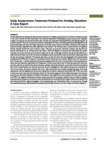
Fig. 1 - MRI showing a massive left hepatolithiasis.
©
C
IC
Ed iz
io ni
In
In January 2007 the Authors observed a 68 yrs old man (w 65 kg; h 177 cm) suffering since 20 yrs from cholelithiasis with recurrent biliary pain but never jaundice, cholangitis or pancreatitis. No other disease was referred for. In December 2006 the patient was admitted for acute pancreatitis; amylases were 1706 UL (n.r.=28-100), slight increase of bilirubin 1,7 mg/dl (n.r.= 0.2-1.1) and increase of cholestatic indexes ( gGT x4, AP x2). A cholangio-MRI described left (II,III, IV segments) massive intrahepatic lithiasis with some branches of the VII segment involved, cefalic and body acute pancreatitis, scleroatrophic cholecistytis and normal size of the common bile duct with several stones. The left biliary tree above the confluence was dilated (Fig 1). Following medical therapy a new cholangio-MRI was performed and a 6cm fluid collection in the body of the pancreas with Wirsung duct dilatation was described as well as a persistent dilatation of the left biliary tree. After a month patient’s general situation was better, no pain or fever were present and amilasys, lipasis, bilirubin and transaminasis were normal. Cholestasis indexes were instead still abnormal. Tumour markers ( CEA, CA 19.9, CA 125) were in the normal range as well. Patient referred to an other istitution for hepatic surgery, refused the proposed surgery (left hepatectomy with caudate resection). He therefore underwent to transhepatic percutaneous cholangiography: the biliary tree was approached through the hepatic branch of the III segment. A dilatation of left main biliary duct at the confluence with a 10mm baloon was performed; removal of the stones from the main branches and pneumatic dilatation with a 10mm baloon of the papilla with removal of choledocal stones followed. Then cholecistostomy was performed to remove the cystic stones. Eventually an external-internal transhepatic percutaneous drain (14F) of the left biliary tree was left in situ. Subsequent repeated cholangiographic sessions using MTBE (metil ter butil etere) and occluding the distal baloon in the choledocus, allowed the selective chateterism of all biliary ducts with complete removal of the stones (Fig 2). Successively injection of saline in the cleaned ducts (30-50 cc- at 12-15 cc/sec) with an automatic device was performed. One month later patient underwent laparoscopic cholecistectomy, the biliary drainage being still in situ. Strong adhesions were described between the liver and the abdominal wall and of the gallbladder with the duodenum and large omentum. Hepatic biopsies at the V segment were performed as well as resection of a 2 cm nodule (with a stone inside) of the free margin of the III segment. Histology confirmed intrahepatic lithiasis with chronic cholecistytis. Discharge occurred 48 hours later with prescription of antibiotic therapy (amoxicillin/clavulanic acid 1grx3/die), selected on antibiogram positive for Klebsiella pneumoniae. Three weeks after the operation (3 months from the initial diagnosis) the patient underwent transhepatic percutaneous cholangiography showing a complete clearance of the biliary tree and a stricture of the Wirsung duct. It was dilated at the confluence with a 3.5 mm baloon and the biliary drainage was removed. MRI control showed a complete resolution of the intraheaptic lithiasis, a normal biliary tree and no trace of the peri pancreatic fluid collection (Fig 3).
al i
Case report
Discussion Intrahepatic lithiasis or hepatolithiasis is characte-
Fig. 2 - Cholangiographic control after stones removal.
rised by the presence of stones above the biliary confluence; it is prevalent in the Far East but increasingly occurring also in Western Countries such as USA (1). First description was from Vachell and Stevens in 1906 (2). Stones formation within biliary duct without associated disease is defined as primary, frequently observed in eastern populations where it is considered endemic, being rare in the West. Frequent is the association with strictures and deformation of the biliary tree: this causes recurrent cholangitis up to biliary cirrhosis with portal hypertension. Biliary sepsis, hepatic abscesses and cholangiocarcinoma are considered potential complications. The origin of a secondary intrahepatic lithiasis may derive from biliary stagnation above a benign or malignant duct stricture; it also occurs along with cystic di425
0457 6 Integrated_Conzo:-
29-09-2011
8:27
Pagina 426
G. Conzo et al.
Fig. 3 - Postoperative control after laparoscopic cholecystectomy.
©
C
IC
Ed iz
io ni
In
latation of the biliary tree. In the secondary forms, more frequent in the western literature, a retrograde migration of stones from the gallbladder to the main ducts is observed, sometimes also the choledocus is involved. Associate strictures are often described (40-80%), sometimes multiple and responsible of relapses (3,4); it is difficult to prove if they cause or are induced by the lithiasis. Some Caroli’s cases present with intrahepatic lithiasis. The left biliary tree is often involved and for unkown reasons notably in the initial phase of the disease (5). In the Chen (6) series of 103 hepatic resections for hepatolithiasis, 86% was in the left biliary tree. Tsunoda proposed a classification of the disease in 4 types on the basis of the site of the stones and the presence of strictures and dilatations (7). Hepatolithiasis is frequent in the eastern countries with incidence varying from 4,1% of Japan to 47,3% of Taiwan (8) ; high rates of relapses are described. Western countries show instead rates of incidence from 0,6 to 1,3% (9,10). Thus two different types of diseases are described and different are the approaches to be used. The primary intrahepatic lithiasis, typical in eastern countries and of unclear etiology, may derive from a recurrent bacterial infection of the bile duct with a bacterial traslocation secondary to a lesion of the intestinal mucosa. It may be related to poor hygienic conditions and probably to specific contents of the diet, often rich in carbohydrates and poor in proteins. Additional cause factors are represented by genic proneness, parasitic infections, congenital abnormalities (11). The association of biliary stagnation and infection is determinant
te
rn az
io n
al i
in the stone formation and moreover a specific enzime activity is able to precipitate conjugate bilirubin with calcium salts. Thus the primary type occurs irrespectively of sex in young patients, often less than 30 yrs of age, and may cause a subtle and asymptomatic evolution to cirrhosis or presents with the typical symptoms (biliary colic, fever, jaundice, cholangitis). In secondary forms patients are older and have a clinical history of biliary colics by cholelithiasis. It is reported also the association between hepatolithiasis and cholangiocarcinoma, maybe related to the chronic cholangitis, with a variable incidence from 2,3 and 10% (12,13) . In such cases diagnosis of the nature of the strictures is extremely complex even with MRI or direct cholangiography. Serum CEA may be useful in these patients (14). Hepatic resection in unclear cases is recommended. Early treatment is mandatory to avoid complications. The introduction of cholangio-MRI among the diagnostic tools has given to transhepatic percutaneous or endoscopic approach a therapeutic sense. A multidisciplinary protocol involving surgeon, endoscopist, interventional radiologist is still controversial. Main goals of treatment are the infection eradication, resolution of biliary stasis with adequate reopening of the biliary tree and salvage of healthy hepatic parenchima. There fore each single case has to be dealed with in referral centre for hepatobiliary disease. Surgical options include choledocotomy with associate sphincteroplasty, biliary derivation, hepatic resections up to liver trasplantation in case of severe secondary biliary cirrhosis (15). Surgical resection carries today low morbidity and mortality rates in referral centers and represents a radical solution for lithiasis and related strictures but it can be extremely complex in case of bilateral multi-segmental involvement. Among the derivative procedures associated or not to resection it is particularly interesting the hepatic-jejunostomy with a chance of percutaneous access to a redo-cholangioscopy (16) and also a defunctioned loop anastomised to the duodenum for a possible future endoscopic trans-duodenal access (17). Patients who underwent hepatectomy with complete removal of stones have a better prognosis in case of massive lithiasis and related strictures compared with those treated without surgery. Despite this, the “conservative” approaches, now widespreadly used, show a comparable efficacy in clearing the stones but maybe carry a higher rate of relapse (11,18,). They include: percutaneous trans-hepatic cholangiographic procedures; endoscopic approaches; percutaneous trans-hepatic coledoscopy that is effective in a high number of cases (11,19) and particularly useful to biopsy dubious strictures; laser or hydraulic lithotomy or chemical dissolution. They all allow a resolution of the pathology up to 90% of ca-
426
0457 6 Integrated_Conzo:-
29-09-2011
8:27
Pagina 427
Integrated treatment of secondary hepatolithiasis. Case report
Conclusion
al i
Hepatic fibrosis or atrophy, recurrent cholangitys with topographic alterations of the biliary tree or the suspiciousness of a cholangiocarcinoma are absolute indications to hepatic resection (even a major resection), often a left lobectomy. Hepatectomies also may have unsatisfactory results and require reoperation (28).
te
rn az
io n
In conclusion, treatment of intrahepatic lithiasis is still matter of debate, basically on the comparison between surgery and conservative approaches. Also the role of resection compared to biliary derivation is controversial. Percutaneous approach seems more effective than endoscopic treatment and is well indicated in case of unilateral disease without complex strictures. Biopsy under visual control is the unchallenged advantage of cholangioscopy; however it carries a higher morbidity. Multiple strictures with biliary distortion, hepatic fibrosis or atrophy and a suspected cholangiocarcinoma are absolute indications to resection. Integration of therapeutic protocols in multidiscilinary teams offers the best long term results.
References
io ni
In
ses (20) especially in secondary cases. Jan reports a success rate for the colangioscopic procedure as high as 83.3% with a 14.5% morbidity rate (21). Occluded strictures, acute angulation of biliary ducts and associate biliary cirrhosis are the main causes of failure for percutaneous procedures. The rate of relapse is around 50% for primary lithiasys, notably in patients with multiple occluded strictures in which it raises to 96% of cases (22,23). Treatment of relapse is still controversial. In case of stenosis the indication to resection should be carefully discussed. In our case the wide dilatation of hepatic biliary duct and papilla and biliary stenting by a large external-internal drainage (14F), for more then three months, reduced the risk of relapse related to biliary hypertension. Often different procedures have to be intergrated to optimise results and reduce morbidity: a multidisciplinary approach is recommended. Some Authors propose surgery (resection, derivation) as the first approach leaving conservative procedures for relapse treatment (24,25,26). This is mainly because surgery allows a complete cleaning and reduces the risk of late relapses descripted at 10% after resection and up to 50% after conservative procedures (14,27).
©
C
IC
Ed iz
1. Harris HW, Kumwenda ZL, Sheen-Chen SM, Shah A, Schecter WP. Recurrent pyogenic cholangitis. Am J Surg 1998; 176: 34-7. 2. Vachell HR, Stevens WM. Case of intrahepatic calculi. B.M.J.1906;1:434-436. 3. Fan ST, Wong J. Complications of Hepatolithiasis. J Gastroenterol Hepatol 1992;7:324-7 4. Matsumoto Y, Fujii H, Itakura J, Miura K, Suda K. Congenital dilatation and stricture of the bile duct as cause of primary intrahepatic calculi. Gakkai Zasshi 1996;97:611-7. 5. Koga A, Miyazaki K, Ichimiya H, Nakayama F. Choice of treatment of Hepatolithiasis based on pathological findings. World J Surg 1984;8:36-40. 6. Chen DW, Tung-Ping Poon R, Liu CL et al. Immediate and longterm outcomes of hepatectomy for hepatolithiasis. Surgery 2004; 135:386-393. 7. Tsunoda T, Tsuchiyda R et al . Long-term results of surgical treatment for intrahepatic stones. Jpn J surg 1985;16:455-62. 8. Nakayama F, Koga A. Hepatolithiasis: present status. World J Surg 1994;8:9-14. 9. Lindstrom CG. Frequency of gallstone disease in a well defined Swedish population: a prospective necropsy study in Malmo. Gastroenterology 1977; 12: 341-346. 10. Simi M, Loriga P, Basoli A, et al. Intrahepatic lithiasis: study of thirty-six cases and review of the literature. Am. J. Surg. 1979; 137: 317-322. 11. Yeh YH, Huang MH, Yang JC, Mo LR, Lin J, Yueh SK. Percutaneous transhepatic cholangioscopy and lithotripsy in the treatment of intrahepatic stones: a study with 5 year follow-up. Gastrointest Endosc 1995; 42: 13-8. 12. Chu KM, Lo CM, Liu CL, Fan ST. Malignancy associated with
hepatolithiasis. Hepatogastroenterology 1997; 44: 352-7. 13. Sane S, Mac Callum JD. Primary carcinoma of the liver: cholangiocarcinoma in hepatolithiasis. Am J Pathol 1942; 18:6758. 14. Kim YT, Byun JS, Kim J, Jang YH, Lee WJ, Ryu JK, et al. Factors predicting concurrent cholangiocarcinomas associated with hepatolithiasis. Hepatogastroenterology 2003; 50:8-12. 15. Strong RW, Chew SP, Wall DR, Fawcett J, Lynch SV. Liver Transplantation for hepatolithiasis. Asian J Surg 2002; 25: 180-3. 16. Fang K, Chou TC. Subcutaneous blind loop : a new type of hepaticocholedochojejunostomy for bilateral intrahepatic calculi. Chin. Med. J. 1977; 3:413-418. 17. Monteiro da Cunha JE, Herman P, Machado MCC, et al. A new biliary access technique for the long-term endoscopic management of intrahepatic stones. J Hepatobiliary Pancreat Surg 2002;9:261-264. 18. Jeng KS, Sheen IS, Yang FS. Percutaneous transhepatic cholangioscopy in the treatment of complicated intrahepatic biliary strictures and hepatolithiasis with internal metallic stent. Surg Laparosc Endosc Percutan Tech 2000; 10: 278-83. 19. Cheung MT, Wai SH, Kwok PCH. Percutaneous transhepatic choledochoscopic removal of intrahepatic stones. Br J Surg. 2003; 90: 1409-1415. 20. Lee SK, Seo DW, Myung SJ, Park ET, Lim BC, Kim HJ, et al. Percutaneous transhepatic cholangioscopic treatment for hepatolithiasis: an evaluation of long-term results and risk factors for recurrence. Gastrointest Endosc 2001; 53: 318-23. 21. Jan YY, Chen MF. Percutaneous transhepatic cholangioscopic lithotomy for hepatolithiasis: long-term results. Gastrointest Endosc 1995; 42: 1-5. 22. Otani K, Shimizu S, et al. Comparison of treatments for hepa-
427
0457 6 Integrated_Conzo:-
29-09-2011
8:27
Pagina 428
G. Conzo et al.
al i
Improvement by a systematic approach. Surgery 1991;109:47480. 26. Choi TK, Wong J. Current management of intrahepatic stones. World J Surg 1990; 14:487-91. 27. Uchiyama K, Onishi H. Indications and procedure for treatment of hepatolithiasis. Arch Surg 2002; 137:149:53. 28. Chijiiwa K, Yamashita H, Yoshida J, Kuroki S, Tanaka M. Current management and long-term prognosis of hepatolithiasis. Arch Surg 1995;130:194-97.
©
C
IC
Ed iz
io ni
In
te
rn az
io n
tolithiasis: hepatic resection versus cholangioscopic lithotomy. J Am Coll Surg 1999;189:177-82. 23. Jenk KS, Yang FS, Chiang HJ, Ohta I. Bile duct stents in the management of hepatolithiasis with long-segment intrahepatic biliary strictures. Br J Surg 1992;79:663-6. 24. Herman P, Perini MV, Machado MAC, et al. Liver resection as the definitive treatment for unilateral non-oriental primary intrahepatic lithiasis. Am J Surg 2006;191:460-464. 25. Fan ST, Choi TK, Lo CM et al. Treatment of hepatolithiasis:
428