Volume 11 Number 17 1983
Nucleic Acids Research
Increased expression of a cloned gene by local mutagenesis of its promoter and ribosome binding site
N.Warburton, P.G.Boseley and A.G.Porter Department of Molecular Genetics, Searle Research and Development, Lane End Road, High Wycombe HP12 4HL, UK Received 14 July 1983; Revised and Accepted 11 August 1983
ABSTRACT A strategy for local mutagenesis of DNA has been developed. The lac promoter in phage M13mp9 was replaced with the E.coli trp promoter. A restriction fragment bearing only the trp promoter region was mutagenized with nitrous acid, religated to the unmutagenized vector and transfected into E.coli. Several clones which give darker blue plaques on indicator media, suggesting increased 0-galactosidase synthesis, were selected for DNA sequencing. One clone has a G—-A transition on the 3' side of the 'Pribnow box1 which results in a constitutive promoter. Two clones have different point mutations (C—-T and T — - C) between the Shine-Dalgarno sequence and initiation codon which raise expression of B-galactosidase two-fold. A secondary structure model suggests that the latter two mutations could exert their effect by destabilizing base-pairing of the lac Z coding region with the ribosome binding site (RBS), thereby allowing easier access to ribosomes. Support for the model comes from the finding that neither of the RBS mutations increase expression of a different downstream gene which forms no obvious secondary structure with the RBS region, whether or not the mutations are present. These results strengthen the hypothesis that secondary structure masking is a major determinant of RBS strength.
INTRODUCTION The DNA sequences which are capable of specifying RNA polymerase (1,2) or ribosome (3,4,5) attachment in prokaryotes are quite short, in both cases around 1OO nucleotides or less. Within these regions, blocks of nucleotides which are important for interaction with the polymerase or ribosome, such as the promoter -35 and -10 regions and the Shine-Dalgarno (S-D) (2,6) sequence of ribosome binding sites have been identified by a combination of approaches. These include protection from nuclease digestion (7), analysis of spontaneous mutants (8,9,10), biochemical methods (7,11,12,13) and deletion mapping (1,5,14).
© IRL Press Limited, Oxford, England.
5837
Nucleic Acids Research It appears that the most important features which determine the strength of E.coli promoters are homology with the -35 (TTGACA) and -10 (TATAATG) 'consensus' sequences, and an ideal spacing of 17 nucleotides between these areas (10,14,15,16). Less is known about the determinants of ribosome binding site (RBS) strength for several reasons. First, the initiation of protein synthesis is a more complex process involving the interaction of ribosomal RNAs, proteins and initiation factors with f-met tRNA and mRNA. Second, the known up and down RBS mutations are few and for the most part have been isolated from unrelated cistrons (8,17,18). Third, sequences which influence expression are sometimes found either within or 3' to the coding region (8,18,19), or 5' to the region protected by ribosomes in an initiation complex (20,21,22). Nevertheless, changes in the initiation codon, the S-D to AUG spacing or the number of potential base-pairs between the S-D sequence and the 3' end of 16s rRNA can dramatically affect expression (8,23,24,25,26). In most cases, however, no simple correlation can be made between the amount of protein synthesized and the S-D to AUG spacing or to the number of potential base- pairs between the S-D and 16S rRNA (9,25,26,27,28). In a different approach to elucidating the major determinants of promoter and RBS strength, we have developed a straightforward strategy for local mutagenesis of restriction enzyme fragments bearing a promoter or RBS, or both. The promoter plus gene or promoter alone, is fused in phase to the E.coli lac Z a-fragment region in a phage M13 vector (29). The amount of S-galactosidase (B-gal) being expressed can be estimated from the intensity of blue colour in phage M13 plaques in agar containing 'Xgal' (5-Bromo4-chloroindoxyl0-D-galactoside) (30). Thus, plates containing thousands of plaques can be rapidly screened for promoter or RBS mutants which express more or less 8-gal fusion protein simply by looking for darker or paler blue plaques. In this paper we describe the method and suggest how it might be generally applied. We also discuss how two different point mutations in the vicinity of the RBS could act to increase expression of the cloned lac Z gene.
5838
Nucleic Acids Research MATERIALS AND METHODS Materials The strain used throughout was E.coll K12 JM101 (A(lac-pro) , SupE, thi, F'traD36, proAB, Iacl9, zAM15 ) , the host for bacteriophage M13 (29) . Phages M13mp8 and 9 were gifts from Dr. N.L. Brown before they were commercially available. All restriction endonucleases and other DNA modifying enzymes were from New England Biolabs or Bethesda Research Laboratories (BRL) and were used in conditions based on the manufacturers' recommendations. Radiochemicals were of the highest specific activity available in stabilized aqueous solution from Amersham International. Low melting temperature (LMT) agarose was from BRL or Sigma; sodium nitrite from Hopkins and Williams; 5-Brorao-4-Chloroindoxyl-8-D-galactoside (Xgal) and isopropyl-S-D-thiogalactoside (IPTG) were from Sigma. Sephacryl S-300 was from Pharmacia. Preparation of DNA Double-stranded phage replicative form (RF) DNA was prepared on a small or large scale from 7 hr. or overnight cultures of infected JM101 by the rapid alkali method (31). The RFs were banded in CsCl gradients. Following pancreatic RNase digestion, each DNA was passed through a column of sephacryl S-3OO (0.8 x 30cm) in 0.2M NaCl, O.lmM EDTA and 2OmM Tris HC1 pH7.5, and the excluded peak retained. This procedure removes all contaminating RNA as well as impurities that sometimes inhibit restriction enzymes. 20yg dsDNA was digested with 40 units each of EcoRI plus PstI (or Avail in the case of pMAN-102; figure 1 ) , and the large vector fragment separated from the 870 (or 765) bp fragment by electrophoresis in a horizontal low melting temperature (1%) agarose minigel (Uniscience, Cambridge) in 25raM Tris-borate (pH8.3), 0.5mM EDTA. In order to extract the vector fragment and the 870 (and 765) bp fragments from the gel, the slices were melted at 65°C, diluted with 2 volumes of 5OmM tris-HCl pH8.0, 0.5mM EDTA, 0.5M NaCl, extracted 3 times with phenol pH7.5, then ether extracted 3 times and finally precipitated with EtOH. Nitrous acid mutagenesis and ligation The purified Pstl-EcoRI 870 bp dsDNA fragment containing trp
5839
Nucleic Acids Research EcgRI
EcoRI
l
Pst I
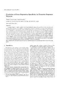
la Figure 1 Vector constructions. I, cut M13mp9 with Avail and Hindlll, polymerase I fill in and ligate (1= lac promoterdeleted ) . la, cut M13mp8 with EcoRI and BamHI, insert 168 bp EcoRI/BamHI human urogastrone gene (40). I-*-II , cut H13mp9 AlacP/O with PstI and EcoRI, insert 870 bp trp promoter/RBS from pWT571 Pstl/EcoRI digest (38,39). I I + n i , cut M13 5PR-00 with EcoRI and Bgll, isolate large fragment and insert 331 bp containing complete human urogastrone gene derived from M13mp8 URO (la). III+IV, cut M13 571/URO with BamHI and cut partially with BgJ.II (at urogastrone codon 47) then self-ligate.
promoter and RBS (isolated as above) was incubated in 70, 210 or 63OmM NaNO-, 0.2mM spermine, 33mM NaOAc pH4.6 in a volume of 25Oyl at 3O°C for 90 min. The pH drop converts NaNO 2 to HN0 2 (32). The reaction was stopped by converting the HN0 2 back to NaNO 2 with lOOpl of 1M Tris-HCl pH8.O, then ethanol precipitated. Equimolar amounts of vector (2.5yg) and HNO2-treated 870 bp (or 765 bp) promoter/RBS fragments were incubated in 66mM Tris-HCl pH7.5, 5mM MgCl,, 2QmM DTT, lmM ATP and 12 units T 4 DNA ligase in a final volume of 0.6ml at 2O°C for 17 hrs. In the DNA segregation experiments, both large and small DNA fragments were kept in 1% LMT agarose and added directly to the ligation mix (up to a maximum of 0.25% agarose in the ligation). 5840
Nucleic Acids Research Transfection 0.4% and 4% of the ligation was added directly to CaCl 2 " treated E.coli JM101 and plated to check the efficiency of the ligation reaction (29). Half of the ligation mix was then added to 0.3ml E.coli JM101 in 80ml CaCl 2 (equivalent to 2.8ml cells in YT at an 0 D 6 0 Q of 0.7) and after taking up DNA, the transformed cells were added to 5ml YT media and grown with shaking at 37°C overnight. The phage titre was measured to determine the dilution giving 4 2 approximately 2-4 x 10 phage plaques on (530cm ) megaplates containing YT agar (Nunc). The additional growth cycle in liquid culture ensures segregation of mutant and non-mutant types. Nucleotide sequencing The 5'-ends of EcoRI-digested candidate DNAs were labelled by the polynucleotide kinase exchange reaction, then recut with Pstl. The 870 bp labelled fragments were purified in 6% polyacrylamide (0.3% bisacrylamide) gels and the sequence from the EcoRI site (through the RBS and trp promoter) was determined by the chemical method of Maxam & Gilbert (33). B-galactosidase assay E.coli JM101 cells were grown at 37°C in minimal media (0.5% casamino acids, 20mM glucose, lmM MgSO. , O.lmM CaCl- and lpg/ml vitamin B 1 ) to an 0 D 6 0 0 of 0.2. They were infected with mutant or parent phage, grown to an 0D,^_ of 0.4 and induced with IPTG OLAJ
at 0.5mM (to induce the lac promoters) and indole acrylic acid (IAA) at 20yg/ml. IAA competes with tryptophan in binding to trp repressor. The resultant complex is unable to bind to DNA, thus preventing repression of genes under trp control (34,35). After 30 minutes of further growth, 2.5ml of culture was removed and cooled. An 0D.._ reading was taken and the cells bUU were then mixed with 2.5ml of induced but uninfected cells to supplement the infected cells with chromosomal 8-gal R-fragment (29). After centrifugation, the cells were resuspended in lml Z buffer (36), then lysed by the addition of 50yl CHC1 3 and 25pl 0.1% SDS, vortexing for 10 sec. The assay was started by the addition of 0.2ml of 4mg/ml ONPG (o-nitro-phenyl-fi-D-galactoside, a colourless compound cleaved by B-galactosidase to the yellow o-nitrophenol). After sufficient yellow colour had developed 5841
Nucleic Acids Research (to give an 0D._ between 0.1 and 2.0) the reaction was stopped by the addition of 0.5ml of 1M Na 2 CO 3 and the reaction time was noted. Further 2.5ml aliquots of culture were taken at intervals during growth (Figure 3 ) . After centrifugation of 0NPG/Na 2 C0, treated samples, 0D, 2 and O D 5 5 Q readings were taken (36) . RESULTS Vector Construction Both the E.coli lac and trp promoters have been used to ensure good expression of cloned genes in prokaryotes (26,37). We chose the trp promoter region for local mutagenesis for two main reasons. First, a series of useful vectors based on the trp promoter lacking the attenuator have previously been constructed (38,39). One of these vectors, pWT571, contains the entire trp promoter and leader polypeptide RBS within a 107 nucleotide stretch (34). This minimizes the number of nucleotides that have to be sequenced to identify each mutant. Second, in pWT571 a unique EcoRI cloning site immediately follows the initiation codon (39) (Figure 2 ) . This facilitates both the isolation of only the promoter/RBS fragment for subsequent nitrous acid mutagenesis (Figure 1 ) , and the insertion of different downstream genes (see later). A summary of the construction of the phage M13-based vectors M13 5PR-00 and M13 pMAN-102 is given in Figure 1 (vectors II and IV, respectively). M13 5PR-OO expresses the B-galactosidase (B-gal) a-fragment under the control of only the trp promoter. M13 pMAN-102 is identical to M13 5PR-00, except that 142 out of the 159 nucleotides of the human urogastrone gene immediately follows the trp leader RBS at the EcoRI site in such a way that a hybrid urogastrone-B-gal protein is expressed (40). Gene fusions with lac Z have been extensively used to monitor or detect gene expression by different promoters, control regions or ribosome binding sites under different conditions (30,40,41, 42,43,44). Usually the hybrid protein retains B-gal activity as indicated by pale blue (or red) coloured plaques (or colonies) on indicator plates. Although the chromosomal trp promoter is normally repressed in the presence of tryptophan (34) , in cells
5842
Nucleic Acids Research Parent 5PR-00
5PR-05
5PR-01
5PR-04
AGTC CC
AGTC CC
AGTC CC
AGTC CC
TAT
a^.
A
A
A C T
(-05) A x
-20
HP? I 7 ;10
I L
Transcription
—" +1
+1
°
(-01) C )•
• S-D j+20
(-04) T «
t j
5-GAACTAGTTAACTAGTACGCAAGTTCACGTAAAAAGGGTATCGACAATGA'ATTCACTTGATCAATTGATCATGCGTTCAAGTGCATT TTTCCCATAGCTGTTACTTAAGT
3'
Figure 2 Nucleotide sequence of the proraoter/RBS of M13 5PR-00 and positions of mutations. The underlinings of the upper (coding) strand indicate (from left to right), the Pribnow (or -10) box, the S-D sequence and the initiation codon (34) . The two-fold symmetry around the promoter -10 region is indicated (from -5 to -18 relative to the transcription start site). The autoradiographs show the nucleotide sequence of each mutant derived from the unique EcoRl site. infected with 5PR-OO or pMAN-102 the high copy number of 200-3OO for the phage replicative form results in partial derepression of trp promoters.
Thus, plaques of both M13 5PR-00 and M13 5843
Nucleic Acids Research pMAN-102
(Figure 1) are a pale blue in rich media containing
Xgal. Local mutagenesis of trp promoter/RBS The steps to isolating up promoter or RBS mutants may be summarized as follows: (1) Isolation of a restriction fragment containing only the promoter/RBS region by digestion of the vector with two different single cutting restriction enzymes. (2) Incubation of the fragment with nitrous acid (HNOj); and subsequent removal of the mutagen. (3) Ligation of the treated fragment to the unmutagenized large vector fragment. (4) Transfection of competent E.coli cells and preparation of a culture supernatant containing M13 phage. (5) Plating out of 4 (>2 x 10 ) M13 phage in YT agar. (6) Selection of darker blue plaques, plaque purification and determination of the nucleotide sequence of the promoter/RBS. In the first experiment, the 870 bp Pstl-EcoRI trp promoter fragment from 5PR-00 (Figure 1) was treated with H N 0 2 (32,45). This fragment contains the 107 bp trp promoter and leader polypeptide R B S , preceded by 763 unrelated nucleotides originally derived from the a m p r gene of pBR322 (38,39,46). The mutagenized fragment was then ligated to the untreated vector, transfected into E.coli JM101 and grown overnight in liquid media. From the 4 culture supernatant, greater than 2 x 1 0 phage plaques were grown in YT agar (see Methods) and inspected for altered intensity of blue colour (Table 1 ) .
Table 1 Plate
HNO2 (tnM)
4 dark blue plaques
Plaque number and colour Plaque colour (Numbers per plate)
Colourless
VPB
PB
>2xloJ
MB
DB
0 0 2 2 4 1 2 0 A restriction fragment containing the trp promoter/RBS was mutagenized with nitrous acid, ligated to vector and the DNA transfected into E.coli JMlOl (see Methods). Equivalent amounts of phage were plated out onto YT agar containing Xgal to assess colour intensity. VPB, very pale blue; PB, pale blue (equivalent to 5PR-00 parental colour); MB, mid-blue; DB, dark blue. 1 2 3 4
5844
0 70 210 630
250 225 2OO
0 3 0 0
>2xlO^ >2xlO< >2xlO
Nucleic Acids Research (designated 5PR-01, -02, -03 and -04) and one slightly darker blue plaque (5PR-05) were chosen for nucleotide sequence analysis and B-galactosidase assays. It was noted that the blue intensities of 5PR-01 to -04 were several-fold greater than that of the parent 5PR-00, while the intensity of 5PR-05 was at least two-fold greater than that of the parent (Table 1 ) . DNA sequence analysis of mutants To locate the mutation(s) responsible for the increased expression of B-gal, the DNA sequence of the 107 nucleotide trp promoter/RBS region was determined from the unique EcoRI site adjacent to the lac Z initiation codon (Figures 1 and 2 ) . The darker blue mutants 5PR-01, -02 and -03 all have the same nucleotide transition mutation (T_—^C) between the S-D and the initiation codon (Figure 2 ) . These may either be sister clones or could result from distinct mutagenic events at the same position on different fragments. 5PR-04 has a G—-T transition in the same region, which is closer to the initiation codon (Figure 2 ) .
The 5PR-01
"-04 series therefore appear to be
ribosome binding site mutants (9). The slightly darker blue mutant 5PR-05 has a G — • A transition two nucleotides to the 3 1 side of the 'Pribnow Box', and before the transcribed portion of the operon (Figure 2 ) . The position of this mutation suggests that promoter function is affected (34,35). No other differences could be seen between the parent and mutant clones in a 120 nucleotide stretch of DNA upstream from the EcoRI site. This stretch contains the original 107 nucleotide trp promoter/RBS fragment (39) and the boundary with the amp gene of pBR322 (Figure 1 ) . Upstream, the remainder of the 870 bp small fragment is unrelated 6-lactamase coding sequence which does not contain a promoter, nor is it transcribed by readthrough from an upstream phage promoter (46,47). These observations strongly suggest that each point mutation in 5PR-01, -04 and -05 is responsible for the increase in expression. B-galactosidase assays In order to quantitate the increase in B-gal synthesis for each mutant, ^n vitro assays of B~gal activity were performed throughout log and stationary phases (36).
Figure 3 shows that
the yield of B-gal for both the -01 and -04 mutants was at least
5845
Nucleic Acids Research two-fold higher than that of the parent. In some experiments the -01 mutant synthesized four to five fold more B-gal than the parent during stationary phase (not shown). In contrast the -05 putative promoter mutant synthesized virtually the same amount of £S-gal as the parent at all times (Figure 3 ) . The apparent discrepancy between the -05 plaque colour and B-gal assay can be rationalized as follows. The -05 promoter mutation (G
»A, Figure 2) is in the same position as a
known partially constitutive trp promoter mutation (35). Although the known mutation is a different nucleotide substitution (a G - T transversion) , it is likely that the -05 mutant is similarly partially constitutive for B-gal and not a genuine promoter up mutation. In rich media a constitutive mutation would further derepress the trp promoter and give a darker blue plaque colour, as was observed. On the other hand, the B-gal assays were performed on extracts of cells grown with IAA in the absence of tryptophan, conditions of no repressor binding in which only genuine promoter up mutations would be seen (34,35). DNA segregation analysis In order to determine whether the 5PR-01, -04 and -05 mutations alone were responsible for increased 6-gal expression in rich media, a DNA exchange experiment was performed. The 870 bp Pstl-EcoRI promoter/RBS fragments of 5PR-01, -04 and -05 (Figure 1 ) , were ligated to the vector fragment of the parent. At the same time, the promoter/RBS fragment of the parent was ligated to the vector fragment of each mutant. After transfection of E.coli JM101 and plating in rich media, the resultant plaque colour intensities clearly corresponded to the colour intensities of the recombinants from which the promoter/RBS fragments had been derived. Since in each case the mutant phenotype segregated with the promoter/RBS fragment, it may be concluded that the observed single point mutations in 5PR-01, -04 and -05 (Figure 2) are alone responsible for the increased B-gal expression in rich media (Figure 3 ) . The 5PR-01 and 5PR-04 mutations could exert their effect by increasing the affinity of the ribosome binding area of the mRNA with one or more ribosomal components (such as initiation
5846
Nucleic Acids Research
1 2 3 4 Tims (hours after IAA induction)
Figure 3 B-galactosidase assays of M13 5PR-00 and three mutants. $-gal activity was assayed as described in Methods and is expressed in Miller units (36). Note that the activities are corrected for 0Dfif)Q fluctuations. (•) , M13 5PR-00; (0), M13 5PR-05; (D) , M13 5PR-04; (A), M13 5PR-01.
factors, protein or RNA) or fMet-tRNA (9). Alternatively, the -01 and -04 mutations could change the secondary (or tertiary) structure of the whole ribosorae binding area including the proximal part of the lac Z coding region (25,49). To distinguish between these possibilities, the promoter/ RBS regions of 5PR-01, -04 and -05 were inserted in front of a human urogastrone - lac Z hybrid gene (40), in the vector pMAN102 (Figure 1 ) . In this clone 142 nucleotides of the synthetic gene for human urogastrone lie between the unique EcoRI site and the lac Z coding region of 5PR-00 such that a hybrid urogastrone-6-gal prctein is synthesized under trp control (Figure 1 ) . Both pMAN-102 and 5PR-00 therefore have identical control regions up to the EcoRI site (Figure 1) and give pale blue plaques in rich media. However, PstI is not unique in pMAN-102 and so the unique Avail site 115 bp closer to the trp promoter/RBS region was used along with EcoRI, to generate slightly shorter (765 bp) promoter/RBS fragments from 5PR-01 , -04 and -05 and pMAN-102 (Figure 1 ) . In a reciprocal DNA exchange experiment similar to the one just described, it was found that the 5PR-05 promoter mutation 5847
Nucleic Acids Research was also effective in further derepressing the synthesis of urogastrone-8-gal in rich media, as expected (not shown). In contrast, neither the -01 nor the -04 RBS mutations altered the expression level of the urogastrone-8-gal hybrid protein as judged by plaque colour. These results indicate that the expression of the 5PR-01 and -04 mutations is probably dependent upon neighbouring downstream lac Z coding sequences (8,25,49). DISCUSSION The selection for up mutations as described in this paper is analogous to the method of maximizing gene expression in which the structure and distance between promoter and gene is varied by limited exonuclease digestion (30). The major differences here are that a mutagen, nitrous acid, is reacted with the promoter/RBS, and the indicator gene is cloned in phage M13 (29,33,48). This approach to local rautagenesis is also similar to that of Hirose et. al. (50). However, the method described here is much more straightforward as the need to use ssDNA for the mutagenesis is circumvented by including spermine, which appears to increase the efficiency of the mutagenesis of dsDNA (45). Also this method does not require DNA sequencing as a first screen for mutant clones. The mechanism and specifity of nitrous acid as a mutagen has been extensively reviewed by Zimmerman (32). Nitrous acid was chosen for the mutagenesis of dsDNA for two reasons. First, both G:C—>-A:T and A:T—>-G:C transitions are known to be induced (32). There are, therefore, a good number of mutational possibilities as the mispairing event could occur with either the coding or non-coding strand. This is in contrast to the method using ssDNA as the target for mutagenesis (50). Second, nitrous acid is a convenient mutagen in that it can be completely removed by raising the pH above 7.0. We have not observed mutations resulting in an altered level of gene expression located outside the area originally treated with nitrous acid. This is in contrast to hydroxylamine treatment of the gal promoter region, where only ~2 0% of the mutations were subsequently found within the promoter/RBS (48).
5848
Nucleic Acids Research The reason for the difference is not known, but may be related to the fact that a different mutagen and host/vector system was used. In order to employ local nitrous acid rautagenesis of promoter regions as a general method, the scheme outlined in Figure 1 is easily modified. The lac promoter-deleted versions of M13 mp8 or 9 (29) are cut with two different restriction enzymes at the start of lac Z and a promoter plus gene inserted, either as the complete gene (equivalent to construction III, figure 1) or directly as a lac Z fusion (cf. pMAN-102). If the fusion cannot be made directly, a restriction site between the gene and the lac Z coding region is opened (eg BamHIinconstructicn III, Figure 1 ) . The ends are trimmed by very limited Bal-31 digestion and religated. Selection for pale blue plaques will give the desired fusion (equivalent to pMAN-102 in Figure 10). Following these manipulations, a unique restriction site will be left upstream of the promoter for subsequent fragment isolation and nitrous acid mutagenesis. However, the whole method has the disadvantage that if a second unique site close to the initiation codon is not present, either the promoter plus gene must be specifically mutagenized or the site must be introduced. For the latter, site-directed mutagenesis is the most likely route since the unique site can be selected for (27,51). The 5PR-01 and 5PR-04 mutations occur between the proposed S-D sequence and the initiation codon, at positions +19 and + 25 of the mRNA, respectively (Figure 2 ) . Neither of these mutations appear to affect the S-D interaction directly (6), nor are they likely to affect transcription initiation as they are found well inside the transcript, and outside the region in the DNA recognized by RNA polymerase (1). These mutations do not alter the S-D to AUG spacing (24,25) and do not appear to significantly alter preferred nucleotide usage (9,52). It has been suggested that termination codons may be important determinants in translation initiation (53). Although the -04 mutation introduces a second TAA termination codon in the vicinity of the RBS, the -01 mutation neither destroys nor introduces any of the three termination codons (Figure 2 ) . Thus the -01 and -04 mutations must affect some other aspect of
5849
Nucleic Acids Research 21 tip.-
M1-3-5PR-04 U
H13-5PR-01
Figure 4 Possible stable secondary structure for the 51 terminal region of M13 5PR-00 mRNA. The positions of the RBS mutations are arrowed. The proposed Shine-Dalgarno (S-D) sequence is boxed and the initiation codon is underlined. Numbering is from the 5' end of the mRNA (34). translation initiation. A clue to the mode of action of these mutations comes from the DNA segregation experiments. It was clearly demonstrated that the -01 and -04 mutations increase expression of B-gal in 5PR-00 but not urogastrone-B-gal in pMAN-102, even though both clones have identical control regions (Figure 2 ) . This strongly suggests that the phenotypic expression of the -01 and -04 mutations is dependent upon the nature of the downstream coding sequences. Other investigators have postulated or provided evidence that mRNA secondary structure masking effects are important in translation initiation (3,9,19,25,49,54). In most cases, local secondary structure has been considered since isolated
5850
Nucleic Acids Research RBS fragments from ~3 5 to ~120 nucleotides rebind well to ribosomes (3,4,5). In some mRNAs, however, long range interactions are involved (19). Using calculations to determine the stability of intramolecular base-paired regions in RNA (55,56) a possible secondary structure for the initiating region of the 5PR-00 (parent) mRNA can be proposed (Figure 4 ) . The structure has a free energy (AG at 25°C) of -9 kcal. In this model, the first part of the lac Z coding region is paired with 51 noncoding sequences. The initiation codon is unpaired in a hairpin loop, thought to be important for efficient translation initiation (9), while most of the S-D sequence is base-paired in a stem region (Figure 4 ) . Both the 5PR-01 and -04 mutations adversely affect the overall stability of the proposed structure, reducing the calculated free energies (AG at 25°C) from -9 kcal to -6.8 kcal and -6.4 kcal, respectively (55). When the same analysis is performed with the initiator region of the urogastrone-lac Z hybrid gene, there is a striking lack of local secondary structure at least in the 5' terminal 90 nucleotides, whether or not the -01 and -04 mutations are present (not shown). Taken together, these observations are consistent with the -01 and -04 mutations changing the local mRNA secondary structure so as to make the initiator AUG and S-D sequence more accessible to ribosomes, leading to an increased rate of translation initiation. This conclusion is in line with other evidence which points to the degree of masking of the initiator region by RNA secondary structure as an important determinant of ribosome binding site strength (3,19,25,49). However, it is unlikely that a simple proportional relationship exists between accessibility and RBS strength, since there are other determinants such as the S-D sequence and AUG. Also, ribosomes usually rebind only to isolated initiator fragments of a certain minimum length, which varies from site to site (3,4,5). With the techniques of local DNA mutagenesis described here and elsewhere (48,50,51) multiple mutations can now be induced in a single binding site or any regulatory region of DNA. Hypotheses, such as those involving secondary structure models, can then be tested by introducing specific single nucleotide
5851
Nucleic Acids Research changes with the powerful technique of oligonucleotide-directed mutagenesis (27,51). ACKNOWLEDGEMENTS We wish to thank Dr. N.L. Brown for M13 mp8 and 9, Dr. J. Adair for critical reading of the manuscript, Mr. J. Hobbs for photography, Mrs. J. Rogers and Mrs. D. Granshaw for typing the manuscript and Dr. B.M. Richards for excellent research facilities. REFERENCES 1. Brown, K.D., Bennett, G.N., Lee, F., Schweingruber, M.E. and Yanofsky, C. (1978) J. Mol. Biol. _1_2J_, 153-177. 2. Rosenberg, M. and Court, D. (1979) Ann. Rev. Genet. 13, 319-353. 3. Steitz, J.A. (1973) Proc. Natl. Acad. Sci. USA 2£/ 26052609. 4. Porter, A.G. and Hindley, J.H. (1973) FEBS Lett. 3_3» 339342. 5. Borisova, G.P., Volkova, T.M. Berzin, U., Rosenthal, G. and Gren, E.J. (1979) Nucleic Acids Res. ^, 1761-1764. 6. Shine, J. and Dalgarno, L. (1975) Nature ^5_4, 34-38. 7. Siebenlist, U., Simpson, R.B. and Gilbert, W. (1980) Cell 2£, 269-281. 8. Dunn, J.J., Buzash-Pollert, E. and Studier, F.W. (1978) Proc. Natl. Acad. Sci. USA 7_5/ 2741-2745. 9. Gold, L., Pribnow, D., Schneider, T., Shinedling, S., Singer, B.S. and Stormo, G. (1981) Ann. Rev. Microbiol. 35, 365-403. 10. Youderian, P., Bouvier, S. and Susskind, M.M. (1982) Cell 2£, 843-853. 11. Steitz, J.A. and Jakes, K. (1975) Proc. Natl, Acad. Sci. USA T2, 4734-4738. 12. Taniguchi, T. and Weissmann, C. (1978) Nature 275, 770-772. 13. Neilson, T., Kofoid, E.C. and Ganoza, M.C. (19F5T Nucleic Acids Res., Symposium No. 7, 313-323. 14. Mandecki, W. and Reznikoff, W. (1982) Nucleic Acids Res. 10, 903-912. 15. Gilbert, W. (1976) in RNA Polymerase, Losick, R. and Chamberlin, M. Eds. pp 193-205, Cold Spring Harbor Laboratory, New York. 16. Calos, M.P. and Miller, J.H. (1981) Mol. Gen. Genet. 183, 559-560. 17. Taniguchi, T. and Weissmann, C. (1978) J. Mol. Biol. 118 533-565. 18. Atkins, J.F., Steitz, J.A., Anderson, C.W. and Model, P. (1979) Cell J_8, 247-256. 19. Min Jou, W., Haegeman, G., Ysebaert, M. and Fiers, W. (1972) Nature _23J7, 82-88. 20. Cannistraro, V.J. and Kennell, D. (1979) Nature 2J2, 407409.
5852
Nucleic Acids Research 21. 22. 23. 24. 25. 26. 27. 28. 29. 30. 31. 32. 33. 34. 35. 36. 37. 38. 39. 40.
41. 42. 4 3. 44. 45. 46. 47. 48. 49.
Queen, C. and Rosenberg, M. (1981) Cell 2^, 241-249. Roberts, T.M., Kacich, R. and Ptashne, M. (1979) Proc. Natl. Acad. Sci. USA 7j>, 760-764. Belin, D., Hedgpeth, J., Selzer, G.B. and Epstein, R.H. (1979) Proc. Natl. Acad. Sci. USA ]_6, 700-704. Chang, A.C.Y., Erlich, H.A., Gunsalus, R.P., Nunberg, J.H., Kaufman, R.J., Schimke, R.T. and Cohen, S.N. Proc. Natl. Acad. Sci. USA 22' 1442-1446. Gheysen, D., Iserentant, D., Derom, C. and Fiers, W. (1982) Gene V7, 55-63. Shepard, H.M., Yelverton, E. and Goeddel, D.V. (1982) DNA I, 125-131 . Gillam, S., Astell, C.R. and Smith, M. (1980) Gene V2_, 129-137. Rosa, M.D. (1981) J. Mol. Biol. 147, 55-71. Messing, J. and Vieira, J. (1982) Gene J_9, 269-276. Guarente, L., Lauer, G., Roberts, T.M. and Ptashne, M. (1980) Cell ^ 0 , 543-553. Birnboim, H.C., and Doly, J. (1979) Nucleic Acids Res. 1_, 1513-1523. Zimmerman, F.K. (1977) Mutation Res. Vi_, 127-148. Maxam, A. and Gilbert, W. (1977) Proc. Natl. Acad. Sci. USA 7_4, 560-564. Crawford, I.P. and Stauffer, G.V. (1980) Ann. Rev. Biochem. ^ 3 , 163-195. Bennett, G.N. and Yanofsky, C. (1978) J. Mol. Biol. 121, 174-192. Miller, J.H. (1972) in Experiments in Molecular Genetics pp. 352-355, Cold Spring Harbor Laboratory, New York. Goeddel, D.V., Heyneker, H.L., Hozumi, T., Arentzen, R., Itakura, K., Yansura, D.G., Ross, M.J., Miozzari, G., Crea, R. and Seeburg, P.H. (1979) Nature £8J[, 544-548. Tacon, W. (1981) PhD. Thesis, University of Sussex, U.K. Tacon, W., Bonass, W.A., Jenkins, B. and Emtage, J.S. (1983) Gene (in press). Smith, J., Cook, E., Fotheringham, I., Pheby, S., Derbyshire, R. , Eaton, M.A.W., Doel, M., Lilley, D.M.J., Pardon, J.F., Patel, T., Lewis, H. and Bell, L.D. (1982) Nucleic Acids Res. H ) , 4467-4481. Casadaban, M.J. and Cohen, S.N. (1980) J. Mol. Biol. 138, 179-207. Casadaban, M.J., Chou, J. and Cohen, N. (1980) J. Bact. 143, 971-980. Koenen, M., Ruther, U. and Muller-Hill, B. (1982) EMBO J. 1, 509-512. Rosner, A., Gorecki, M. and Aviv, H. (1982) Z. Naturforsch. _3J7, 441-444. Sandri-Goldin, R.M., Levine, M. and Glorioso, J.C. (1981) J. Virol. J3§, 41-49. Sutcliffe, J.G. (1978) Proc. Natl. Acad. Sci. USA 75, 3737-3741. Van Wezenbeek, P.M.G.F., Hulsebos, T.J.M. and Schoenmakers, J.G.G. (1980) Gene 21- 129-148. Busby, S., Irani, M. and de Crombrugghe, B. (1982) J. Mol. Biol. JJ?_4, 197-209. Hall, M.N., Gabay, J., DebarbouillS, M. and Schwartz, M. (1982) Nature 295, 616-618. 5853
Nucleic Acids Research 50. 51. 52. 53. 54. 55. 56.
5854
Hirose, S., Takeuchi, K and Suzuki, Y. (1982) Proc. Natl. Acad. Sci. USA 21, 7258-7262. Shortle, D., Di Maio, D. and Nathens, D. (1981) Ann. Rev. Genet. ±5, 265-294. Scherer, G.F.E., Walkinshaw, M.D., Arnott, S. and Morre\ D.J. (1980) Nucleic Acids. Res. 8, 3895-3907. Atkins, J.F. (1979) Nucleic Acids Res. 1_, 1035-1041. Lodish, H.F. (1970) J. Mol. Biol. _50, 689-702. Tinoco, I., Borer, P.N., Dengler, B., Levine, M.D., Uhlenbeck, O.C., Crothers, D.M. and Gralla, J. (1973) Nature New Biology 2A6^, 40-41. Borer, P.N., Dengler, B., Tinoco, I. and Uhlenbeck, O.C. (1974) J. Mol. Biol. 86, 843-853.