JASN Express. Published on March 16, 2005 as doi: 10.1681/ASN.2004030219
Genome-Wide Scan in a Novel IgA Nephropathy Model Identifies a Susceptibility Locus on Murine Chromosome 10, in a Region Syntenic to Human IGAN1 on Chromosome 6q22–23 Hitoshi Suzuki,* Yusuke Suzuki,* Takahiro Yamanaka,* Sachiko Hirose,† Hiroyuki Nishimura,‡ Junichi Toei,‡ Satoshi Horikoshi,* and Yasuhiko Tomino* *Division of Nephrology, Department of Internal Medicine, and †Second Department of Pathology, Juntendo University School of Medicine, Tokyo; and ‡Toin Human Science and Technology Center, Toin University of Yokohama, Yokohama, Japan Genetic factors are considered to be involved in the initiation and progression of IgA nephropathy (IgAN) on the basis of racial differences in the prevalence and familial aggregation. The ddY mouse is a spontaneous animal model of human IgAN, with a highly variable incidence and extent of glomerular injury as a result of the heterogeneous background, resembling the human situation. It was hypothesized that susceptibility genes for IgAN can be detected by a genome-wide scan using this model. First, serial renal biopsies were performed at 20, 40, and 60 wk of age in 361 ddY mice. The ddY mice were classified into three groups on the basis of the onset of glomerular injury: Early onset at 20 wk (31.9%), late onset at 40 wk (37.9%), and quiescent even at 60 wk (30.2%). The severity of glomerular lesions in both onset groups correlated with the intensity of glomerular IgA deposition but not with serum IgA level. The genome-wide scan with 270 microsatellite markers identified three chromosomal regions on chromosomes 1, 9, and 10, which were significantly associated with the glomerular injuries. Surprisingly, the peak marker D10MIT86 on chromosome 10 is located on the region syntenic to human 6q22–23 with IGAN1, which is the responsible candidate of familial IgAN. In addition, D1MIT16 on chromosome 1 was very closely located at the locus of selectin gene, which is a known candidate of human IgAN. In conclusion, the three-group ddY mouse model can be a useful tool for identifying the susceptibility genes and also to examine their roles in the pathogenesis of IgAN. J Am Soc Nephrol 16: ???–???, 2005. doi: 10.1681/ASN.2004030219
I
gA nephropathy (IgAN) is the most common form of primary glomerulonephritis in the world and frequently progresses to end-stage renal failure (1). Although specific therapy for IgAN is urgently required, the etiopathogenesis of IgAN remains unclear. The large number of studies conducted during the past three decades since the first description of IgAN by Berger and Hinglais (2) have identified many abnormalities in IgAN. Recent studies also examined the roles of abnormalities in the IgA molecule itself, such as negative charge and poor glycosylation, and some of IgA receptors, in the pathogenesis of IgAN (3– 6). However, the molecular mechanisms underlying glomerular IgA deposition and progression are poorly understood. Therefore, a new approach or a novel disease model is needed for the elucidation. Previous studies reported a higher risk of identical twins to IgAN and several cases of familial IgAN (7,8). Of note, linkage analysis of familial IgAN revealed a close association with the trait 6q22–23 (IGAN1) in this disease (9), although no proteins
Received March 20, 2004. Accepted January 13, 2005. Published online ahead of print. Publication date available at www.jasn.org. Address correspondence to: Dr. Yasuhiko Tomino, Division of Nephrology, Department of Internal Medicine, Juntendo University School of Medicine 2-1-1 Hongo, Bunkyo-ku, Tokyo 113-8421, Japan. Phone: 81-3-5802-1064; Fax: 81-33813-1183; E-mail:
[email protected] Copyright © 2005 by the American Society of Nephrology
coded by IGAN1 have been identified. Moreover, certain candidate genes associated with IgAN, such as human leukocyte antigen, MHC, uteroglobin (UG), selectin, and angiotensinconverting enzyme (ACE) genes (10 –17), were also reported. These findings suggested that genetic background could contribute to the disease susceptibility (18 –21). Because a combination of various genes and environmental factors is critical in the incidence of IgAN, the interrelation between the susceptibility locus and disease phenotypes should be assessed using appropriate models. To date, several animal models of human IgAN have been used, including the UG knockout mouse, UG antisense transgenic mouse (22), minks with Aleutian disease (23), experimental mouse model by Sendai virus (24), the soluble CD89 transgenic mouse (25), and ddY mouse. It has been suggested that UG prevents the formation of IgA-fibronectin complexes with subsequent mesangial deposition as an anti-inflammatory protein in mice (22). However, Coppo et al. (26) reported that UG in human IgAN does not function in the same way as in transgenic mice. However, the ddY mouse is an animal model of spontaneous IgAN, and the glomerular lesions in these mice include mesangial proliferation and extracellular matrix expansion with paramesangial IgA depositions similar to those found in human IgAN (27). However, this mouse model has a serious flaw with respect to human IgAN in that the extent of the ISSN: 1046-6673/1604-0001
2
Journal of the American Society of Nephrology
disease is highly variable because the affected mice were originally maintained as outbred animals (28 –30). However, we considered this mouse to be appropriate for our experiment because this clinical feature based on the heterogeneous genetic background resembles human IgAN. Therefore, it was presumed that we could study the regulatory genes that are responsible for the onset and/or progression of IgAN by genome-wide genetic association study between ddY mice with or without glomerular injury as determined by serial renal biopsies. In this study, we performed serial renal biopsies on each of the 361 ddY mice and found that ddY mice could be categorized into three groups on the basis of the onset of glomerular injury: The early-onset group, the late-onset group, and the quiescent group. Then, we designed a genome-wide association study between the early-onset and the quiescent groups to detect the susceptibility genes for IgAN. The identified genes should be investigated in the future to assess their role in the pathogenesis of IgAN. The results also point to the importance of ddY mice as a useful tool for the verification of those genes.
Materials and Methods Mice A total of 361 female ddY mice at 10 wk of age (SLC Japan, Shizuoka, Japan) were maintained at the animal facility of Juntendo University and provided with regular chow (MF; Oriental Yeast, Tokyo, Japan) and water ad libitum in a specific-pathogen-free room. The controls were 22 age-matched female BALB/c mice. The experimental protocol was approved by the Ethics Review Committee for Animal Experimentation of Juntendo University School of Medicine.
Serum and Urinary Analyses Blood samples were obtained from the orbital venous plexus using capillary tubes every 10 wk. The serum samples were stored at ⫺80°C before use. The serum levels of IgA were measured by single radioimmunodiffusion (SRL, Tokyo, Japan). Urinary albumin was measured using an ELISA kit (Albuwell; Exocell, Philadelphia, PA). The severity of hematuria was determined by reagent strips (Multistix; BayerSankyo, Tokyo, Japan). Urinalysis was also performed every 10 wk. Serum and urine samples from BALB/c mice were used as controls.
Serial Renal Biopsies Renal biopsies were performed at 20, 40, and 60 wk of age. The right kidney was examined at 20 and 60 wk of age, and the left kidney was used for the biopsy at 40 wk of age. Pentobarbital sodium (0.2 mg/g body wt) was used for anesthesia. Before the operation, the back hair was shaved and the skin was disinfected with a povidone-iodine solution. An incision of approximately 10 mm was made in the lower back, and the kidney was pulled out using small forceps. A slice of the lower pole of the kidney was removed using a scalpel.
Histologic Examination of Renal Tissues For light microscopy, the specimens were fixed in 10% neutral phosphate-buffered formalin, embedded in paraffin, and sliced at 2 m. The slices were stained with hematoxylin/eosin and periodic acid-Schiff. Snap-frozen 4-m-thick renal sections were used for immunofluorescence with FITC-conjugated goat anti-mouse IgA (BD Biosciences, Pharmingen, San Diego, CA) and goat anti-mouse complement 3 (C3; Cappel Research Reagents, Costa Mesa, CA) antisera. For electron
J Am Soc Nephrol 16: ???–???, 2005
microscopy, the specimens were fixed in phosphate-buffered glutaraldehyde for 2 h, postfixed in 2% osmium tetroxide for 2 h, and then embedded in Epon resin after dehydration. Ultrathin sections were sliced at 70 nm, stained with 4% uranyl acetate and lead citrate, and then examined under an electron microscope (Hitachi 7100, Tokyo, Japan).
Genotyping of Each ddY Mouse Genomic DNA was obtained from mouse tails by the QIAamp DNA mini kit (Qiagen, Valencia, CA). Genotyping using 270 microsatellite markers was performed via PCR with fluorescence-labeled oligonucleotides (Perkin Elmer-Cetus, Foster City, CA) and subsequent ABI 3700 sequencer (Applied Biosystems, Foster City, CA). Electrophoretic profiles were analyzed by Genescan and Genotyper software (Applied Biosystems).
Phenotyping of Individual ddY Mice The biopsy specimens were evaluated in a triple-blind manner by two nephrologists and one pathologist. The biopsy specimens that contained ⬎30 glomeruli were used for histopathologic analysis. All biopsies were analyzed quantitatively to determine the percentages of glomeruli with (1) segmental and global sclerosis and/or (2) mesangial cell proliferation and/or (3) increase in mesangial matrix. All biopsies were scored semiquantitatively for the percentages of the aforementioned lesioned glomeruli (score 0, 0%; score 1, 1 to 24%; score 2, 25 to 49%; and score 3, ⱖ50% of all glomeruli) (31,32). To determine the cutoff criteria of the histology score, we defined renal injury as renal lesions with a histology score of more than the mean ⫾2 SD of the score of BALB/c mice. In addition to the glomerular score, we classified ddY mice into two groups on the basis of the serum IgA level: High and low serum IgA groups. Mice with serum IgA levels of more than the mean ⫾2 SD of BALB/c mice at 30 wk of age (170 mg/dl) were classified as the high serum IgA group.
Statistical Analyses Mice were categorized into two groups by phenotypes and into three groups by genotypes (homozygotes with respect to allele 1; allele 2 and heterozygote with respect to alleles 1 and 2). The 2 ⫻ 3 contingency tables were tested for the 2 statistics. For the chromosomal regions with significant association, allele frequencies were also compared between cases and controls. To obtain the empirical significance levels associated with the 2 statistics observed in the genome-wide association studies, we conducted permutation tests by permutating the phenotype data of 361 ddY mice with fixed genotype data. A total of 10,000 replicates were tested. Alternatively, segregation of 270 microsatellite markers in randomly mated ddY mice (37 families with 10 offspring) were simulated with the SIMULATE program (33,34) using the observed allele frequencies and the recombination frequencies between the markers obtained from the database (http://www.broad.mit.edu/ cgi-bin/mouse/index). Affection status was randomly assigned to the progeny, and a total of 10,000 replicates of the progeny were tested for the frequencies of type I errors. Confidence intervals for the empirical significance levels were calculated by BINOM program (35). Correlations between the different parameters were analyzed by the t test. ANOVA was used to determine differences in the characteristics among groups. Data are expressed as mean ⫾ SD or median values. P ⬍ 0.05 was considered significant. All statistical analyses were performed using the Macintosh version of StatView 5.0 software (Abacus Concepts, Berkeley, CA).
J Am Soc Nephrol 16: ???–???, 2005
Genome-Wide Scan for Murine IgA Nephropathy
3
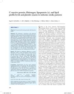
Results Categorization of ddY Mice into Three Pathologic Groups We examined the onset and progression of glomerular injury by serial renal biopsies in 361 female ddY mice. The extent of histopathologic changes was quantified by analyzing all biopsies (Figure 1A). The score of renal injury correlated with the level of albuminuria (data not shown). The histology score of BALB/c mice was 1.42 ⫾ 0.97. Thus, ddY mice with histology scores of ⱖ4.0 (mean ⫾ 2 SD of BALB/c mice) were regarded to have IgAN. The ddY mice could be classified into three groups on the basis of the onset of glomerular injury. We defined these groups as follows: Early onset (31.9% of the total number of mice), late onset (37.9%), and quiescent (30.2%). The scoring of glomerular injury at each age in the three groups is shown in Figure 1B. In the early-onset group, glomerular injury including mesangial cell proliferation and segmental expansion of mesangial matrix could be detected at 20 wk of age. At 40 wk of age, this group of mice showed progression of mesangial matrix expansion from focal segmental to diffuse global lesions. At 60 wk of age, many mice were found to have progressed to diffuse global glomerulosclerosis with glomerular enlargement (Figure 2A). The deposition of IgA in glomerular mesangial areas gradually increased from 20 to 40 wk of age. Moderate IgA deposition was observed not only in the glomerular mesangial areas but also in the capillary walls at 60 wk of age (Figure 2B). In the late-onset group, the same type of glomerular injury observed in the early-onset group at 20 wk of age was detected from 40 wk of age (Figure 2A). Although this group did not show significant glomerular IgA deposition at 20 wk of age, this was detected in association with glomerular injury (Figure 2B). Granular deposition of IgA and C3 was co-localized in glomerular mesangial areas in the early- and late-onset groups (Figure 2B). Electron microscopy showed electron-dense deposits mainly in the glomerular mesangial areas in both groups (Figure 2C). In contrast, no significant glomerular lesion or IgA deposition was observed in the quiescent group even at 60 wk of age or in the control BALB/c mice (Figure 2, A and B).
High Variability of Clinical Parameters in ddY Mice
Figure 1. Box-and-whisker plots of glomerular injury scores of all ddY mice, early-onset, late-onset, and quiescent groups. (A) Renal samples were scored semiquantitatively for renal injury at 20, 40, and 60 wk of age. Score of renal injury varied widely at each age. (B) Cases with scores of ⬎4 were regarded as glomerular injury (see Materials and Methods section). Mice with glomerular injury at 20 wk of age were classified as the early-onset group. Mice with renal injury first detected at 40 wk of age were classified as the late-onset group. The quiescent group did not show significant glomerular injury even at 60 wk of age.
The serum concentration of IgA in ddY mice ranged from 45 to 720 mg/dl at 20 wk of age. Although levels of serum IgA varied widely, the mean level gradually increased with aging (Figure 3A). The serum concentration of IgA of ddY mice was significantly higher than that of BALB/c mice at each age (Figure 3A). At 20 wk of age, 28% of ddY mice had high serum levels of IgA (⬎170 mg/dl: mean ⫾ 2 SD of BALB/c mice). However, half of the ddY mice had normal serum concentration of IgA during the experiment, matching those of BALB/c mice. The urinary albumin concentration in BALB/c mice was 11.6 ⫾ 2.6 mg/dl even at 60 wk of age, whereas that in ddY mice increased with aging from 14.4 ⫾ 12.8 at 20 wk to 16.7 ⫾ 11.1 at 30, 42.9 ⫾ 106.7 at 40, 45.2 ⫾ 85.1 at 50, and 54.1 ⫾ 121.3 mg/dl at 60 wk of age (Figure 3B). Significantly higher levels of albuminuria were noted in 29% of ddY mice compared with BALB/c mice at 20 wk of age. Approximately 30% of ddY mice showed no significant elevation of albuminuria even at 60 wk
4
Journal of the American Society of Nephrology
J Am Soc Nephrol 16: ???–???, 2005
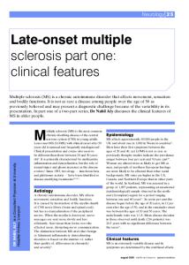
Figure 2. (A) Renal histopathology of the early-onset, late-onset, and quiescent groups of ddY mice. In the early-onset group, expansion of the glomerular mesangial matrix with paramesangial deposits was observed at 20 wk of age. These changes worsened with aging. In the late-onset group, the same type of glomerular injury was detected from 40 wk of age. In the quiescent group, no significant glomerulonephritis, as seen in BALB/c mice, was observed during the experiment. (B) Glomerular IgA and complement 3 (C3) depositions in each group of ddY mice. In the early-onset group, mesangial IgA deposition was seen at 20 wk of age. At 40 wk of age, depositions of mesangial IgA and C3 could be detected in the early- and late-onset groups. Glomerular IgA and C3 depositions were not observed during the experiment in the quiescent group. (C) Electron-dense deposits were observed mainly in the glomerular mesangial areas. Electron microscopy showed marked expansion of the glomerular mesangial matrix and mesangial electron-dense deposits. GBM, glomerular basement membrane; Po, podocyte; EN, endothelial cells; MC, mesangial cells; ED, electron-dense deposits. Magnification, ⫻400 in A and B; ⫻3200 in C.
J Am Soc Nephrol 16: ???–???, 2005
Genome-Wide Scan for Murine IgA Nephropathy
5
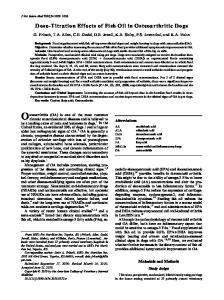
Figure 3. (A) Serum levels of IgA in ddY and BALB/c mice. The serum level of IgA of ddY mice (n ⫽ 361) was higher than that of BALB/c mice (n ⫽ 22) at each age. The mean IgA values increased with aging in ddY mice. (B) Urinary albumin concentrations in ddY and BALB/c mice. The levels of urinary albumin of ddY mice were significantly higher than those of BALB/c mice from 40 wk of age. Data are presented as mean ⫾ SD. *P ⬍ 0.01.
of age. Although these rates of albuminuria were correlated with the incidence of glomerular injury, ddY mice did not show hematuria throughout the experiments.
Lack of Correlation between Glomerular Injury Associated with IgA Deposition and Serum IgA Levels The incidence of glomerular injury correlated significantly with the level of urinary albumin (P ⫽ 0.0031; Figure 4A) and the intensity of glomerular IgA deposition (P ⫽ 0.002; Figure 4B) in both the early- and late-onset groups. However, serum concentration of IgA did not correlate with the incidence of glomerular injury in any of the groups (P ⫽ 0.818; Figure 4A). Moreover, there was no significant correlation between serum IgA levels and the intensity of glomerular IgA deposition in any of the ddY groups (P ⫽ 0.8622; Figure 4C).
Detection of Susceptibility Loci of Renal Injury by GenomeWide Genetic Association Study in ddY Mice A genome-wide scan using microsatellite markers with an average resolution of 8 cM (270 markers) was performed in each ddY mouse. Nearly 97% of these microsatellite markers were biallelic. Only six microsatellite markers had three alleles. Because the frequencies of the least frequent third alleles of these markers were not significantly different between cases and controls, mice with major two alleles were analyzed for these markers. First, we analyzed the association between the early-onset group (n ⫽ 115) and the quiescent group (n ⫽ 109). The data of 2 tests at given marker loci in ddY mice are shown in Table 1 and Figure 5. The marker loci with significant association to renal injury were distributed on chromosomes 1, 9, and 10. The marker loci with the highest 2 values in each chromosome were D1MIT16 at 87.2 cM on chromosome 1 (2 ⫽ 16.645, P ⫽ 0.00029, genome-wide significant P ⫽ 0.0054), D9MIT252 at 17.0
cM on chromosome 9 (2 ⫽ 22.260, P ⫽ 0.0001, genome-wide significant P ⬍ 0.0001), and D10MIT86 at 17.0 cM on chromosome 10 (2 ⫽ 18.945, P ⫽ 0.00022, genome-wide significant P ⫽ 0.0054; Table 1, Figure 5, top three panels). Table 1 shows the flanking markers of each peak and their positions. The peak marker D10MIT86 on chromosome 10 is located on the region syntenic to human 6q22–23 with IGAN1, which is responsible for the susceptibility to familial IgAN. In addition, the peak marker D1MIT16 at 87.2 cM on chromosome 1 is located close to the selectin gene (sele) at 86.6 cM. Next, we performed an association study between ddY mice with glomerular injury at 40 wk of age, including the early- and late-onset groups, and the quiescent ddY mice. The 2 values of each marker loci were lower than the previous data from the early-onset group alone shown in Figure 5; i.e., D1MIT16 at 87.2 cM on chromosome 1 (2 ⫽ 9.9, P ⫽ 0.01285), D9MIT252 at 17.0 cM on chromosome 9 (2 ⫽ 21.3, P ⫽ 0.00019), D10MIT86 at 17.0 cM on chromosome 10 (2 ⫽ 5.2, P ⫽ 0.019), and D1MIT216 at 49.7 cM on chromosome 1 (2 ⫽ 7.9, P ⫽ 0.017). These results suggested that the early-onset group could have a stronger genetic background with regard to the onset of glomerular injury than the late-onset group. Therefore, the association study between the early-onset and quiescent groups seems to be more useful for analysis of the susceptibility gene in this disease. As indicated above, mice that had serum IgA levels that exceeded the mean ⫾ 2 SD of BALB/c mice at 30 wk of age (170 mg/dl) were regarded as the high serum IgA group. A high serum level of IgA was significantly associated with D12MIT20 at 58.0 cM on chromosome 12 (2 ⫽ 17.1, P ⫽ 0.00018, genomewide significant P ⫽ 0.0014; Figure 5, bottom). The flanking markers are shown in Table 1. The marker D12MIT20 was closely located at the gene of the Ig heavy chain (Igh). Serum
6
Journal of the American Society of Nephrology
J Am Soc Nephrol 16: ???–???, 2005
Figure 4. (A) Urinary albumin concentrations but not serum IgA concentrations correlated with glomerular injury. The incidence of glomerular injury was associated with albuminuria (P ⫽ 0.0031). In the quiescent group of ddY mice, no significant albuminuria was observed, similar to the control mice. There was no significant correlation between serum IgA levels and the incidence of glomerular injury in each group (P ⫽ 0.8185). (B) Glomerular injury is associated with glomerular IgA deposition. In the earlyand late-onset groups, glomerular IgA depositions were detected in association with glomerular injury (P ⫽ 0.002). (C) Intensity of glomerular IgA deposition does not correlate with the serum levels of IgA. There was no significant correlation between serum IgA levels and intensity of glomerular IgA deposition (P ⫽ 0.8622). Data are mean ⫾ SD. *P ⬍ 0.001. IgA levels did not show significant association with the susceptibility loci of glomerular injury.
Discussion In this study, we demonstrated that the ddY mice could be classified into three groups during the course of the disease—the early-onset group (31.9%), the late-onset group (37.9%), and the quiescent group (30.2%)— on the basis of serial renal biopsies. Because genetic background may influence the disease susceptibility in this model, we performed a genome-wide genetic association study for the onset of renal injury or high levels of serum IgA. Our results identified three major chromosomal intervals— chromosomes 1, 9, and 10 —that significantly contributed to glomerular injury with IgA deposition. Surprising, the significantly associated marker locus D10MIT86 on chromosome 10 is located
on the region syntenic to human 6q22–23 known to be involved in the susceptibility to human familial IgAN (9). In addition, D1MIT16 on chromosome 1 was very close to the locus of the selectin gene, which was also previously reported as a candidate gene for IgAN in Japanese patients (15). However, a significant association with high levels of serum IgA was also identified at the D12MIT20 locus, which is located very close to the Igh gene. The IGAN1 is located in the 6.5-cM interval. The protein associated with onset and/or progression of IgAN underlying in this interval has not yet been identified. In this study, the mouse chromosomal region syntenic to human IGAN1 was well associated with mesangioproliferative glomerular injury with IgA deposition but not hematuria. In addition, it was more strongly associated with renal injury at 20 wk of age (2 ⫽ 16.4, P ⫽ 0.00028) than renal injury at 40 wk of age (2 ⫽ 9.6, P ⫽
J Am Soc Nephrol 16: ???–???, 2005
Genome-Wide Scan for Murine IgA Nephropathy
Table 1. Flanking markers of each peak on chromosomes 1, 9, 10, and 12 and their positions Position (cM)
Chromosome 83.4 85 86.6 87.2 87.8 87.9 88.3 89 91.3 Chromosome 12.0 14.0 15.0 17.0 18.0 19.0 20.0 21.0 Chromosome 7.0 9.0 11.0 12.0 15.0 16.0 17.0 19.0 21.0 22.0 Chromosome 53.0 55.0 56.0 57.9 58.0 59.0 61.0
Marker
2 Value
P Value
D1MIT268 399 314 16 35 15 37 145 112
2.501 7.508 13.101 16.544 14.500 11.487 7.609 2.111 1.399
0.0924 0.0307 0.0047 0.00029 0.0046 0.0046 0.0214 0.3228 0.2998
88 325 297 252 205 128 206 285
2.711 9.263 15.120 22.123 17.199 7.172 2.840 0.829
0.0761 0.0740 0.0005 0.0001 0.0066 0.0113 0.0298 0.0551
D10MIT50 51 169 282 124 2 86 105 3 85
5.108 9.366 10.089 10.522 14.051 14.687 16.358 10.010 6.778 5.579
0.0404 0.0057 0.0037 0.0004 0.0008 0.0002 0.00022 0.0057 0.0114 0.0518
16 17 133 196 20 150 144
3.489 8.704 10.503 16.439 17.079 13.236 2.545
0.0220 0.0086 0.0032 0.0006 0.00018 0.0002 0.0127
1
9
10
12
0.01443). Therefore, the candidate protein associated with the onset but not the progression of the mesangioproliferative glomerulonephritis may exist in the area of IGAN1. However, all mice in the early-onset group did not have this locus, and 25% of the mice in this group were not related to IGAN1. In human familial IgAN, only 60% of patients are linked to IGAN1 (9), suggesting that other genes may be involved in the induction of the disease. Indeed, this genetic association study demonstrated other susceptibility loci, including a locus closely located to the selectin gene. In general, the genetic background has a greater influence on younger affected groups than on older affected groups. In the late-onset group, the value of
7
association with glomerular injury was significant only at D9MIT252 on chromosome 9, indicating that the candidate gene in this region may have more contribution to the pathogenesis of glomerular injury. Further studies using the ddY mouse model are required to assess their gene interaction and protein analysis. In this study, the susceptibility locus of high serum IgA levels was identified at D12MIT20 on chromosome 12, which was located very close to the Igh gene. A previous study using the “HIGA” mice, which are generated by selective breeding of ddY mice with high serum IgA levels (36), reported that the susceptibility locus of serum polymeric IgA was also around the Igh gene by QTL mapping (37). However, we also confirmed that the onset and the degree of renal injury in HIGA mice were highly variable. Considered together, these results suggest that the regulatory genes for renal injury and high serum IgA levels are likely to be different. Although every inflamed glomerulus in the early- and late-onset groups showed glomerular IgA deposition, only half of the animals of both groups had high serum IgA levels. Moreover, high serum IgA levels in this model did not correlate with the intensity of IgA deposition or the severity of glomerular injury. These findings indicate that effector mechanisms with IgA rather than serum IgA level may be critical for the incidence of this disease. Studies by other investigators and our group demonstrated that the Fc receptor (FcR) is one of the crucial effector molecules that provide a critical linkage between immunoglobulins and renal injury (25,38 – 40). Therefore, the FcR (i.e., Fc␣/) or other IgA receptors (e.g., asialoglycoprotein receptor, transferrin receptors) should be assessed in this model (41– 46). Because several reports have demonstrated disease susceptibility alleles within the Igh locus in human IgAN (47–50), Igh-based IgA alteration could modify the effector function, such as systemic clearance, and should also be examined carefully in this disease. These findings, based on our analytical method of grouping ddY mice, clearly demonstrated that the clinical and pathologic features of this model resemble those in human IgAN. However, there are some different features between mouse and human IgAN. For example, hematuria is completely absent in the mouse model. The molecular structure of murine IgA differs from that of human IgA. Mice have only one form of IgA that lacks a hinge region. Human IgA is mostly monomeric, whereas murine IgA is mostly polymeric. Moreover, human IgA has O- and N-glycans, whereas murine IgA has only Nglycans. In this regard, abnormal undergalactosylation of the hinge lesion of IgA1 and hyporesponsive mucosal immune system have been discussed as one of the major mechanisms underlying the development of human IgAN (51). Thus, murine models are not appropriate for investigating the disease phenotype because of the undergalactosylation. However, as shown in this study, some critical features of human IgAN, such as mesangioproliferative glomerular injury and mesangial IgA deposition, are detected even in the murine IgAN, suggesting that some common mechanisms may underlie both human and mouse IgAN. In this sense, our findings, especially those of the genetic analyses, may be important. These findings that the syntenic regions of IGAN1 and selectin gene, which are candi-
8
Journal of the American Society of Nephrology
J Am Soc Nephrol 16: ???–???, 2005
Figure 5. Susceptibility loci for renal injury on mouse chromosomes 1, 9, and 10 at 20 wk of age. Upper solid line at 2 ⫽ 14.4 indicates the significant threshold, and the lower dotted line at 2 ⫽ 10.0 indicates the suggestive thresholds, which were calculated by permutation analysis. On chromosome 1, the maximal 2 value was 16.5 (P ⫽ 0.00029, genome-wide significant P ⫽ 0.0054) at the D1MIT16 marker locus, which is very close to the selectin gene (Sele). The peak marker D10MIT86 is located at 17.0 cM on chromosome 10 (2 ⫽ 16.4, P ⫽ 0.00028, genome-wide significant P ⫽ 0.0054), which is located on the region syntenic to human 6q22–23 with IGAN1 locus, which is responsible for the susceptibility to familial IgAN. The associations for high serum IgA levels on mouse chromosome 12 are shown on the last panel. A significant association to marker locus was noted at the D12MIT20 locus at 58.0 cM (2 ⫽ 17.1, P ⫽ 0.00018, genome-wide significant P ⫽ 0.0014), which is very close to the gene of the Ig heavy chain (Igh). Upper solid line at 2 ⫽ 14.4 indicates the significant threshold, and the lower dotted line at 2 ⫽ 10.0 indicates the suggestive thresholds, which were calculated by permutation analysis. *P: genome-wide significant P value.
J Am Soc Nephrol 16: ???–???, 2005
Genome-Wide Scan for Murine IgA Nephropathy
9
Table 2. Empirical significance levels associated with the observed 2 statistics for renal disease Marker
D1MIT16 D9MIT252 D10MIT86 D12MIT20
2
Permutation test
Simulation
Lower End Point
P Value
Upper End Point
Lower End Point
P Value
Upper End Point
0.0041 0.00037 0.0041 0.0008
0.0054 ⬍0.0001 0.0054 0.0014
0.007 0 0.007 0.0023
0.031 0.0016 0.031 0.025
0.035 0.0025 0.035 0.028
0.039 0.0037 0.039 0.031
16.7 22.3 18.9 17.1
Empirical significance levels with 95% confidence intervals. dates for human IgAN, are major genetic determinants of murine IgAN, indicate that the underlying mechanisms are independent of undergalactosylation. Although the molecular mechanism regulated by IGAN1 is still not defined, selectin has been already discussed as a candidate molecule in the pathogenesis of human IgAN. For example, upregulation of l-selectin expression on peripheral lymphocytes has been already reported in patients with IgAN (52). However, the study included only a small group of patients and the results have not yet been confirmed by other investigators, emphasizing the need for further studies to clarify the role of this molecule in this disease. In general, l-selectin is a well-characterized homing receptor (53). l-selectin expressed on T cells and other leukocytes is mainly involved in the adhesion of T cells to the high endothelial venules of peripheral lymph nodes and is thought to mediate the adhesion of naı¨ve T cells to high endothelial venules and subsequent extravasation in peripheral lymph nodes (54). Although the precise role of l-selectin in lymphocyte migration to sites of inflammation is still controversial, previous studies emphasized the potential role of lselectin in the pathogenesis of inflammation (55– 60). On the basis of our findings in this and previous studies (60), we hypothesize that l-selectin may play a role in the pathogenesis of murine IgAN through T cell homing. Functional analysis studies using the ddY model are warranted. In conclusion, our mouse model can be useful for genomewide genetic association study to identify susceptibility genes that contribute to certain aspects of IgAN. In addition, the molecular mechanisms that are based on those candidate genes and specific therapy of IgAN can be partly verified using this grouped ddY mouse. In this regard, selective breeding of the early-onset group may provide a more appropriate model. Moreover, active modification study to induce hematuria in this animal is also required for a better understanding of the pathogenesis of human IgAN.
Acknowledgments Part of this study was supported by research funds from the Special Study Group on Progressive Glomerular Disease; Ministry of Health, Labor and Welfare of Japan; the Japan Intractable Diseases Research Foundation; and the Study Group on IgA Nephropathy.
References 1. Floege J, Feehaly J: IgA nephropathy: Recent developments. J Am Soc Nephrol 11: 2395–2403, 2000
2. Berger J, Hinglais N: Les Depots intercapillaries d’IgA-IgG. J Urol Nephrol 74: 694 – 695, 1968 3. D’Amico G: Pathogenesis of immunoglobulin A nephropathy. Curr Opin Nephrol Hypertens 7: 247–250, 1998 4. Hiki Y, Kokubo T, Iwase H, Masaki Y, Sano T, Tanaka A, Toma K, Hotta K, Kobayashi Y: Underglycosylation of IgA1 hinge plays a certain role for its glomerular deposition in IgA nephropathy. J Am Soc Nephrol 10: 760 –769, 1999 5. Tomana M, Novak J, Julian BA, Matousovic K, Konecny K, Mestecky J: Circulating immune complexes in IgA nephropathy consist of IgA1 with galactose-deficient hinge region and antiglycan antibodies. J Clin Invest 104: 73– 81, 1999 6. Novak J, Vu HL, Novak L, Julian BA, Mestecky J, Tomana M: Interactions of human mesangial cells with IgA and IgA-containing immune complexes. Kidney Int 62: 465– 475, 2002 7. Julian BA, Quiggins PA, Thompson JS, Woodford SY, Gleason K, Wyatt RJ: Familial IgA nephropathy. Evidence of an inherited mechanism of disease. N Engl J Med 312: 202–208, 1985 8. Scolari F, Amoroso A, Savoldi S, Prati E, Scaini P, Manganoni A, Borelli I, Mazzola G, Canale L, Sacchi G, Miglietti N, Cristinelli L, Curtoni ES, Binda PL, Bonomelli D, Maiorca R: Familial occurrence of primary glomerulonephritis: Evidence for a role of genetic factors. Nephrol Dial Transplant 7: 587–596, 1992 9. Gharavi AG, Yan Y, Scolari F, Schena FP, Frasca GM, Ghiggeri GM, Cooper K, Amoroso A, Viola BF, Battini G, Caridi G, Canova C, Farhi A, Subramanian V, NelsonWilliams C, Woodford S, Julian BA, Wyatt RJ, Lifton RP: IgA nephropathy, the most common cause of glomerulonephritis, is linked to 6q22–23. Nat Genet 26: 354 –357, 2000 10. Fennessy M, Hitman GA, Moore RH, Metcalfe K, Medcraft J, Sinico RA, Mustonen JT, D’Amico G: HLA-DQ gene polymorphism in primary IgA nephropathy in three European populations. Kidney Int 49: 477– 480, 1996 11. Akiyama F, Tanaka T, Yamada R, Ohnishi Y, Tsunoda T, Maeda S, Takei T, Obara W, Ito K, Honda K, Uchida K, Tsuchiya K, Nitta K, Yumura W, Nihei H, Ujiie T, Nagane Y, Miyano S, Suzuki Y, Fujioka T, Narita I, Gejyo F, Nakamura Y: Single-nucleotide polymorphism in the class II region of the major histocompatibility complex in Japanese patients with immunoglobulin A nephropathy. J Hum Genet 47: 532–538, 2002 12. Kim YS, Kang D, Kwon DY, Park WY, Kim H, Lee DS, Lim CS, Han JS, Kim S, Lee JS: Uteroglobin gene polymorphism affect the progression of immunoglobulin A nephropathy
10
13.
14.
15.
16.
17.
18. 19.
20. 21. 22.
23.
24.
25.
26.
27.
Journal of the American Society of Nephrology
by modulating the level of uteroglobin expression. Pharmacogenetics 11: 299 –305, 2001 Narita I, Saito N, Goto S, Jin S, Omori K, Sakatsume M, Gejyo F: Role of uteroglobin G38A polymorphism in the progression of IgA nephropathy in Japanese patients. Kidney Int 61: 1853–1858, 2002 Menegatti E, Nardacchione A, Alpa M, Agnes C, Rossi D, Chiara M, Modena V, Sena LM, Roccatello D: Polymorphism of uteroglobin gene in systemic lupus erythematosus and IgA nephropathy. Lab Invest 82: 543–546, 2002 Takei T, Iida A, Nitta K, Tanaka T, Ohnishi Y, Yamada R, Maeda S, Tsunoda T, Takeoka S, Ito K, Honda K, Uchida K, Tsuchiya K, Suzuki Y, Fujioka T, Ujiie T, Nagane Y, Miyano S, Narita I, Gejyo F, Nihei H, Nakamura Y: Association between single-nucleotide polymorphisms in selectin genes and immunoglobulin A nephropathy. Am J Hum Genet 70: 781–786, 2002 Schena FP, D’Altri C, Cerullo G, Manno C, Gesualdo L: ACE gene polymorphism and IgA nephropathy: An ethnically homogeneous study and a meta-analysis. Kidney Int 60: 732–740, 2001 Narita I, Goto S, Saito N, Song J, Ajiro J, Sato F, Saga D, Kondo D, Akazawa K, Sakatsume M, Gejyo F: Interaction between ACE and ADD1 gene polymorphisms in the progression of IgA nephropathy. Hypertension 42: 304 –309, 2003 Schena FP: Immunogenetic aspects of primary IgA nephropathy. Kidney Int 48: 1998 –2013, 1995 Hsu SIH, Ramirez SB, Winn MP, Bonventre JV, Owen WF: Evidence for genetic factors in the development and progression of IgA nephropathy. Kidney Int 57: 1818 –1835, 2000 Galla JH: Molecular genetics in IgA nephropathy. Nephron 88: 107–112, 2001 Scolari F: Inherited forms of IgA nephropathy. J Nephrol 16: 317–320, 2003 Zheng F, Kundu GC, Zhang Z, Ward J, Demayo F, Mukherjee AB: Uteroglobin is essential in preventing immunoglobulin A nephropathy in mice. Nat Med 5: 1018 – 1025, 1999 Portis JL, Coe JE: Deposition of IgA in renal glomeruli of mink affected with Aleutian disease. Am J Pathol 96: 227– 236, 1979 Jessen RH, Emancipator SN, Jacobs GH, Nedrud JG: Experimental IgA-IgG nephropathy induced by a viral respiratory pathogen. Dependence on antigen form and immune status. Lab Invest 67: 379 –386, 1992 Launary P, Grossetete B, Acros-Fajardo M, Gaudin E, Torres SP, Beaudoin L, Patey-Mariaud de Serre N, Lehuen A, Monteiro RC: Fc␣ receptor (CD89) mediates the development of immunoglobulin A (IgA) nephropathy (Berger’s disease). Evidence for pathogenic soluble receptor-IgA complexes in patients and CD89 transgenic mice. J Exp Med 17: 1999 –2009, 2000 Coppo R, Chiesa M, Cirina P, Peruzzi L, Amore A; European IgACE Study Group: In human IgA nephropathy uteroglobin does not play the role inferred from transgenic mice. Am J Kidney Dis 40: 495–503, 2002 Imai H, Nakamoto Y, Asakura K, Miki K, Yasuda T, Miura AB: Spontaneous glomerular IgA deposition in ddY mice: An animal model of IgA nephritis. Kidney Int 27: 756 –761, 1985
J Am Soc Nephrol 16: ???–???, 2005
28. Takeuchi E, Doi T, Shimada T, Muso E, Maruyama N, Yoshida H: Retroviral gp70 antigen in spontaneous mesangial glomerulonephritis of ddY mice. Kidney Int 35: 638 – 646, 1989 29. Shimizu M, Tomino Y, Abe M, Shirai T, Koide H: Retroviral envelope glycoprotein (gp70) is not a prerequisite for pathogenesis of primary immunoglobulin A nephropathy in ddY mice. Nephron 62: 328 –331, 1992 30. Tomino Y, Shimizu M, Koide H, Abe M, Shirai T: Effect of monoclonal antibody CD4 on glomerulonephritis of ddY mice, a spontaneous animal model of IgA nephropathy. Am J Kidney Dis 21: 427– 432, 1993 31. Katafuchi R, Kiyoshi Y, Oh Y: Glomerular score as a prognosticator in IgA nephropathy: Its usefulness and limitation. Clin Nephrol 1: 1– 8, 1998 32. Ballardie FW, Roberts IS: Controlled prospective trial of prednisolone and cytotoxics in progressive IgA nephropathy. J Am Soc Nephrol 13: 142–148, 2002 33. Terwilliger JD, Speer M, Ott J: Chromosome-based method for rapid computer simulation in human genetic linkage analysis. Genet Epidemiol 10: 217–224, 1993 34. Terwilliger JD, Ott J: Handbook of Human Genetic Linkage, The Johns Hopkins University Press, Baltimore, 1994 35. Ott J: Analysis of Human Genetic Linkage, 3rd Ed., The Johns Hopkins University Press, Baltimore, 1999 36. Miyawaki S, Muso E, Takeuchi E, Matsushima H, Shibata Y, Sasayama S, Yoshida H: Selective breeding for high serum IgA levels from noninbred ddY mice: Isolation of a strain with an early onset of glomerular IgA deposition. Nephron 76: 201–207, 1997 37. Oida E, Nogaki F, Kobayashi I, Kamata T, Ono T, Miyawaki S, Serikawa T, Yoshida H, Kita T, Muso E: Quantitative trait loci (QTL) analysis reveals a close linkage between the hinge region and trimeric IgA dominancy in a high IgA strain (HIGA) of ddY mice. Eur J Immunol 34: 2200 –2208, 2004 38. Suzuki Y, Shirato I, Okumura K, Ravetch JV, Takai T, Tomino Y, Ra C: Distinct contribution of Fc receptors and angiotensin II-dependent pathways in anti-GBM glomerulonephritis. Kidney Int 54: 1166 –1174, 1998 39. Suzuki Y, Gomez-Guerrero C, Shirato I, Lopez- Franco O, Hernandez-Vargas P, Sanjuan G, Ruiz-Ortega M, Sugaya T, Okumura K, Tomino Y, Ra C, Egido J: Susceptibility to T cell-mediated injury in immune complex disease is linked to local activation of renin-angiotensin system: The role of NF-AT pathway. J Immunol 15: 4136 – 4146, 2002 40. Suzuki Y, Gomez-Guerrero C, Shirato I, Lopez-Franco O, Gallego-Delgado J, Sanjuan G, Lazaro A, Hernandez-Vargas P, Okumura K, Tomino Y, Ra C, Egido J: Pre-existing glomerular immune complexes induce polymorphonuclear cell recruitment through an Fc receptor-dependent respiratory burst: potential role in the perpetuation of immune nephritis. J Immunol 15: 3243–3253, 2003 41. Monteiro RC, Van De Winkel JG: IgA Fc receptors. Annu Rev Immunol 21: 177–204, 2003 42. McDonald KJ, Cameron AJ, Allen JM, Jardine AG: Expression of Fc alpha/mu receptor by human mesangial cells: A candidate receptor for immune complex deposition in IgA nephropathy. Biochem Biophys Res Commun 11: 438 – 442, 2002 43. Shimizu Y, Honda S, Yotsumoto K, Tahara-Hanaoka S, Eyre HJ, Sutherland GR, Endoh Y, Shibuya K, Koyama A,
J Am Soc Nephrol 16: ???–???, 2005
44.
45.
46.
47.
48.
49.
50.
51.
Nakauchi H, Shibuya A: Fc(alpha)/mu receptor is a single gene-family member closely related to polymeric immunoglobulin receptor encoded on Chromosome 1. Immunogenetics 53: 709 –711, 2001 Leung JC, Tsang AW, Chan DT, Lai KN: Absence of CD89, polymeric immunoglobulin receptor, and asialoglycoprotein receptor on human mesangial cells. J Am Soc Nephrol 11: 241–249, 2000 Haddad E, Moura IC, Arcos-Fajardo M, Macher MA, Baudouin V, Alberti C, Loirat C, Monteiro RC, Peuchmaur M: Enhanced expression of the CD71 mesangial IgA1 receptor in Berger disease and Henoch-Schonlein nephritis: Association between CD71 expression and IgA deposits. J Am Soc Nephrol 14: 327–337, 2003 Moura IC, Arcos-Fajardo M, Sadaka C, Leroy V, Benhamou M, Novak J, Vrtovsnik F, Haddad E, Chintalacharuvu KR, Monteiro RC: Glycosylation and size of IgA1 are essential for interaction with mesangial transferrin receptor in IgA nephropathy. J Am Soc Nephrol 15: 622– 634, 2004 Yano N, Asakura K, Endoh M, Abe Y, Nomoto Y, Sakai H, Kurokawa K, Tsukamoto H: Polymorphism in the I␣1 germ line transcript regulatory region and IgA productivity in patients with IgAN. J Immunol 160: 4936 – 4942, 1998 Moore RH: Immunoglobulin heavy chain switch region gene polymorphisms in glomerulonephritis. Kidney Int 38: 332–336, 1990 Aupetit C, Drouet M, Pinaud E, Denizot Y, Aldigier JC, Bridoux F, Cogne M: Alleles of the alpha1 immunoglobulin gene 3⬘ enhancer control evolution of IgA nephropathy toward renal failure. Kidney Int 58: 966 –971, 2000 Drouet M, Aupetit C, Denizot Y, Bois M, Bridoux F, Aldigier JC, Cogne M: Analysis of three genetic markers in IgA nephropathy patients from a single region. Clin Nephrol 57: 253–260, 2002 Barratt J, Bailey EM, Buck KS, Mailley J, Moayyedi P,
Genome-Wide Scan for Murine IgA Nephropathy
52.
53.
54.
55.
56.
57.
58.
59. 60.
11
Feehally J, Turney JH, Crabtree JE, Allen AC: Exaggerated systemic antibody response to mucosal Helicobacter pylori infection in IgA nephropathy. Am J Kidney Dis 33: 1049 – 1057, 1999 Kennel-de March A, Bene MC, Renoult E, Kessler M, Faure GC, Kolopp-Sarda MN: Enhanced expression of L-selectin on peripheral blood lymphocytes from patients with IgA nephropathy. Clin Exp Immunol 115: 542–546, 1999 Gallatin WM, Weissman IL, Butcher EC: A cell-surface molecule involved in organ-specific homing of lymphocytes. Nature 304: 30 –34, 1983 Butcher EC: Leukocyte-endothelial cell recognition: Three (or more) steps to specificity and diversity. Cell 67: 1033– 1036, 1991 Dawson J, Sedgwick AD, Edwards JC, Lees P: The monoclonal antibody MEL-14 can block lymphocyte migration into a site of chronic inflammation. Eur J Immunol 22: 1647–1650, 1992 Arbones ML, Ord DC, Ley K, Ratech H, Maynard-Curry C, Otten G, Capon DJ, Tedder TF: Lymphocyte homing and leukocyte rolling and migration are impaired in L-selectindeficient mice. Immunity 1: 247–260, 1994 Lange-Sperandio B, Cachat F, Thornhill BA, Chevalier RL: Selectin mediate macrophage infiltration in obstructive nephropathy in newborn mice. Kidney Int 61: 516 –524, 2002 Dadfar E, Lundahl J, Jacobson SH: Monocyte adhesion molecule expression in interstitial inflammation in patients with renal failure. Nephrol Dial Transplant 19: 614 – 622, 2004 Rigby S, Dailey MO: Traffic of L-selectin-negative T cells to sites of inflammation. Eur J Immunol 30: 98 –107, 2000 Kanamaru Y, Nakao A, Mamura M, Suzuki Y, Shirato I, Okumura K, Tomino Y, Ra C: Blockade of TGF-beta signaling in T cells prevents the development of experimental glomerulonephritis. J Immunol 166: 2818 –2823, 2001