Systematic Review
A Systematic Review of Laryngomalacia and Acid Reflux Trevor T. Hartl, MD1, and Neil K. Chadha, MBChB, MPHe, FRCS1
No sponsorships or competing interests have been disclosed for this article.
Abstract Objective. To identify and appraise the evidence for an association between laryngomalacia (LM) and acid reflux through a systematic review of the existing literature. Data Sources. MEDLINE, EMBASE, the Cochrane Library, Google Scholar, and collected additional publications cited in bibliographies. Review Methods. Literature search by both authors with structured criteria to select studies evaluated for systematic review. The Oxford Centre for Evidence-Based Medicine (CEBM) guidelines were applied to assess study quality of evidence. Results. Twenty-seven studies, representing 1295 neonates with LM, were included. Levels of evidence varied from CEBM level 2a (n = 1) to 4 (n = 23). Although reflux definitions were diverse, overall reflux prevalence in this group was 59% (pooled odds ratio [OR] of 4 controlled studies = 1.15, P = .67). Further evidence supporting an association between reflux and LM included the ubiquity of acid reflux using dual-probe pH monitoring in children with LM (2 studies; n = 84), the increased prevalence of reflux in severe as compared with mild LM (3 studies; n = 237; pooled OR = 9.86, P \ .0001), case series and reports of LM improvement with antireflux therapy (6 studies; n = 275), and histological evidence of refluxrelated laryngeal inflammation in children with LM (2 studies; n = 18). Conclusion. The literature shows a coexistence between acid reflux and LM, but the evidence for a causal association is limited. In view of the widespread use of antireflux treatment in LM, a randomized controlled trial of antireflux medication vs placebo appears justified.
Keywords laryngomalacia, reflux, gastroesophageal reflux, GERD, LPR, laryngopharyngeal reflux, systematic review, meta-analysis Received September 5, 2011; revised April 18, 2012; accepted June 5, 2012.
Otolaryngology– Head and Neck Surgery 147(4) 619–626 Ó American Academy of Otolaryngology—Head and Neck Surgery Foundation 2012 Reprints and permission: sagepub.com/journalsPermissions.nav DOI: 10.1177/0194599812452833 http://otojournal.org
L
aryngomalacia (LM) is the most common cause of stridor in infancy and the most common laryngeal anomaly of the newborn.1 It is characterized by the prolapse of flaccid supraglottic structures inward during inspiration, which can result in upper airway obstruction. Laryngomalacia is widely described as having an association with acid reflux, and empiric treatment with antireflux medication in the management of LM is common. To date, there has been no systematic review of the evidence for a role of acid reflux in the etiology of LM. The aim of this study is to systematically identify and appraise the evidence for this disease association and to explore the potential role for the treatment of reflux in the management of LM.
Methods Search Strategy Published studies on reflux and LM were identified by using a comprehensive search strategy in MEDLINE, EMBASE, the Cochrane Library, and Google Scholar. The search terms used included laryngomalacia(s), gastro esophageal reflux, and the following derivatives: larynx chondromalacia(s), gastrooesophageal reflux, gastroesophageal reflux, gastrooesophageal reflux, reflux, GER, GERD, laryngopharyngeal reflux, and LPR. These terms were combined using Boolean operators. Searches were not restricted by language. Reference lists from identified articles were searched and cross-referenced to obtain further relevant articles. Authors were contacted for missing data where required.
Criteria for Inclusion The identified studies were assessed for eligibility independently by 2 authors and included only if they explored an 1
Otolaryngology, Head & Neck Surgery, University of British Columbia, Vancouver, BC, Canada This article was presented at the 2011 AAO-HNSF Annual Meeting & OTO EXPO; September 11-14, 2011; San Francisco, California. Corresponding Author: Trevor T. Hartl, MD, Resident in Otolaryngology, Head & Neck Surgery, University of British Columbia, BC Children’s Hospital, 4480 Oak Street, Vancouver, BC V6H 3V4, Canada Email:
[email protected]
Otolaryngology–Head and Neck Surgery 147(4)
Identification
620
Records identified through database searching (n = 122 )
Additional records identified through other sources (n = 7)
Eligibility
Screening
Records after duplicates removed (n = 129)
Records screened (n = 41)
Records excluded (n = 88)
Full-text articles assessed for eligibility (n = 41)
Full-text articles excluded, with reasons (n = 14 )
Included
Studies included in qualitative synthesis (n = 27 )
Studies included in quantitative synthesis (meta-analysis) (n = 6 )
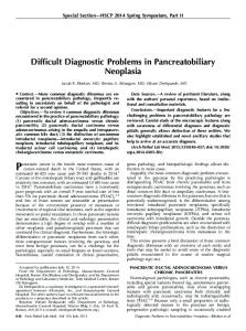
Figure 1. Study selection.
association between reflux and LM in infants. Studies were excluded from meta-analysis if they presented duplicate data.
Retrospective chart review data collection studies were excluded from this assessment.
Assessment of Quality
Analysis
The methodological quality of identified observational studies was assessed using the Oxford Center for EvidenceBased Medicine (CEBM) March 2009 Levels of Evidence.2 The CEBM table was applied to each appraised article, using the ‘‘Therapy/Prevention, Aetiology/Harm’’ category for interpreting study quality.
Data were extracted from the included studies independently by 2 authors, using data forms and outcome measures developed a priori. Descriptive statistics were extracted or calculated for outcomes (where possible), and a qualitative synthesis of the results was undertaken (see Figure 1). Where possible, pooled odds ratios (ORs) and/ or pooled relative risks were calculated with 95% confidence intervals (CIs), using established methods for metaanalysis.3 Study heterogeneity was investigated informally by comparing the studies and statistically using the Cochran Q statistic. Where there was no evidence of heterogeneity, a combined OR was calculated using the Mantel-Haenszel method under the assumption of fixed effects (ie, that the underlying true exposure effect is the same in each study). Where there was evidence of heterogeneity, a combined OR was calculated using the DerSimonian-Laird method under the assumption of random effects (ie, that each study has its own true exposure effect and there is a random distribution of these true exposure effects around a central effect).3
Risk of Bias in Individual Studies Risk of selection and information bias was evaluated at the study level, and a quality assessment score was applied, ranging from 0 to 4, considering the following: 1. Sample selection (consecutive or not: 1 = yes; 0 = no or nonstated) 2. Eligibility criteria (1 = stated; 0 = unclear or nonstated) 3. Method of reflux diagnosis (1 = validated; 0 = nonvalidated or unspecified) 4. Method of laryngomalacia diagnosis (1 = direct or fiber-optic laryngoscopy; 0 = physical examination or unspecified)
Hartl and Chadha
621
Results Study Selection
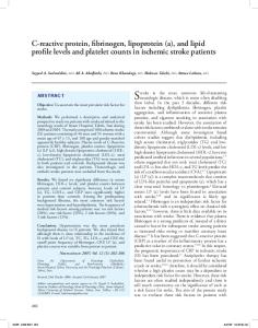
Four studies in our review reported reflux prevalence in patients with LM as well as several other respiratory diagnoses (using barium swallow studies, single-channel 24hour pH probe studies, and esophageal biopsies), enabling the calculation and meta-analysis of odds ratios (reflux vs other respiratory diagnoses; see Figure 2). The overall effect is not statistically significant; reflux was not demonstrated to be more prevalent in infants with LM compared with matched children with other respiratory diagnoses.
Initial searches identified a total of 129 studies. After review of the study titles and abstracts, 41 studies were considered potentially relevant to the study, and the full-text article was obtained. After detailed review of these studies and hand searching their reference lists to obtain further potentially relevant studies, a total of 27 studies met the inclusion criteria (explored the association between reflux and LM) and were thus incorporated in the systematic review. Selected articles consisted of 2 case reports, 16 retrospective reviews, 5 prospective reviews, 2 case-controlled studies, 1 cohort study, and 1 literature review. Quality assessment scores were as follows: 2 studies scored 1, 1 study scored 2, 3 studies scored 3, and 1 study scored 4 (Table 1). Twenty retrospective case series were excluded from this assessment. One study described consecutive samples, and eligibility criteria were clear in 5 studies.
Levels of Evidence The Oxford CEBM Levels of Evidence tools2 were applied, revealing a generally low level of evidence in the literature describing the association of reflux with LM. Twenty-three studies rated level 4, 3 studies rated level 3b, and 1 cohort study rated level 2b. These levels of evidence are defined by the CEBM as follows: 1. 1a = systematic review of randomized controlled trials (RCTs) 2. 1b = individual RCT 3. 1c = all-or-none case series 4. 2a = systematic review (with homogeneity) of cohort studies 5. 2b = individual cohort study 6. 2c = outcomes research 7. 3a = systematic review (with homogeneity) of case control studies 8. 3b = individual case control study 9. 4 = case series (and poor-quality cohort and case control studies) 10. 5 = expert opinion These CEBM numbers enable an assessment of the potential utility of evidence provided (based on their study design), whereas the quality of individual studies may be better reflected by the assessment of bias reflected in the quality score provided in Table 1.
Prevalence of Acid Reflux in Infants with Laryngomalacia Twenty-five of the 27 studies included in our review reported the prevalence of acid reflux in infants with LM. In total, 1295 neonates were represented in these studies, with an overall prevalence of 59%.
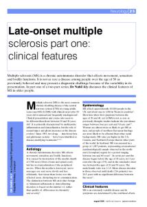
Reflux as a Cofactor in Laryngomalacia Symptom Severity Twelve studies in our review representing 313 infants with LM showed a 65% prevalence of reflux among patients with severe LM, 6% higher than the overall prevalence across all 25 studies. Study quality, severity criteria, reflux definitions, and study methodology varied widely, and there were no controlled studies. A subgroup of 3 studies was identified in which patients with LM were subdivided according to severity (mild, moderate, and severe). Odds ratios were calculated, and meta-analysis revealed that patients suffering from moderate to severe LM were 9.86 times more likely to have reflux than patients with a mild form of the disease (P \ .0001) (see Figure 3).
Laryngomalacia Improvement with Antireflux Therapy Although several articles discussed the probability of LM symptom improvement with antireflux therapies (including feeding modifications, upright positioning, pharmacotherapy, and surgical fundoplication), 6 studies reported antireflux treatment outcomes for symptoms of LM. Findings, summarized in Table 2, show weak and inconsistent evidence for benefit in these patients.
Histological Evidence of Reflux-Related Inflammation in Laryngomalacia Two studies investigating a histological association between these 2 disease entities were found and reviewed, each a retrospective review of 9 patients with severe laryngomalacia, characterizing supraglottic tissue biopsies under the microscope. Both studies found a mild intraepithelial inflammatory infiltrate in patients also diagnosed with reflux. These inflammatory changes—even in severely symptomatic patients—did not appear significant enough to be responsible for gross morphologic changes characteristic of LM, suggesting that acid reflux, although coexistent in most of these patients, was an unlikely causative agent. A summary of their findings is provided in Table 3. Neither study used controls, limiting our ability to draw conclusions about what inflammatory changes may be considered pathologic or specifically reflux related.
Discussion Reflux Diagnosis Problems The definition of reflux varied widely among study authors. The importance of this problem cannot be understated, as it
622
13/27 (48)
19/28 (68) 3/6 (50)
3/4 (75) 2/2 (100) 3/9 (33) 18/24 (75) 24/24 (100) 103/297 (35) 1/1 (100) 17/39 (44) 65/96 (68)
11/23 (47) 141/169 (83)
7/12 (58) 21/41 (51)
21/28 (75) 11/26 (42)
Giannoni 199816
Groblewski 200917 Hadfield 200318
Halstead 199919 Henry 198220 Iyer 199921 Manning 200522 Matthews 19994 Nussbaum 199023 Orenstein 198324 Polonovski 199025 Roger 199526
Senders 200127 Thompson 20075
Ungkanont 199828 Yellon 200129
Yellon 200030 Yuen 200631
Biopsy revealing esophagitis ‘‘Symptoms and laryngoscopy findings of supraglottic edema and/or erythema’’ ‘‘Signs and symptoms, along with an edematous and erythematous larynx’’
None specified Positive barium swallow Barium reflux documented in upper esophagus and pH \4 for .8% of time on single-probe pH Twenty-hour single-probe pH study; positive if pH \4 for .4% of the study duration, or Euler and Byrne score .50 Positive barium swallow and/or single-probe 24-hour pH study Contrast esophagography, pH-metry, esophageal endoscopy with biopsy, or ‘‘symptoms’’ Positive 24-hour single-probe pH (n = 20) and/or barium swallow (n = 13) None specified Positive 24-hour single-probe pH study with a calculated reflux index (RI) for preoperative/postoperative comparison Positive single-probe 24-hour pH study Barium swallow, milk radionucleotide scan ‘‘Clinical details’’ Positive ‘‘contrast study or pH probe’’ (n = 6) or ‘‘clinical diagnosis’’ (n = 12) Dual-probe 24-hour pH, with .1 episode of .4 seconds of pH \4 None specified None specified Positive barium swallow or endoscopy Hx and occasionally 24-hour pH monitoring ‘‘Symptoms of GER’’ Positive barium swallow (127/157) and/or esophagoscopy (20/41) and/or 24-hour dual-probe pH (60/60) Barium swallow, UGI study, single-probe 24-hour pH study None specified
Reflux Definition
Abbreviations: GER, gastroesophageal reflux; Hx, history; NA, not applicable; UGI, upper gastrointestinal.
89/148 (60) 769/1295 (59) 203/313 (65)
7/9 (78) 132/201 (65.6)
Chandra 200114 Dickson 200915
Zoumalan 200732 Overall prevalence Prevalence with severe LM
11/18 (61)
Bouchard 199913
Prevalence, No. (%)
11/16 (69) 16/20 (80) 21/27 (78)
10
Alexiou 2010 Belmont 198411 Bibi 200112
Lead Author
Table 1. Literature Reporting Prevalence of Reflux in Infants with Laryngomalacia (LM)
aryepiglottoplasty supraglottoplasty
aryepiglottoplasty aryepiglottoplasty
Not reported
Not reported Variable with 2 subgroups; 12 of 21 severe Not reported Not reported
Severe LM, requiring supraglottoplasty Variable, with 3 subgroups; 32 of 38 severe
Not reported Severe Severe LM, requiring Severe LM, requiring Not reported Not reported Severe, 1 patient Severe LM, requiring Severe LM, requiring
Severe LM, requiring supraglottoplasty Severe LM, requiring aryepiglottoplasty
Not reported
Severe LM, requiring supraglottoplasty Not reported
Not reported Not reported Not reported; includes LM, tracheomalacia, and both Not reported
Disease Severity
NA
1 NA
NA 1
NA 3
NA NA NA 2 NA NA NA NA NA
NA 3
4
NA NA
NA
NA NA 3
Quality Score
Hartl and Chadha
623
Figure 2. Forest plot of reflux prevalence in patients with laryngomalacia (LM) vs patients with other respiratory diagnoses. Each study effect estimate is shown by a solid black square, the size of which represents the weight that study exerts in the meta-analysis. The pooled estimate is marked with a solid diamond. The 95% confidence intervals (CIs) are displayed as horizontal lines.
Figure 3. Forest plot of reflux prevalence among patients with mild laryngomalacia (LM) vs those with moderate to severe disease. CI, confidence interval.
undermines all attempts at comparative or summative analysis. This diagnostic heterogeneity is represented in Table 1. Furthermore, some studies focused specifically on gastroesophageal reflux disease (GERD) findings, whereas others investigated the contribution of laryngopharyngeal reflux (LPR). Among those studies performing dual-probe pH monitoring to diagnose reflux, an important congruency was demonstrated; both Matthews et al4 (24 patients) and Thompson5 (60 patients) showed that when reflux is defined using 24-hour dual-probe pH manometry (Thompson’s criterion was reported as ‘‘one or more episodes of pH \4 for .4 seconds’’), 100% of infants with LM achieved the diagnosis. Little et al, in their prospective study of 222 children (birth to 16 years) with various respiratory diagnoses, using 24-hour dual-probe pH monitoring, helped to explain the importance of assessing for LPR using the additional pharyngeal probe: 76/168 had pharyngeal reflux despite having normal esophageal acid exposure . . . had the pharyngeal probe not been used, 46% of the children with documented extraesophageal (pharyngeal) reflux would have been falsely presumed to have normal reflux parameters.6 The dual-probe pH data appear to show that either LPR has the stronger association with LM or dual-probe manometry—with a low threshold of exposure to diagnose reflux—is a more sensitive, if not necessarily specific, test. Rabinowitz et al, in their 2003 study of 28 pediatric patients with otolaryngologic symptoms, went further, concluding,
Standard distal pH probe monitoring alone gives a false negative result in a substantial proportion of the infants and children with ENT symptoms being evaluated for GER. Beyond its value in clinical practice, UER testing should be employed in research studies that evaluate the impact of GER therapy on ENT symptoms.7 Still missing from all pediatric reflux data (dual-probe pH and otherwise) currently in the literature, perhaps due to the ethical implications, is a representation of true controls (reflux findings in a population of ‘‘normal’’ infants) to help determine the relative significance of ‘‘pathologic’’ findings. It seems imperative to confirm that acid reflux, as diagnosed by dual pH probes, is aphysiologic in infants; although these authors considered 1 episode of pharyngeal reflux to be pathologic, this may not be true. Also missing in the reflux literature is a validated reflux findings score for the pediatric population.
Symptom Severity and a Potential ‘‘Biologic Gradient’’ Relationship The meaning of the combined prevalence data is further diminished by the variable disease severity represented in each study population. But when we focus only on studies of infants with severe LM (vs studies including a variable disease presentation), we see that the prevalence of reflux appears higher. Still, conclusions drawn from this summation of data are weak because the study quality, measurements, and methods vary widely. A more useful aggregation was possible with the 3 studies that each grouped patients with LM according to
624
Otolaryngology–Head and Neck Surgery 147(4)
Table 2. Evidence for Laryngomalacia (LM) Improvement with Antireflux Therapy Lead Author
Reflux Prevalence, No. (%)
Study Method
Evidence for Response to Treatment
Belmont 198411
16/20 (80)
Retrospective review, no controls
Halstead 199919
3/4 (75)
Retrospective review, no controls
Henry 198220
2/2 (100)
Case report, no controls
Polonovski 199025
17/39 (44)
Retrospective review, no controls
Thompson 20075
141/169 (8)
Prospective review, no controls
Yellon 200129
21/41 (51)
Retrospective review, no controls
Two patients with severe LM underwent fundoplication for excessive reflux, with successful acid suppression but no relief of stridor Three patients with severe LM treated with antireflux ‘‘medication’’; 2 had apneas resolve, and the third required fundoplication; his apneas resolved, and his stridor improved Two patients with severe LM underwent fundoplication with successful acid suppression and complete disappearance of stridor Seventeen patients with severe LM were treated for an undefined period with positioning, thickened food, ‘‘and drugs such as metoclopramide;’’ in patients with esophagitis, antacids were given; no respiratory symptom improvement One hundred fifty-seven patients with moderate to severe LM received GERD treatment for a mean of 7.3 months; after 6 months of therapy, coughing and choking symptoms improved in 88.7% of patients, and regurgitation improved in 69.3%; improvement in LM symptoms was not reported Eight of 9 patients with moderate LM showed ‘‘clinical improvement of stridor following medical treatment of GERD;’’ 17 children with severe LM showed no improvement with medical treatment of GERD.
Abbreviation: GERD, gastroesophageal reflux disease.
Table 3. Histologic Evidence of Reflux in Laryngomalacia (LM) Lead Author
Prevalence, No. (%)
Study Method
Evidence
Chandra 200114
7/9 (78)
Retrospective review, no controls
Iyer 199921
3/9 (33)
Retrospective review, no controls
Nine patients with severe LM underwent supraglottoplasty 1 biopsy; shows submucosal edema and lymphatic dilation; ‘‘A paucity of inflammatory cells suggests that refluxate may not directly exacerbate airway compromise through inflammatory mechanisms’’ Nine patients with severe LM underwent aryepiglottoplasty 1 biopsy; a common finding was intraepithelial eosinophils, consistent with the idea that reflux, to a lesser or greater degree, may be universal in LM
severity. This meta-analysis may represent the most compelling new evidence for an association this review was able to identify: an apparent biologic gradient; greater exposure may lead to greater incidence/severity of the
effect. Of course, the reverse association is equally likely (it is possible that the greater negative inspiratory effort required of infants with laryngomalacia to breathe generates a greater difference between the intra-abdominal
Hartl and Chadha pressure and the intrathoracic pressure, permitting more reflux events to occur in the event of inevitable transient relaxations of the lower esophageal sphincter). Although still weak in isolation, this is 1 of the 9 criteria listed in the Bradford-Hill criteria of causation—an oft-referred, though nonvalidated, effort to determine cause-effect relationships.8
Treatment Outcome Data Are Inconsistent and Uncontrolled Without consistent modalities of therapy, uniform and accurate methods of diagnosis, objective disease end points to determine improvement, and statistical analysis, a metaanalysis of treatment outcome data is of little practical use. Thompson’s 2007 study,5 the most scientifically rigorous of the 6 studies reporting outcomes of reflux treatment on symptoms of LM, warrants special attention. In it, the author suggests that antireflux therapy may reduce symptoms of LM, particularly in patients with mild disease, as compared with moderate or severe disease. The findings are based on large numbers of patients and are statistically significant. However, end points were subjective parent reports of improved symptoms of coughing, choking, and regurgitation. Two potential difficulties with interpretation exist here. The first relates to unblinded, subjective reporting; perhaps the parents, even if they could reliably report a change in these symptoms over a period of months, demonstrated bias. And since the study personnel were also unblinded, further potential for expectations bias is present. Second, reductions in stridor or apneas—symptoms more specific to LM—were not reported, introducing the possibility that antireflux medication may have simply treated reflux effectively (not LM). Furthermore, the most consistent improvements were reported after 6 months of therapy. In 2010, in a publication titled ‘‘LM: Factors That Influence Disease Severity and Outcomes of Management,’’ Thompson9 commented on the natural history of LM in infants, noting, ‘‘Symptoms worsen at 4-8 months, improve between 8 and 12 months, and usually resolve by 12-18 months of age.’’ The mean age at diagnosis for patients in the 2007 study was older than 3 months (102.8 days). In light of this, and considering that there were no controls (infants with LM not receiving antireflux therapy), it seems possible that much of the reported symptom improvements in this study reflect the natural history of the disease.
Conclusion Although an association between reflux and laryngomalacia is commonly cited, and with more than 2 dozen studies in the literature suggesting that one may cause the other, convincing data are lacking. Reflux appears to occur often in the setting of LM, but a lack of consensus in diagnostic criteria for reflux and inconsistent, low-quality study methods limit our ability to meta-analyze available data. Twenty-four-hour dual-probe pH monitoring shows promise as a sensitive diagnostic tool, and where it is available, the authors support its use as the primary diagnostic modality in infants with LM and suspected reflux—especially in the context of a scientific study. But there are currently no controlled studies to
625 correlate pH data to pathologic reflux findings in infants. Reflux may occur more often in patients suffering from more severe LM symptoms, but this kind of dose-response relationship requires further definition. There exists no histological evidence confirming a role for reflux in the etiology of LM. In view of the widespread use of antireflux treatment in infants with laryngomalacia and the paucity of quality studies on this disease association, there is a need for further investigation of the role of reflux in LM, and a welldesigned randomized controlled trial of antireflux medication vs placebo is justified. Author Contributions Trevor T. Hartl, lead investigator, primary author; Neil K. Chadha, principal investigator, coauthor.
Disclosures Competing interests: None. Sponsorships: None. Funding source: None.
References 1. Holinger LD. Etiology of stridor in the neonate, infant, and child. Ann Otol. 1980;89:397-400. 2. OCEBM Levels of Evidence Working Group. The Oxford 2011 Levels of Evidence. Oxford Centre for Evidence-Based Medicine. http://www.cebm.net/index.aspx?o=5653 3. Blettner M, Sauerbrei W, Schlehofer B, et al. Traditional reviews, meta-analyses and pooled analyses in epidemiology. Int J Epidemiol. 1999;28:1-9. 4. Matthews BL, Little JP, Mcguirt WF Jr, Koufman JA. Reflux in infants with laryngomalacia: results of 24-hour doubleprobe pH monitoring. Otolaryngol Head Neck Surg. 1999;120: 860-864. 5. Thompson DM. Abnormal sensorimotor integrative function of the larynx in congenital laryngomalacia: a new theory of etiology. Laryngoscope. 2007;117(6, pt 2)(suppl 114):1-33. 6. Little JP, Matthews BL, Glock MS, et al. Extraesophageal pediatric reflux: 24-hour double-probe pH monitoring of 222 children. Ann Otol Rhinol Laryngol Suppl. 1997;169:7. 7. Rabinowitz SS, Piecuch S, Jibaly R, Goldsmith A, Schwarz SM. Optimizing the diagnosis of gastroesophageal reflux in children with otolaryngologic symptoms. Int J Pediatr Otorhinolaryngol. 2003;67:625. 8. Hill AB. The environment and disease: association or causation? Proc R Soc Med. 1965;58:295-300. 9. Thompson DM. LM: factors that influence disease severity and outcomes of management. Curr Opin Otolaryngol Head Neck Surg. 2010;18:564-570. 10. Alexiou S, Rakoczy KJ, Maupin KD. Bronchoalveolar lavage pepsin activity and lipid laden macrophages in infants with laryngomalacia and gastroesophageal reflux. Am J Respir Crit Care Med. 2010;181:A6235. 11. Belmont JR, Grundfast K. Congenital laryngeal stridor (laryngomalacia): etiologic factors and associated disorders. Ann Otol Rhinol Laryngol. 1984;93:430-437.
626 12. Bibi H, Khvolis E, Shoseyov D, et al. The prevalence of gastroesophageal reflux in children with tracheomalacia and laryngomalacia. Chest. 2001;119:409-413. 13. Bouchard S, Lallier M, Yazbeck S, Bensoussan A. The otolaryngologic manifestations of gastroesophageal reflux: when is a pH study indicated? J Pediatr Surg. 1999;34:1053-1056. 14. Chandra RK, Gerber ME, Holinger LD. Histological insight into the pathogenesis of severe laryngomalacia. Int J Pediatr Otorhinolaryngol. 2001;61:31-38. 15. Dickson JM, Richter GT, Meinzen-Derr J, Rutter MJ, Thompson DM. Secondary airway lesions in infants with laryngomalacia. Ann Otol Rhinol Laryngol. 2009;118:37-43. 16. Giannoni C, Sulek M, Friedman EM, Duncan NO III. Gastroesophageal reflux association with laryngomalacia: a prospective study. Int J Pediatr Otorhinolaryngol. 1998;43:11-20. 17. Groblewski JC, Shah RK, Zalzal GH. Microdebrider-assisted supraglottoplasty for laryngomalacia. Ann Otol Rhinol Laryngol. 2009;118:592-597. 18. Hadfield PJ, Albert DM, Bailey CM, Lindley K, Pierro A. The effect of aryepiglottoplasty for laryngomalacia on gastrooesophageal reflux. Int J Pediatr Otorhinolaryngol. 2003;67: 11-14. 19. Halstead LA. Role of gastroesophageal reflux in pediatric upper airway disorders. Otolaryngol Head Neck Surg. 1999; 120:208-214. 20. Henry RL, Mellis CM. Resolution of inspiratory stridor after fundoplication: case report. Aust Paediatr J. 1982;18:126-127. 21. Iyer VK, Pearman K, Raafat F. Laryngeal mucosal histology in laryngomalacia: the evidence for gastro-oesophageal reflux laryngitis. Int J Pediatr Otorhinolaryngol. 1999;49:225-230. 22. Manning SC, Inglis AF, Mouzakes J, Carron J, Perkins JA. Laryngeal anatomic differences in pediatric patients with
Otolaryngology–Head and Neck Surgery 147(4)
23. 24. 25.
26.
27.
28.
29.
30.
31.
32.
severe laryngomalacia. Arch Otolaryngol Head Neck Surg. 2005;131:340-343. Nussbaum E, Maggi JC. Laryngomalacia in children. Chest. 1990;98:942-944. Orenstein SR, Orenstein DM, Whitington PF. Gastroesophageal reflux causing stridor. Chest. 1983;84:301-302. Polonovski JM, Contencin P, Francois M, Viala P, Narcy P. Aryepiglottic fold excision for the treatment of severe laryngomalacia. Ann Otol Rhinol Laryngol. 1990;99:625-627. Roger G, Denoyelle F, Triglia JM, Garabedian EN. Severe laryngomalacia: surgical indications and results in 115 patients. Laryngoscope. 1995;105:1111-1117. Senders CW, Navarrete EG. Laser supraglottoplasty for laryngomalacia: are specific anatomical defects more influential than associated anomalies on outcome? Int J Pediatr Otorhinolaryngol. 2001;57:235-244. Ungkanont K, Friedman EM, Sulek M. A retrospective analysis of airway endoscopy in patients less than 1-month old. Laryngoscope. 1998;108:1724-1728. Yellon RF, Goldberg H. Update on gastroesophageal reflux disease in pediatric airway disorders. Am J Med. 2001; 111(suppl 8A):78S-84S. Yellon RF, Coticchia J, Dixit S. Esophageal biopsy for the diagnosis of gastroesophageal reflux-associated otolaryngologic problems in children [review]. Am J Med. 2000;108(suppl 4a):131S-138S. Yuen HW, Tan HK, Balakrishnan A. Synchronous airway lesions and associated anomalies in children with laryngomalacia evaluated with rigid endoscopy. Int J Pediatr Otorhinolaryngol. 2006;70:1779-1784. Zoumalan R, Maddalozzo J, Holinger LD. Etiology of stridor in infants. Ann Otol Rhinol Laryngol. 2007;116:329-334.