Human apolipoprotein C-I expression in mice impairs learning and memory functions Karlygash Abildayeva,* Jimmy F. P. Berbe´e,†,§ Arjan Blokland,** Paula J. Jansen,* Frans J. Hoek,†† Onno Meijer,§§ Dieter Lu¨tjohann,*** Thomas Gautier,††† Thierry Pillot,§§§ Jan De Vente,* Louis M. Havekes,§ Frans C. S. Ramaekers,* Folkert Kuipers,††† Patrick C. N. Rensen,†,§ and Monique Mulder1,* Department of Molecular Cell Biology and Department of Basic Neurosciences, European Graduate School of Neuroscience,* Faculty of Psychology,** Institute Brain and Behavior, University of Maastricht, Maastricht; Department of General Internal Medicine, Endocrinology and Metabolic Diseases,† Leiden University Medical Center, and Department of Biomedical Research,§ TNO-Quality of Life, Gaubius Laboratory, Leiden; Department of Clinical Chemistry,†† Academic Medical Center, Amsterdam; Department of Medical Pharmacology,§§ Leiden Amsterdam Center for Drug Research and Leiden Medical Center, Leiden; Department of Pediatrics,††† University Medical Center Groningen, Groningen, The Netherlands; Department of Clinical Pharmacology,*** University of Bonn, Bonn, Germany; and Laboratoire de Me´decine et The´rapeutique Mole´culaire,§§§ Lipidomix, Institut National Polytechnique de Lorraine, Nancy, France
Abstract The H2 allele of APOC1, giving rise to increased gene expression of apolipoprotein C-I (apoC-I), is in genetic disequilibrium with the APOE4 allele and may provide a major risk factor for Alzheimer’s disease (AD). We found that apoC-I protein is present in astrocytes and endothelial cells within hippocampal regions in both human control and AD brains. Interestingly, apoC-I colocalized with b-amyloid (Ab) in plaques in AD brains, and in vitro experiments revealed that aggregation of Ab was delayed in the presence of apoC-I. Moreover, apoC-I was found to exacerbate the soluble Ab oligomer-induced neuronal death. To establish a potential role for apoC-I in cognitive functions, we used human (h) APOC11/0 transgenic mice that express APOC1 mRNA throughout their brains and apoC-I protein in astrocytes and endothelial cells. The hAPOC11/0 mice displayed impaired hippocampal-dependent learning and memory functions compared with their wild-type littermates, as judged from their performance in the object recognition task (P 5 0.012) and in the Morris water maze task (P 5 0.010). ApoC-I may affect learning as a result of its inhibitory properties toward apoE-dependent lipid metabolism. However, no differences in brain mRNA or protein levels of endogenous apoE were detected between transgenic and wild-type mice. In conclusion, human apoC-I expression impairs cognitive functions in mice independent of apoE expression, which supports the potential of a modulatory role for apoC-I during the development of AD.—Abildayeva, K., J. F. P. Berbe´e, A. Blokland, P. J. Jansen, F. J. Hoek, O. Meijer, D. Lu¨tjohann, T. Gautier, T. Pillot, J. De Vente, L. M. Havekes, F. C. S. Ramaekers, F. Kuipers, P. C. N. Rensen, and M. Mulder. Human apolipo-
protein C-I expression in mice impairs learning and memory functions. J. Lipid Res. 2008. 49: 856–869. Supplementary key words Alzheimer’s disease & apolipoprotein E & object recognition task & Morris water maze task & b-amyloid
After the identification of apolipoprotein E4 (apoE4) as a major risk factor for the development of sporadic, lateonset Alzheimer’s disease (AD) (1, 2), efforts have been initiated to identify additional lipid-related susceptibility genes for this disease (3). Interestingly, APOE4 is in genetic linkage disequilibrium with the HpaI restriction polymorphism in the promoter region of APOC1, which is localized 5 kb downstream of the APOE gene on chromosome 19. Although apolipoprotein C-I (apoC-I) is expressed mainly in the liver, substantial expression has also been detected in other tissues, including the brain (4). The HpaI polymorphism (the so-called H2 allele) leads to a highly significant, 1.5-fold increase in APOC1 gene transcription (5) and was reported to be associated with AD (6–12). Moreover, the H2 allele of APOC1 was found to be associated with impaired memory and frontal lobe function (13) and with the loss of hippocampal volumes in humans (14). Although apoE4 is a well-established and strong risk factor of AD in Caucasians and in European-Americans, it is not associated with AD in African-Americans. Interestingly, the frequency of APOC1 H2 with apoE4 is 0.85 in
Manuscript received 9 May 2007 and in revised form 12 November 2007 and in re-revised form 17 December 2007. Published, JLR Papers in Press, December 26, 2007. DOI 10.1194/jlr.M700518-JLR200
1
To whom correspondence should be addressed. e-mail:
[email protected]
Copyright D 2008 by the American Society for Biochemistry and Molecular Biology, Inc.
856
Journal of Lipid Research
Volume 49, 2008
This article is available online at http://www.jlr.org
European-Americans but only 0.55 in African-Americans, whereas the frequency of APOC1 H2 with apoE3 is 0.02 in European-Americans and 0.08 in African-Americans (5). This has led to the hypothesis that the H2 allele of APOC1 may be an independent or additional risk factor for AD (8, 9, 15, 16) and that not apoE4 but rather apoC-I modulates the pathogenesis of AD (17). The physiological roles of apoC-I have not yet been established in detail. Studies with mice overexpressing human apoC-I (18) revealed gene dose-dependent effects on circulating levels of triglycerides (TGs), FFAs, and total cholesterol (19, 20). This may be attributable to the interference of apoC-I with apoE-dependent clearance of TG-
rich lipoproteins by the low density lipoprotein receptor family in the liver (21–23) and by modulation of the activity of enzymes involved in plasma lipid metabolism (24, 25). The presence of relatively high levels of apoC-I within the brain (4) suggests a role for this apolipoprotein in brain lipid metabolism. Disturbances in brain lipid metabolism can lead to cognitive impairments in humans and in rodents, indicating that a well-regulated brain lipid metabolism is necessary for normal brain functioning (26–30). Therefore, we examined the localization of apoC-I in postmortem brains of AD- and control patients, the interaction of apoC-I with b-amyloid (Ab) in vitro, and assessed learning and memory functions as well as indices of brain
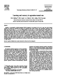
Fig. 1. Immunohistochemical analysis of human control and Alzheimer’s disease (AD) hippocampal CA1 sections. A: Apolipoprotein C-I (apoC-I) and glial fibrillary acidic protein (GFAP). B: ApoC-I and von Willebrand factor. C: Double staining for both apoC-I and b-amyloid (Ab) or for apoC-I and apoE. Bars 5 25 mm.
ApoC-I: role in learning and memory
857
Fig. 1.—Continued.
lipid metabolism in transgenic mice hemizygous for human apoC-I (hAPOC11/0 ) and their wild-type littermates.
MATERIALS AND METHODS Immunohistochemistry Postmortem human brain samples were obtained from the Netherlands Brain Bank (Amsterdam, The Netherlands). Brains were characterized with respect to their neuropathological status following criteria established by Khachaturian (31). Samples from nondemented subjects displaying no neuropathological changes related to AD or any other neurodegenerative disorder were designated as controls. Immunohistochemical procedures were similar to those used in mouse brain. Mice were anesthetized and perfused transcardially with PBS followed by Somogyi fixative as described (32). After removal, the brain was transected midsagittally, and the lateral block was postfixed in the same fixative (4jC, overnight) and cryoprotected in sucrose (10% for 24 h followed by 30% for 1–2 weeks) in 0.1 M phosphate buffer. The cryostat sections (35 mm) were incubated overnight at 4jC with the following primary antibodies: goat anti-human apoC1 (Academy Bio-Medical Co., Houston, TX), rabbit anti-glial fibrillary acidic protein (GFAP) (DAKO), rabbit anti-amyloid b (Ab) (Calbiochem Immunochemicals, San Diego, Ca), rabbit anti-von Willebrand factor (Abcam, Cambridge, UK), rabbit antihuman apoE (DAKO), and goat anti-mouse apoE (Santa Cruz Biotechnology, Inc., Santa Cruz, CA); or for 1 h at room temperature with biotinylated Lycopersicon esculentum (tomato) lectin (Vector Laboratories, Inc., Burlingame, CA). Subsequently, sections were incubated for 1 h at room temperature with the
858
Journal of Lipid Research
Volume 49, 2008
following antibodies: biotinylated donkey anti-goat and subsequently with avidin-biotin solution of the Vectastain ABC Elite Kit (Vector Laboratories), FITC-conjugated donkey anti-goat (Santa Cruz Biotechnology), or Texas Red-conjugated donkey anti-rabbit (Abcam). Sections were also incubated with donkey anti-rabbit biotin (Jackson ImmunoResearch Laboratories, Inc., West Grove, PA) followed by avidin-Texas Red (Vector Laboratories). Autofluorescence was blocked by incubation for 75 s in 0.3% Sudan Black (Sigma) solution in ethanol. The samples were imaged using a Bio-Rad MRC600 confocal microscope (Bio-Rad Laboratories, Ltd., Hemel Hempstead, UK).
In situ hybridization In situ hybridization with human APOC1-specific end-labeled oligonucleotide probes was performed on 20 mm cryostat sections as described (33). One hundred microliters of the riboprobe mix (murine probe, 5¶-CTTCAGTTTATCCGGTATGCTCTCCAATGTTCCGGACAAATCC-3¶; mismatch murine probe, 5¶-CgTCAGTgTATCCtGTATGaTCTCCcATGTTaCGGACcAATCC-3¶; human probe 1, 5¶-TGTGTTTCCAAACTCCTTCAGCTTATCCAAGGCACTGGAGACG-3¶; mismatch human probe 1, 5¶-TtTGTTTaCAAACgCCTTCcGCTTAgCCAAGtCACTGtAGACG-3¶) was applied to each slide, which was then covered with a standard microscopic coverslip and put in a moist chamber for overnight hybridization at 53jC. The next day, the slides were washed, treated with RNase A, dehydrated in a graded ethanol series, and put in a cassette, and X-Omat AR film (Kodak, Rochester, NY) was exposed to the sections for 14 days.
Animals Transgenic mice with high expression of human APOC1 in the liver and a moderate expression in the brain were generated as
Fig. 1.—Continued.
described previously (19, 34). Female human apoC-I (hAPOC11/0 ) transgenic mice (n 5 12) and their wild-type littermates (n 5 14) were bred at TNO-Quality of Life in Leiden, The Netherlands. Mice were housed under standard conditions in conventional cages and provided a standard chow diet and water ad libitum; lights were on from 7:00 AM to 7:00 PM in a temperature- and humidity-controlled room. The ethical board of Maastricht University approved all of the experimental procedures.
Behavioral testing Twelve month old mice were subjected to behavioral tests between 9:00 AM and 3:00 PM. The experimenters were unaware of the genetic background of the mice. There were no differences in the results in the mice tested early or late on the days of the experiment. The object recognition task measures whether a mouse remembers an object it has explored in a previous learning trial. The
task was performed in a circular arena with a diameter of 49 cm, as described previously (35). Eight days before being subjected to the Morris water escape task, the mice were housed individually. The spatial discrimination performance of the mice was assessed as described previously (35, 36).
Phospholipid profile determination One milliliter of a chloroform-methanol (2:1) extract of total brain homogenates was dried under a nitrogen stream, and the extracted lipids were dissolved in methanol-chloroform (2:1) for separation and quantification by high-performance thin-layer chromatography (37).
Sterol profile determination Serum and brains were snap-frozen in liquid nitrogen. Brains were spun in a speed vacuum dryer (12 mbar) (Savant AES 1000)
ApoC-I: role in learning and memory
859
Fig. 2. ApoC-I peptides inhibit Ab aggregation in vitro. A: Ab(1-40) peptide (100 mM, closed squares) was incubated at 37jC in PBS (pH 7.4) for the indicated times in the absence or presence of apoC-I (closed triangles) or apoA-I (open circles) at a molar ratio of 1:100. The aggregation was monitored as a function of time by measuring the turbidity of the solution at 400 nm against a blank containing only buffer and apoC-I or apoA-I. B: Ab(1-40) peptide (100 mM) was incubated at 37jC in PBS (pH 7.4) for 16 h in the absence of presence of apoC-I or apoA-I at a molar ratio of 1:100. Th-T, thioflavin-T. Data are means 6 SEM.
for 48 h to relate individual sterol concentrations to dry weight. The sterols were extracted from 100 ml of serum in the same manner. Levels of cholesterol were determined in a gas chromatographflame ionization detector, and levels of plant sterols (campesterol and sitosterol), cholesterol precursors (lanosterol, lathosterol, and desmosterol), and cholesterol metabolites [24(S)-hydroxycholesterol and cholestanol] were determined using gas chromatography-mass spectrometry as described previously (38).
Fig. 3. ApoC-I exacerbates soluble Ab oligomer-induced neuronal death. Cortical mouse neurons were incubated for 24 h with 1 mM soluble Ab(1-40) oligomers in the absence or presence of apoC-I or apoA-I at an apolipoprotein/Ab peptide molar ratio of 1:100, as described previously (80). Soluble Ab oligomers and apolipoproteins were mixed and incubated at 37jC for 10 min before cell exposure. Soluble Ab oligomer-induced cell death was monitored using a Live/Dead Viability/Cytotoxicity Kit (A) and the 3-(4,5dimethyl-thiazol-2-yl)-2,5-diphenyltetrazolium bromide (MTT) assay (B). Data are means 6 SEM of three different experiments with four determinations each and are normalized to the effect of vehicle, designated as 100%. Statistical differences among the subgroups for each condition were determined by ANOVA followed by Scheffe’s post hoc test (P , 0.05). No significant difference was found between apolipoprotein-treated and control cells in the absence of soluble Ab oligomers.
Quantitative real-time (QRT)-PCR was performed using an Applied Biosystems 7700 sequence detector with 1.6.3 software (Perkin-Elmer Corp., Foster City, CA) as described previously (39) with modifications (40). Primer sequences are available upon request. Primers were obtained from Invitrogen. Fluorogenic probes, labeled with 6-carboxy-fluorescein and 6-carboxytetramethyl-rhodamine, were made by Eurogentec (Seraing, Belgium).
RNA isolation and quantitative real-time PCR procedures Total RNA was isolated using the Trizol method (Invitrogen, Breda, The Netherlands) according to the manufacturer’s instructions. The integrity of RNA was checked by agarose gel electrophoresis, and RNA concentration was measured spectrophotometrically (NanoDrop; Witec AG, Littau, Germany).
860
Journal of Lipid Research
Volume 49, 2008
Thioflavin-T fluorimetric assay The fibril formation of Ab(1-40) peptide was measured by a thioflavin-T binding assay as described previously (41, 42). Both apoA-I and apoC-I were purified from human plasma lipoproteins as described (43). The apolipoproteins were nonlipidated.
Fig. 4. Detection of human (h) APOC1 mRNA in the brains of hAPOC11/0 mice and wild-type (WT) littermates by in situ hybridization. The presence of hAPOC1 mRNA is seen throughout the brain of the hAPOC11/0 mice, whereas wild-type mice are negative.
Neuronal cell culture studies Primary cultures of cerebral cortical neurons were prepared from embryonic day 15 C57BL/6J mouse fetuses as described previously (44). Briefly, dissociated cells were plated at 105 cells/ cm2 in plastic dishes or on glass coverslips precoated overnight with poly-DL-ornithine (1.5 or 15 mg/ml, respectively). The cells were cultured in a chemically defined DMEM/F12 serum-free medium and supplemented with insulin (500 nM), putrescine (60 mM), sodium selenite (30 nM), transferrin (100 mM), progesterone (100 nM), and 0.1% (w/v) ovalbumin. Cultures were maintained at 35jC in a humidified 6% CO2 atmosphere. After 6–7 days in vitro, the cellular population was determined to be at least 95% neuronal by immunostaining, as described previously (45). Cortical neurons were treated in the absence or presence of APOC1 at 6–7 days in vitro for the indicated times with soluble oligomers of Ab(1-40), prepared as described (44).
Neuronal viability and apoptosis Cell viability was initially determined by morphological observation. Ab neurotoxicity was also assessed quantitatively by monitoring mitochondrial reduction activity using the 3-(4,5dimethyl-thiazol-2-yl)-2,5-diphenyltetrazolium bromide assay as described previously (44). The simultaneous determination of live and dead cells was achieved with the Live/Dead Viability/ Cytotoxicity Kit (Roche Molecular Biochemicals, Paris, France) according to the manufacturer’s instructions.
Statistics Group differences in behavioral tests were analyzed using a one- or two-factor ANOVA (group or group and day, respectively). In case of nonnormal distribution of the data, the data were transformed logarithmically. To determine the spatial bias in the probe trial of the Morris task, the time the mice spent
Fig. 5. Immunohistochemistry of a hAPOC11/0 and a wild-type brain stained for human apoC-I (A), and immunohistochemistry of representative hippocampal sections of hAPOC11/0 mice double labeled for GFAP and human apoC-I (B), for human apoC-I and von Willebrand factor (C), and for apoC-I and tomato lectin (D). Bars 5 25 mm.
in the different quadrants was evaluated. Group differences in sterol/ phospholipid analysis, body weight, and temperature changes were evaluated using a t-test. Statistical significance in QRT-PCR experiments was determined by comparing means using an unpaired Student’s t-test and a Mann-Whitney U-test.
RESULTS We performed an immunohistochemical analysis of apoC-I on human hippocampal sections (CA1 region) of control and AD patients. ApoC-I protein was detectable primarily in astrocytes in control and AD brains (Fig. 1A), as is evident from its colocalization with GFAP. In addition, in the AD as well as in the control brains, apoC-I was detected in endothelial cells lining the blood vessels, as demonstrated by its colocalization with von Willebrand factor (Fig. 1B). Interestingly, we observed that apoC-I staining overlapped with the staining for Ab protein, the key protein of senile plaques that represent one of the hallmarks of AD (Fig. 1C). However, not all Ab-positive plaques were positive for apoC-I. Moreover, human apoC-I colocalized with apoE in the same plaques in the brain of AD patients (Fig. 1C). To examine whether apoC-I might interfere directly with the aggregation of Ab, we incubated Ab in the absence or presence of apoC-I or apoA-I peptides and ApoC-I: role in learning and memory
861
Fig. 5.—Continued.
862
Journal of Lipid Research
Volume 49, 2008
Fig. 5.—Continued.
measured the aggregate formation by thioflavin-T fluorescence. Figure 2A shows the time course and the extent of aggregate formation, illustrating the different kinetics of aggregate formation for Ab peptide alone and in association with apoC-I or apoA-I peptides. Aggregate formation for Ab peptide alone as well as with apoA-I started after 110 min, whereas for the mixture of Ab and apoC-I, peptide initiation of aggregation was delayed up to 840 min. Figure 2B clearly shows reduced Ab(1–40) fibril formation
Fig. 6. hAPOC11/0 mice (n 5 9) display impaired performance in the object recognition task compared with wild-type (WT) littermates (n 5 14). One hour after a first trial using two identical objects, mice were subjected to a second trial with dissimilar objects. In this second trial, the discrimination index was determined from the time spent exploring the new object relative to the familiar object. Data represent means 6 SEM. * P 5 0.012.
in samples containing apoC-I peptides, whereas the presence of apoA-I peptide did not prevent fibril formation. ApoC-I did not affect the viability of cortical murine primary neurons (Fig. 3A). However, apoC-I was found to exacerbate Ab(1-40)-induced neuronal cell death (Fig. 3A, B). This modifying effect of apoC-I on Ab(1-40)-induced neuronal cell death was only observed if apoC-I was preincubated with Ab(1-40) and not when apoC-I was preincubated with the neurons for 24 h and Ab(1-40) was added after subsequent washing of the cells (data not shown). This indicated the requirement of physical interaction between the two peptides. This effect appears to be specific for soluble Ab(1-40), because apoC-I did not modify the toxicity of preaggregated Ab peptide (data not shown). Next, we examined whether learning and memory functions are affected by the expression of human apoC-I, using hAPOC11/0 transgenic mice as an experimental model. In situ hybridization experiments demonstrated that human APOC1 mRNA is present throughout the brain of hAPOC11/0 transgenic mice. The specificity of the probe for human apoC-I was shown by the absence of staining in brains of wild-type mice (Fig. 4). Human apoC-I protein could be demonstrated immunohistochemically in the hippocampus of hAPOC11/0 transgenic mice, whereas no immunopositive cells were observed in the hippocampus of wild-type mice (Fig. 5A). To verify whether the distribution of human apoC-I over the specific brain cell ApoC-I: role in learning and memory
863
Fig. 7. hAPOC11/0 mice (n 5 9) display slightly impaired spatial learning in the Morris water escape task compared with wild-type littermates (n 5 14) as measured by distance to the platform (A) and escape latency (B). The hAPOC11/0 mice tended to swim slower, as measured by average swimming speed (C). Twenty-four hours after this trial, the mice were given a probe trial, in which the distance (D) and time spent in the quadrants (E; target quadrant and annulus) were measured. Data represent means 6 SEM. * P , 0.01.
types in the mice reflects that in humans, we first performed a double staining with both anti-human apoC-I and anti-GFAP antibodies. Indeed, apoC-I was expressed in astrocytes, as shown by the colocalization of human apoC-I and GFAP in the hippocampal region (Fig. 5B). Moreover, it was detected in endothelial cells, as revealed by double staining for human apoC-I and von Willebrand TABLE 1. Brain phospholipid levels of hAPOC11/0 and wild-type mice Wild Type (n 5 14)
Phospholipid
Lysophosphatidylcholine Sphingomyelin Phosphatidylcholine Lysophosphatidylethanolamine Phosphatidylserine Phosphatidlyinositol Phosphatidylethanolamine Total
6.7 6 6.5 6 96.8 6 37.9 6 60.3 6 8.1 6 140.2 6 356.5 6
2.3 1.3 10.0 16.1 8.2 1.5 15.6 29.2
hAPOC11/0 (n 5 12)
6.9 6 7.3 6 99.0 6 38.2 6 61.7 6 7.9 6 142.5 6 363.0 6
Values represent means 6 SD, expressed in nmol/mg brain.
864
Journal of Lipid Research
Volume 49, 2008
1.7 1.2 10.2 16.1 10.3 1.4 16.6 30.8
factor (Fig. 5C). More specifically, human apoC-I was detected at the basolateral side of endothelial cells stained with anti-tomato lectin, a marker for microglia that also stains endothelial cells of blood vessels (46, 47) (Fig. 5D). The hAPOC11/0 transgenic and wild-type mice that participated in this study were littermates with a highly homogeneous genetic background, obtained by back-crossing to the C57Bl/6 background at least 10 times. The hAPOC11/0 mice appeared healthy, and there were no gross differences between the hAPOC11/0 transgenic mice and their wild-type littermates with respect to overall appearance, behavior in the cage, handling response, or body weight. The weight of the mice was 26.0 6 1.7 g for the hAPOC11/0 transgenic mice and 25.0 6 1.8 g for their wild-type littermates before the beginning of the behavioral experiments at the age of 12 months. To examine the effect of human apoC-I overexpression on memory functions, hAPOC11/0 mice and wild-type littermates were subjected to the object recognition task. One hour after a first trial using two identical objects, the wild-
TABLE 2. Serum and brain sterol levels in hAPOC11/0 and wild-type mice Serum Sterol
Cholesterol Lathosterol Lanosterol Desmosterol 24(S)-Hydroxycholesterol Cholestanol Campesterol Sitosterol
Wild Type (n 5 14)
57.1 6 8.0 15.1 6 10.5 53.2 6 14.1 76.5 6 7.9 268 6 0.9 76.1 6 35.9 1.4 6 0.3 0.2 6 0.05
Brain hAPOC11/0 (n 5 11)
152.2 133.2 168.7 165.3 271.7 421.6 4.9 0.8
6 6 6 6 6 6 6 6
14.9a (mg/dl) 29.9a (mg/ml) 42.4a (mg/ml) 24.5a (mg/ml) 1.8a (mg/ml) 91.8a (mg/ml) 9.2a (mg/dl) 0.1a (mg/dl)
Wild Type (n 5 14)
61.6 6 2.3 1,624 6 148.5 78.1 6 16.5 485.9 6 57.5 164.0 6 18.3 169.5 6 29.0 74.5 6 12.9 11.2 6 1.2
hAPOC11/0 (n 5 12)
63.2 6 1.5 (mg/ml) 1,727 6 241.6 (ng/ml) 75.2 6 13.6 (ng/ml) 537.1 6 79.2 (ng/ml) 155.1 6 11.0 (ng/ml) 185.8 6 32.8 (ng/ml) 87.8 6 16.8a (ng/ml) 12.4 6 1.5a (ng/ml)
Values represent means 6 SD. a Significant differences (P , 0.001).
type mice were able to discriminate between the familiar and the new object (Fig. 6). In contrast, the hAPOC11/0 mice failed to discriminate between the objects, as their discrimination index, which is a measure for selective exploration of a new object, did not differ from zero (hAPOC11/0 vs. wild-type, P 5 0.012). This indicates that human APOC1 expression in mice significantly impairs object memory. hAPOC11/0 and wild-type littermates were also subjected to the Morris water escape task to examine their spatial learning. Mice were subjected to a swimming trial, in which the platform was visible in a circular water tank. Subsequently, the platform was submerged and mice were subjected to the spatial discrimination training trial, consisting of four sessions per day on 4 successive days. Although the absolute differences between the wild-type and hAPOC11/0 mice on the measures distance swum and escape latency were relatively small, statistical analysis revealed reliable differences on both measures (both P , 0.01) (Fig. 7A, B). This indicates that hAPOC11/0 mice acquired this task less well than the wild-type mice. There was no significant difference in average swimming speed between the groups (Fig. 7C), although the hAPOC11/0 mice swam slower than the wild-type mice on the 2nd day (P , 0.05) (Fig. 7C). In the probe trial, the hAPOC11/0 mice swam a shorter distance in the target quadrant (P , 0.05) (Fig. 7D). There was only a marginal difference between the two groups for the time spent in the target quadrant (P , 0.09) (Fig. 7E). These data suggest a modest impairment in spatial learning and memory in hAPOC11/0 mice. Because apoC-I is known to affect plasma TG, FFA, and total cholesterol levels, we determined brain phospholipid and sterol profiles to assess whether the presence of human apoC-I affects brain lipid metabolism and may thereby alter brain functioning. In the brain, fatty acids are mostly retained by phospholipids (48–50). Therefore, we measured brain phospholipid levels in hAPOC11/0 transgenic and wild-type mice. No difference in phospholipid profiles could be detected between the two groups of mice (Table 1). As expected, serum sterol levels were increased in the hAPOC11/0 transgenic mice compared with their wild-type littermates (Table 2). Yet, this was not associated with changes in brain sterol levels. As shown in Table 2, the sterol distribution in brains of hAPOC11/0 and wild-type mice was comparable, except for a small but significant
increase in the amount of the plant sterols campesterol (P , 0.05) and sitosterol (P , 0.05). Because APOC1 is encoded by the same gene cluster as APOE and apoE levels in the brain may affect learning and memory processes (51–54), we determined the APOE mRNA levels in the brains of hAPOC11/0 and wild-type mice by QRT-PCR. No significant difference could be detected between the two groups (Table 3). In addition, no differences were detectable in the protein levels of apoE in the brains of the two groups of mice, as determined by Western blotting (data not shown). In addition, assessment of the gene expression profiles of key regulatory, metabolic, and transporter genes involved in cholesterol metabolism [hydroxy-methylglutaryl-coenzyme A reductase (HMG-CoAR), low density lipoprotein receptor, scavenger receptor class B type I, the ABC transporters ABCA1, ABCG1, and ABCG4, LCAT, phospholipid transfer protein, and cholesterol 24-hydroxylase] revealed no significant difference between the two groups of animals except for very limited changes in the levels of HMG-CoA-R and ABCG4 mRNA (Table 3). Expression of genes involved in fatty acid metabolism (fatty acid synthase, acetyl-CoA carboxylase 1, lipoprotein lipase, and stearoyl-CoA desaturase 1) also did not differ between the two groups of mice. Neither were there any significant differences between transgenic mice and wild-type mice in the expression of genes that act as regulators of lipid metabolism [liver X receptor-a (LXRa), the peroxisome proliferator-activated receptors PPARa and PPARg, the sterol-regulatory element binding proteins SREBP-1a, SREBP1c, and SREBP-2, and the carbohydrate response element binding protein], except for a very modest difference in the expression of 9-cis-retinoic acid receptor-a. Also, no differences were detected in the expression of genes involved in cellular stress (heme oxygenase-1 and apoJ) and inflammation (interleukin-6, tumor necrosis factor-a, and inducible nitric oxide synthase) (Table 3). Amyloid precursor protein expression was slightly, but not significantly (P 5 0.09), reduced in the brains of hAPOC11/0 mice. However, in this case, there were six versus four samples.
DISCUSSION Here, we provide evidence underscoring the hypothesis that the H2 allele of APOC1, giving rise to increased ApoC-I: role in learning and memory
865
TABLE 3. mRNA expression levels in brain of hAPOCI 1/0 and wild-type mice Variable
Wild Type (n 5 8)
Gene
Cholesterol metabolism Synthesis HMG-CoA LDL receptor Scavenger receptor class B type I Efflux ABCA1 ABCG1 ABCG4 LCAT Phospholipid transfer protein Apolipoprotein E Catabolism Cholesterol 24-hydroxylase A1 Fatty acid metabolism Synthesis FAS Acetyl-CoA carboxylase-1 Stearoyl-CoA desaturase-1 Uptake LPL Regulators of lipid metabolism 9-cis-Retinoic acid receptor-a Liver X receptor-a Peroxisome proliferator-activated receptor a Peroxisome proliferator-activated receptor g Sterol-regulatory element binding protein-1a Sterol-regulatory element binding protein-1c Sterol-regulatory element binding protein-2 Carbohydrate response element binding protein Cellular stress Heme oxygenase-1 Apolipoprotein Jb Inflammation Interleukin-6 Tumor necrosis factor-a inducible nitric oxide synthase Amyloid precursor proteinb
hAPOC11/0 (n 5 8)
1.0 1.0 1.0 1.0 1.0 1.0 1.0 1.0 1.0 1.0
6 6 6 6 6 6 6 6 6 6
0.16 0.18 0.22 0.16 0.13 0.13 0.18 0.17 0.09 0.17
1.26 1.32 1.29 1.24 1.20 1.23 1.27 1.17 1.17 1.08
6 6 6 6 6 6 6 6 6 6
0.27a 0.34 0.33 0.35 0.24 0.24a 0.37 0.22 0.15 0.21
1.0 1.0 1.0 1.0
6 6 6 6
0.24 0.19 0.26 0.28
1.23 1.14 1.23 1.20
6 6 6 6
0.31 0.29 0.45 0.34
1.0 1.0 1.0 1.0 1.0 1.0 1.0 1.0
6 6 6 6 6 6 6 6
0.12 0.19 0.19 0.18 0.21 0.31 0.21 0.23
1.23 1.21 1.25 1.16 1.11 1.20 1.13 1.22
6 6 6 6 6 6 6 6
0.24a 0.38 0.45 0.26 0.36 0.31 0.28 0.45
1.0 6 0.42 1.0 6 0.33 1.0 1.0 1.0 1.0
6 6 6 6
0.29 0.30 0.27 0.32
1.33 6 0.29 0.81 6 0.25 1.12 0.91 1.16 0.70
6 6 6 6
0.45 0.32 0.45 0.17
Values represent means 6 SD. a Significant difference (Mann-Whitney U-test, P , 0.05). b The sample number for these genes was n 5 6 for the wild type versus n 5 4 for hAPOC11/0.
expression of the gene, is a risk factor for the development of AD (5–9, 16, 55, 56). In hippocampal sections (CA1 region) of both control and AD subjects, apoC-I protein was detected in astrocytes as well as in endothelial cells lining the blood vessels, which is in agreement with previous observations (56). We show that apoC-I colocalizes with Ab and with apoE in senile plaques in AD brains and that the aggregation of Ab is delayed in the presence of apoC-I in vitro. For the first time, we report that the expression of human apoC-I impairs learning and memory functions in mice, using hAPOC11/0 mice that display APOC1 mRNA throughout their brains and human apoC-I protein predominantly in astrocytes and endothelial cells lining the cerebral microvessels. In this regard, it is of interest that in H2 allelic AD patients, who are usually also apoE4 carriers, apoC-I protein levels in the cerebrospinal fluid were found to be significantly higher than in H1 allele carriers (56). We show that apoC-I inhibits Ab(1-40) fibril formation in vitro, whereas incubation with the apoA-I peptide does not, although apoA-I is present in senile plaques (57). Surprisingly, apoC-I was found to exacerbate the Ab(1-40)induced neuronal cell death. This was found only when apoC-I was preincubated with the Ab, suggesting the requirement of direct physical interaction between the two peptides. It is becoming more and more accepted that 866
Journal of Lipid Research
Volume 49, 2008
rather than the plaques, the soluble Ab is associated with impaired cognitive functions. Youssef et al. (58) reported that truncated Ab oligomers induce learning impairment and neuronal apoptosis. Thus, rather than reducing the toxicity of Ab, apoC-I seems to exacerbate it. The involved mechanisms remain to be clarified. Similar to apoC-I, apoE2 and apoE3, but not apoE4, form stable complexes with Ab in vitro (43, 59). The role of apoE in the etiology of AD might be related to a protective role of apoE2 and apoE3 isoforms by complexing Ab and inducing the clearance of soluble Ab from the extracellular space, thus limiting Ab peptide aggregation and neurotoxicity. Therefore, APOE4 carriers may have two additional risk factors for AD, because APOE4 is in genetic disequilibrium with the H2 allele of APOC1 and the H2 allele is associated with increased apoC-I protein levels in the brains of AD patients (56). Compared with their wild-type littermates, the hAPOC11/0 mice displayed impaired hippocampal-dependent memory functions, as indicated by the results obtained from the object recognition task and, to a lesser extent, the Morris water maze task. Learning tested in the spatial Morris water maze task, as applied in the present study, is generally accepted to be highly dependent on the neuronal plasticity of the hippocampus. Previously, it was reported that homozygous hAPOC11/1 mice, but not hemizygous hAPOC11/0 mice, have a thinner
hair coat than their nontransgenic littermates (20). The test results in the water maze task may have been confounded by the mice becoming hypothermic, especially because transgenic female mice were used (60). However, there were no differences in thermoregulation between the hemizygous hAPOC11/0 transgenic mice and their wild-type littermates, as their body temperatures remained comparable when they were kept for up to 6 h at 4jC. The mechanisms underlying the observed effects of human apoC-I expression on cognitive function remain to be clarified. ApoC-I and apoE are produced by the same gene cluster (i.e., APOE/APOC1/APOC2/APOC4) on chromosome 19. ApoE has a major role in cholesterol transport (23, 61), and in the brain it is thought to deliver cholesterol to neurons for the outgrowth of synapses (62–64). ApoC-I inhibits the clearance of lipoproteins by interfering with their apoE-mediated receptor binding (23, 61). Because apoE-knockout mice show impaired learning and memory functions (51–54), human apoC-I may affect cognitive functions by reducing the expression of mouse apoE in the brain or by counteracting the effects of apoE on lipid distribution within the brain. However, no differences were observed in brain apoE mRNA and protein levels between hAPOC11/0 and wild-type mice. Neither were there any differences detected in the distribution of apoE throughout the brain as determined by immunohistochemistry (data not shown). To obtain insight into a potential role of apoC-I in modulating the apoE-dependent transport and distribution of lipids, we determined sterol concentrations in the brain. However, whereas the levels of all sterols were increased in serum of hAPOC11/0 mice, only the levels of campesterol and sitosterol were slightly, but significantly, increased in their brains. The increased circulating sterol levels may explain the increased levels in the brain (65). A very small, but significant, increase in mRNA levels of HMG-CoA was observed in the hAPOC11/0 brains, as well as in mRNA levels of ABCG4. ApoC-I has profound effects on TG and FFA levels in the circulation, as a result of its inhibitory effect on the action of LPL (25). LPL is also associated with neuronal and vascular endothelial cells within the brain (66) and may play a role in regeneration after brain injury and in neurite extension (67–72). In the brain, FAs are retained predominantly in phospholipids. However, we could not detect any effect of the expression of human apoC-I on total brain phospholipid profiles. The possibility remains that the effects of apoC-I on brain sterols and/or phospholipids are restricted to specific brain regions, such as the hippocampus, or specific cell types and therefore are missed in our analysis using whole brain homogenates. APOC1 is a LXR target gene (73, 74). LXRs play a key role in the regulation of cellular cholesterol metabolism (75) and inflammatory processes (76). Two LXR isoforms (i.e., LXRa and LXRb) are expressed in the central nervous system (77), and we recently found that administration of the synthetic LXR agonist T0901317 to C57BL6/J mice induced murine APOC1 expression 2.7-fold in the brain (unpublished data), indicating an important role
for apoC-I in the brain. Thus, naturally occurring LXR agonists, such as 24(S)-hydroxycholesterol, may modulate not only the expression of apoE (78) in the brain but also that of apoC-I. We have reported that apoC-I increases proinflammatory responses in mice and protects against mortality from bacterial infection by enhancing the early antibacterial response (79). Given the fact that AD is an inflammatory disease, it is possible that a chronic increase of apoC-I levels in the brain (e.g., in subjects expressing the H2 allele) may lead to a chronic inflammatory state that contributes to the development of AD. Yet, no effects of human apoC-I on the brain expression of interleukin-6 and tumor necrosis factor-a were found under nonstressed conditions in our study. The amyloid precursor protein expression was reduced slightly in the hAPOC11/0 brains, but no significance (P 5 0.09) was reached, likely as a result of the small sample size (n 5 6 vs. n 5 4). In conclusion, apoC-I is ubiquitously present in the human brain, colocalizes in plaques with Ab, and delays its aggregation in vitro. The expression of human apoC-I in the mouse brain results in impaired learning and memory processes. Therefore, it will be of interest to further investigate whether apoC-I has a modulatory effect on the development of AD. The authors thank Marjo van de Waarenburg, Helma Kuijpers, and Veerle Bieghs for excellent technical assistance. This work was supported by an Internationale Stichting Alzheimer Onderzoek (Grant 03516), by the Marie Curie Fellowship Organization (Quality of Life and Management of Living Resources; Contract QLK6-CT-2000-60042, Fellow Reference No. QLK6GH-00-60042-20), and by the Netherlands Organization for Scientific Research (VIDI Grant 917.36.351 to P.C.N.R.).
REFERENCES 1. Corder, E. H., A. M. Saunders, W. J. Strittmatter, D. E. Schmechel, P. C. Gaskell, G. W. Small, A. D. Roses, J. L. Haines, and M. A. Pericak-Vance. 1993. Gene dose of apolipoprotein E type 4 allele and the risk of Alzheimer’s disease in late onset families. Science. 261: 921–923. 2. Saunders, A. M., W. J. Strittmatter, D. Schmechel, P. H. GeorgeHyslop, M. A. Pericak-Vance, S. H. Joo, B. L. Rosi, J. F. Gusella, D. R. Crapper-MacLachlan, M. J. Alberts, et al. 1993. Association of apolipoprotein E allele epsilon 4 with late-onset familial and sporadic Alzheimer’s disease. Neurology. 43: 1467–1472. 3. St. George-Hyslop, P. H. 2000. Molecular genetics of Alzheimer’s disease. Biol. Psychiatry. 47: 183–199. 4. Lauer, S. J., D. Walker, N. A. Elshourbagy, C. A. Reardon, B. LevyWilson, and J. M. Taylor. 1988. Two copies of the human apolipoprotein C-I gene are linked closely to the apolipoprotein E gene. J. Biol. Chem. 263: 7277–7286. 5. Xu, Y., L. Berglund, R. Ramakrishnan, R. Mayeux, C. Ngai, S. Holleran, B. Tycko, T. Leff, and N. S. Shachter. 1999. A common Hpa I RFLP of apolipoprotein C-I increases gene transcription and exhibits an ethnically distinct pattern of linkage disequilibrium with the alleles of apolipoprotein E. J. Lipid Res. 40: 50–58. 6. Kamino, K., A. Yoshiiwa, Y. Nishiwaki, K. Nagano, H. Yamamoto, T. Kobayashi, Y. Nonomura, H. Yoneda, T. Sakai, M. Imagawa, et al. 1996. Genetic association study between senile dementia of Alzheimer’s type and APOE/C1/C2 gene cluster. Gerontology. 42(Suppl. 1): 12–19. 7. Mullan, M., P. Scibelli, R. Duara, D. Fallin, M. Gold, J. Schinka, J. Hoyne, A. Osborne, S. Sevush, and F. Crawford. 1996. Familial
ApoC-I: role in learning and memory
867
8. 9. 10.
11.
12. 13.
14.
15.
16. 17. 18.
19.
20.
21. 22.
23.
24.
25.
26.
868
and population-based studies of apolipoprotein E and Alzheimer’s disease. Ann. N. Y. Acad. Sci. 802: 16–26. Poduslo, S. E., M. Neal, K. Herring, and J. Shelly. 1998. The apolipoprotein CI A allele as a risk factor for Alzheimer’s disease. Neurochem. Res. 23: 361–367. Poduslo, S. E., M. Neal, and J. Schwankhaus. 1995. A closely linked gene to apolipoprotein E may serve as an additional risk factor for Alzheimer’s disease. Neurosci. Lett. 201: 81–83. Scacchi, R., G. Gambina, M. Ruggeri, M. C. Martini, G. Ferrari, M. Silvestri, R. Schiavon, and R. M. Corbo. 1999. Plasma levels of apolipoprotein E and genetic markers in elderly patients with Alzheimer’s disease. Neurosci. Lett. 259: 33–36. Schellenberg, G. D., M. Boehnke, E. M. Wijsman, D. K. Moore, G. M. Martin, and T. D. Bird. 1992. Genetic association and linkage analysis of the apolipoprotein CII locus and familial Alzheimer’s disease. Ann. Neurol. 31: 223–227. Bertram, L., M. B. McQueen, K. Mullin, D. Blacker, and R. E. Tanzi. 2007. Systematic meta-analyses of Alzheimer disease genetic association studies: the AlzGene database. Nat. Genet. 39: 17–23. Bartres-Faz, D., C. Junque, I. C. Clemente, J. M. Serra-Grabulosa, J. Guardia, A. Lopez-Alomar, J. Sanchez-Aldeguer, J. M. Mercader, N. Bargallo, M. Olondo, et al. 2001. MRI and genetic correlates of cognitive function in elders with memory impairment. Neurobiol. Aging. 22: 449–459. Serra-Grabulosa, J. M., P. Salgado-Pineda, C. Junque, C. SolePadulles, P. Moral, A. Lopez-Alomar, T. Lopez, A. Lopez-Guillen, N. Bargallo, J. M. Mercader, et al. 2003. Apolipoproteins E and C1 and brain morphology in memory impaired elders. Neurogenetics. 4: 141–146. Chartier-Harlin, M. C., M. Parfitt, S. Legrain, J. Perez-Tur, T. Brousseau, A. Evans, C. Berr, O. Vidal, P. Roques, V. Gourlet, et al. 1994. Apolipoprotein E, epsilon 4 allele as a major risk factor for sporadic early and late-onset forms of Alzheimer’s disease: analysis of the 19q13.2 chromosomal region. Hum. Mol. Genet. 3: 569–574. Drigalenko, E., S. Poduslo, and R. Elston. 1998. Interaction of the apolipoprotein E and CI loci in predisposing to late-onset Alzheimer’s disease. Neurology. 51: 131–135. de Knijff, P., and C. M. van Duijn. 1998. Role of APOE in dementia: a critical reappraisal. Haemostasis. 28: 195–201. van der Hoogt, C. C., J. F. Berbee, S. M. Espirito Santo, G. Gerritsen, Y. D. Krom, A. van der Zee, L. M. Havekes, K. W. van Dijk, and P. C. Rensen. 2006. Apolipoprotein CI causes hypertriglyceridemia independent of the very-low-density lipoprotein receptor and apolipoprotein CIII in mice. Biochim. Biophys. Acta. 1761: 213–220. Jong, M. C., V. E. Dahlmans, P. J. van Gorp, K. W. van Dijk, M. L. Breuer, M. H. Hofker, and L. M. Havekes. 1996. In the absence of the low density lipoprotein receptor, human apolipoprotein C1 overexpression in transgenic mice inhibits the hepatic uptake of very low density lipoproteins via a receptor-associated proteinsensitive pathway. J. Clin. Invest. 98: 2259–2267. Jong, M. C., M. J. Gijbels, V. E. Dahlmans, P. J. Gorp, S. J. Koopman, M. Ponec, M. H. Hofker, and L. M. Havekes. 1998. Hyperlipidemia and cutaneous abnormalities in transgenic mice overexpressing human apolipoprotein C1. J. Clin. Invest. 101: 145–152. Jong, M. C., M. H. Hofker, and L. M. Havekes. 1999. Role of ApoCs in lipoprotein metabolism: functional differences between ApoC1, ApoC2, and ApoC3. Arterioscler. Thromb. Vasc. Biol. 19: 472–484. Sehayek, E., and S. Eisenberg. 1991. Mechanisms of inhibition by apolipoprotein C of apolipoprotein E-dependent cellular metabolism of human triglyceride-rich lipoproteins through the low density lipoprotein receptor pathway. J. Biol. Chem. 266: 18259–18267. Weisgraber, K. H., R. W. Mahley, R. C. Kowal, J. Herz, J. L. Goldstein, and M. S. Brown. 1990. Apolipoprotein C-I modulates the interaction of apolipoprotein E with beta-migrating very low density lipoproteins (beta-VLDL) and inhibits binding of betaVLDL to low density lipoprotein receptor-related protein. J. Biol. Chem. 265: 22453–22459. Havel, R. J., C. J. Fielding, T. Olivecrona, V. G. Shore, P. E. Fielding, and T. Egelrud. 1973. Cofactor activity of protein components of human very low density lipoproteins in the hydrolysis of triglycerides by lipoprotein lipase from different sources. Biochemistry. 12: 1828–1833. Berbee, J. F., C. C. van der Hoogt, D. Sundararaman, L. M. Havekes, and P. C. Rensen. 2005. Severe hypertriglyceridemia in human APOC1 transgenic mice is caused by apoC-I-induced inhibition of LPL. J. Lipid Res. 46: 297–306. Lukiw, W. J., M. Pappolla, R. P. Pelaez, and N. G. Bazan. 2005.
Journal of Lipid Research
Volume 49, 2008
27. 28.
29. 30. 31. 32.
33.
34.
35. 36.
37.
38. 39. 40.
41. 42.
43.
44.
45.
46.
47.
Alzheimer’s disease—a dysfunction in cholesterol and lipid metabolism. Cell. Mol. Neurobiol. 25: 475–483. Reiss, A. B. 2005. Cholesterol and apolipoprotein E in Alzheimer’s disease. Am. J. Alzheimers Dis. Other Demen. 20: 91–96. Alessandri, J. M., P. Guesnet, S. Vancassel, P. Astorg, I. Denis, B. Langelier, S. Aid, C. Poumes-Ballihaut, G. Champeil-Potokar, and M. Lavialle. 2004. Polyunsaturated fatty acids in the central nervous system: evolution of concepts and nutritional implications throughout life. Reprod. Nutr. Dev. 44: 509–538. Lane, R. M., and M. R. Farlow. 2005. Lipid homeostasis and apolipoprotein E in the development and progression of Alzheimer’s disease. J. Lipid Res. 46: 949–968. Poirier, J. 2005. Apolipoprotein E, cholesterol transport and synthesis in sporadic Alzheimer’s disease. Neurobiol. Aging. 26: 355–361. Khachaturian, Z. S. 1985. Diagnosis of Alzheimer’s disease. Arch. Neurol. 42: 1097–1105. Somogyi, P., and H. Takagi. 1982. A note on the use of picric acid-paraformaldehyde-glutaraldehyde fixative for correlated light and electron microscopic immunocytochemistry. Neuroscience. 7: 1779–1783. Meijer, O. C., P. J. Steenbergen, and E. R. De Kloet. 2000. Differential expression and regional distribution of steroid receptor coactivators SRC-1 and SRC-2 in brain and pituitary. Endocrinology. 141: 2192–2199. Jong, M. C., J. H. van Ree, V. E. Dahlmans, R. R. Frants, M. H. Hofker, and L. M. Havekes. 1997. Reduced very-low-density lipoprotein fractional catabolic rate in apolipoprotein C1-deficient mice. Biochem. J. 321: 445–450. Blokland, A., E. Geraerts, and M. Been. 2004. A detailed analysis of rats’ spatial memory in a probe trial of a Morris task. Behav. Brain Res. 154: 71–75. Sevre, K., J. D. Lefrandt, G. Nordby, I. Os, M. Mulder, R. O. Gans, M. Rostrup, and A. J. Smit. 2001. Autonomic function in hypertensive and normotensive subjects: the importance of gender. Hypertension. 37: 1351–1356. Weerheim, A. M., A. M. Kolb, A. Sturk, and R. Nieuwland. 2002. Phospholipid composition of cell-derived microparticles determined by one-dimensional high-performance thin-layer chromatography. Anal. Biochem. 302: 191–198. Dzeletovic, S., O. Breuer, E. Lund, and U. Diczfalusy. 1995. Determination of cholesterol oxidation products in human plasma by isotope dilution-mass spectrometry. Anal. Biochem. 225: 73–80. Heid, C. A., J. Stevens, K. J. Livak, and P. M. Williams. 1996. Real time quantitative PCR. Genome Res. 6: 986–994. Plosch, T., T. Kok, V. W. Bloks, M. J. Smit, R. Havinga, G. Chimini, A. K. Groen, and F. Kuipers. 2002. Increased hepatobiliary and fecal cholesterol excretion upon activation of the liver X receptor is independent of ABCA1. J. Biol. Chem. 277: 33870–33877. LeVine, H., 3rd. 1993. Thioflavine T interaction with synthetic Alzheimer’s disease beta-amyloid peptides: detection of amyloid aggregation in solution. Protein Sci. 2: 404–410. Pillot, T., L. Lins, M. Goethals, B. Vanloo, J. Baert, J. Vandekerckhove, M. Rosseneu, and R. Brasseur. 1997. The 118-135 peptide of the human prion protein forms amyloid fibrils and induces liposome fusion. J. Mol. Biol. 274: 381–393. Pillot, T., M. Goethals, B. Vanloo, L. Lins, R. Brasseur, J. Vandekerckhove, and M. Rosseneu. 1997. Specific modulation of the fusogenic properties of the Alzheimer beta-amyloid peptide by apolipoprotein E isoforms. Eur. J. Biochem. 243: 650–659. Fifre, A., I. Sponne, V. Koziel, B. Kriem, F. T. Yen Potin, B. E. Bihain, J. L. Olivier, T. Oster, and T. Pillot. 2006. Microtubuleassociated protein MAP1A, MAP1B, and MAP2 proteolysis during soluble amyloid beta-peptide-induced neuronal apoptosis. Synergistic involvement of calpain and caspase-3. J. Biol. Chem. 281: 229–240. Kriem, B., I. Sponne, A. Fifre, C. Malaplate-Armand, K. Lozac’hPillot, V. Koziel, F. T. Yen-Potin, B. Bihain, T. Oster, J. L. Olivier, and T. Pillot. 2005. Cytosolic phospholipase A2 mediates neuronal apoptosis induced by soluble oligomers of the amyloid-beta peptide. FASEB J. 19: 85–87. Mazzetti, S., S. Frigerio, M. Gelati, A. Salmaggi, and L. VitellaroZuccarello. 2004. Lycopersicon esculentum lectin: an effective and versatile endothelial marker of normal and tumoral blood vessels in the central nervous system. Eur. J. Histochem. 48: 423–428. Rofina, J., I. van Andel, A. M. van Ederen, N. Papaioannou, H. Yamaguchi, and E. Gruys. 2003. Canine counterpart of senile dementia of the Alzheimer type: amyloid plaques near capillaries
48.
49.
50.
51.
52.
53.
54. 55. 56.
57. 58.
59. 60. 61.
62. 63. 64. 65.
but lack of spatial relationship with activated microglia and macrophages. Amyloid. 10: 86–96. Champeil-Potokar, G., I. Denis, B. Goustard-Langelier, J. M. Alessandri, P. Guesnet, and M. Lavialle. 2004. Astrocytes in culture require docosahexaenoic acid to restore the n-3/n-6 polyunsaturated fatty acid balance in their membrane phospholipids. J. Neurosci. Res. 75: 96–106. Bourre, J. M., G. Pascal, G. Durand, M. Masson, O. Dumont, and M. Piciotti. 1984. Alterations in the fatty acid composition of rat brain cells (neurons, astrocytes, and oligodendrocytes) and of subcellular fractions (myelin and synaptosomes) induced by a diet devoid of n-3 fatty acids. J. Neurochem. 43: 342–348. Bourre, J. M., M. Bonneil, J. Chaudiere, M. Clement, O. Dumont, G. Durand, H. Lafont, G. Nalbone, G. Pascal, and M. Piciotti. 1992. Structural and functional importance of dietary polyunsaturated fatty acids in the nervous system. Adv. Exp. Med. Biol. 318: 211–229. Oitzl, M. S., M. Mulder, P. J. Lucassen, L. M. Havekes, J. Grootendorst, and E. R. de Kloet. 1997. Severe learning deficits in apolipoprotein E-knockout mice in a water maze task. Brain Res. 752: 189–196. Zhou, Y., P. D. Elkins, L. A. Howell, D. H. Ryan, and R. B. Harris. 1998. Apolipoprotein-E deficiency results in an altered stress responsiveness in addition to an impaired spatial memory in young mice. Brain Res. 788: 151–159. Grootendorst, J., E. R. de Kloet, S. Dalm, and M. S. Oitzl. 2001. Reversal of cognitive deficit of apolipoprotein E knockout mice after repeated exposure to a common environmental experience. Neuroscience. 108: 237–247. Gordon, I., E. Grauer, I. Genis, E. Sehayek, and D. M. Michaelson. 1995. Memory deficits and cholinergic impairments in apolipoprotein E-deficient mice. Neurosci. Lett. 199: 1–4. Ki, C. S., D. L. Na, D. K. Kim, H. J. Kim, and J. W. Kim. 2002. Genetic association of an apolipoprotein C-I (APOC1) gene polymorphism with late-onset Alzheimer’s disease. Neurosci. Lett. 319: 75–78. Petit-Turcotte, C., S. M. Stohl, U. Beffert, J. S. Cohn, N. Aumont, M. Tremblay, D. Dea, L. Yang, J. Poirier, and N. S. Shachter. 2001. Apolipoprotein C-I expression in the brain in Alzheimer’s disease. Neurobiol. Dis. 8: 953–963. Wisniewski, T., A. A. Golabek, E. Kida, K. E. Wisniewski, and B. Frangione. 1995. Conformational mimicry in Alzheimer’s disease. Role of apolipoproteins in amyloidogenesis. Am. J. Pathol. 147: 238–244. Youssef, I., S. Florent-Bechard, C. Malaplate-Armand, V. Koziel, B. Bihain, J. L. Olivier, B. Leininger-Muller, B. Kriem, T. Oster, and T. Pillot. 2008. N-Truncated amyloid-beta oligomers induce learning impairment and neuronal apoptosis. Neurobiol. Aging. In press. LaDu, M. J., J. R. Lukens, C. A. Reardon, and G. S. Getz. 1997. Association of human, rat, and rabbit apolipoprotein E with betaamyloid. J. Neurosci. Res. 49: 9–18. Iivonen, H., L. Nurminen, M. Harri, H. Tanila, and J. Puolivali. 2003. Hypothermia in mice tested in Morris water maze. Behav. Brain Res. 141: 207–213. Kowal, R. C., J. Herz, K. H. Weisgraber, R. W. Mahley, M. S. Brown, and J. L. Goldstein. 1990. Opposing effects of apolipoproteins E and C on lipoprotein binding to low density lipoprotein receptorrelated protein. J. Biol. Chem. 265: 10771–10779. Pfrieger, F. W. 2003. Role of cholesterol in synapse formation and function. Biochim. Biophys. Acta. 1610: 271–280. Pfrieger, F. W. 2003. Outsourcing in the brain: do neurons depend on cholesterol delivery by astrocytes? Bioessays. 25: 72–78. Pfrieger, F. W. 2003. Cholesterol homeostasis and function in neurons of the central nervous system. Cell. Mol. Life Sci. 60: 1158–1171. Jansen, P. J., D. Lutjohann, K. Abildayeva, T. Vanmierlo, T. Plosch, J. Plat, K. von Bergmann, A. K. Groen, F. C. Ramaekers, F. Kuipers,
66. 67. 68.
69. 70.
71.
72.
73.
74. 75. 76. 77.
78.
79.
80.
et al. 2006. Dietary plant sterols accumulate in the brain. Biochim. Biophys. Acta. 1761: 445–453. Vilaro, S., L. Camps, M. Reina, J. Perez-Clausell, M. Llobera, and T. Olivecrona. 1990. Localization of lipoprotein lipase to discrete areas of the guinea pig brain. Brain Res. 506: 249–253. Blain, J. F., E. Paradis, S. B. Gaudreault, D. Champagne, D. Richard, and J. Poirier. 2004. A role for lipoprotein lipase during synaptic remodeling in the adult mouse brain. Neurobiol. Dis. 15: 510–519. Paradis, E., S. Clavel, P. Julien, M. R. Murthy, F. de Bilbao, D. Arsenijevic, P. Giannakopoulos, P. Vallet, and D. Richard. 2004. Lipoprotein lipase and endothelial lipase expression in mouse brain: regional distribution and selective induction following kainic acid-induced lesion and focal cerebral ischemia. Neurobiol. Dis. 15: 312–325. Huey, P. U., K. C. Waugh, J. Etienne, and R. H. Eckel. 2002. Lipoprotein lipase is expressed in rat sciatic nerve and regulated in response to crush injury. J. Lipid Res. 43: 19–25. Postuma, R. B., R. N. Martins, R. Cappai, K. Beyreuther, C. L. Masters, D. K. Strickland, S. S. Mok, and D. H. Small. 1998. Effects of the amyloid protein precursor of Alzheimer’s disease and other ligands of the LDL receptor-related protein on neurite outgrowth from sympathetic neurons in culture. FEBS Lett. 428: 13–16. Paradis, E., S. Clement, P. Julien, and M. R. Ven Murthy. 2003. Lipoprotein lipase affects the survival and differentiation of neural cells exposed to very low density lipoprotein. J. Biol. Chem. 278: 9698–9705. Paradis, E., P. Julien, and M. R. Ven Murthy. 2004. Requirement for enzymatically active lipoprotein lipase in neuronal differentiation: a site-directed mutagenesis study. Brain Res. Dev. Brain Res. 149: 29–37. Mak, P. A., B. A. Laffitte, C. Desrumaux, S. B. Joseph, L. K. Curtiss, D. J. Mangelsdorf, P. Tontonoz, and P. A. Edwards. 2002. Regulated expression of the apolipoprotein E/C-I/C-IV/C-II gene cluster in murine and human macrophages. A critical role for nuclear liver X receptors alpha and beta. J. Biol. Chem. 277: 31900–31908. Castrillo, A., and P. Tontonoz. 2004. Nuclear receptors in macrophage biology: at the crossroads of lipid metabolism and inflammation. Annu. Rev. Cell Dev. Biol. 20: 455–480. Ulven, S. M., K. T. Dalen, J. A. Gustafsson, and H. I. Nebb. 2005. LXR is crucial in lipid metabolism. Prostaglandins Leukot. Essent. Fatty Acids. 73: 59–63. Zelcer, N., and P. Tontonoz. 2006. Liver X receptors as integrators of metabolic and inflammatory signaling. J. Clin. Invest. 116:607–614. Whitney, K. D., M. A. Watson, J. L. Collins, W. G. Benson, T. M. Stone, M. J. Numerick, T. K. Tippin, J. G. Wilson, D. A. Winegar, and S. A. Kliewer. 2002. Regulation of cholesterol homeostasis by the liver X receptors in the central nervous system. Mol. Endocrinol. 16: 1378–1385. Abildayeva, K., P. J. Jansen, V. Hirsch-Reinshagen, V. W. Bloks, A. H. Bakker, F. C. Ramaekers, J. de Vente, A. K. Groen, C. L. Wellington, F. Kuipers, et al. 2006. 24(S)-Hydroxycholesterol participates in a liver X receptor-controlled pathway in astrocytes that regulates apolipoprotein E-mediated cholesterol efflux. J. Biol. Chem. 281: 12799–12808. Berbee, J. F., C. C. van der Hoogt, R. Kleemann, E. F. Schippers, R. L. Kitchens, J. T. van Dissel, I. A. Bakker-Woudenberg, L. M. Havekes, and P. C. Rensen. 2006. Apolipoprotein CI stimulates the response to lipopolysaccharide and reduces mortality in Gramnegative sepsis. FASEB J. 20: 2162–2164. Drouet, B., A. Fifre, M. Pincon-Raymond, J. Vandekerckhove, M. Rosseneu, J. L. Gueant, J. Chambaz, and T. Pillot. 2001. ApoE protects cortical neurones against neurotoxicity induced by the non-fibrillar C-terminal domain of the amyloid-beta peptide. J. Neurochem. 76: 117–127.
ApoC-I: role in learning and memory
869