Neurobiology of Learning and Memory 96 (2011) 624–636
Contents lists available at SciVerse ScienceDirect
Neurobiology of Learning and Memory journal...
Neurobiology of Learning and Memory 96 (2011) 624–636
Contents lists available at SciVerse ScienceDirect
Neurobiology of Learning and Memory journal homepage: www.elsevier.com/locate/ynlme
Review
The role of the basal ganglia in learning and memory: Insight from Parkinson’s disease Karin Foerde ⇑, Daphna Shohamy ⇑ Dept. of Psychology, 406 Schermerhorn Hall, Columbia University, NY 10027, United States
a r t i c l e
i n f o
Article history: Available online 16 September 2011 Keywords: Striatum Learning Dopamine Parkinson’s disease Hippocampus
a b s t r a c t It has long been known that memory is not a single process. Rather, there are different kinds of memory that are supported by distinct neural systems. This idea stemmed from early findings of dissociable patterns of memory impairments in patients with selective damage to different brain regions. These studies highlighted the role of the basal ganglia in non-declarative memory, such as procedural or habit learning, contrasting it with the known role of the medial temporal lobes in declarative memory. In recent years, major advances across multiple areas of neuroscience have revealed an important role for the basal ganglia in motivation and decision making. These findings have led to new discoveries about the role of the basal ganglia in learning and highlighted the essential role of dopamine in specific forms of learning. Here we review these recent advances with an emphasis on novel discoveries from studies of learning in patients with Parkinson’s disease. We discuss how these findings promote the development of current theories away from accounts that emphasize the verbalizability of the contents of memory and towards a focus on the specific computations carried out by distinct brain regions. Finally, we discuss new challenges that arise in the face of accumulating evidence for dynamic and interconnected memory systems that jointly contribute to learning. Ó 2011 Elsevier Inc. All rights reserved.
1. Introduction Research on the neurobiology of learning and memory has lead to the proposal that different forms of memory are supported by different brain structures. A focus of research within this framework is to determine which structures contribute to which mnemonic processes; for example, double dissociation studies aim to tease apart unique patterns of brain activity in response to different tasks. The division of long-term memory into declarative and non-declarative processes has been one such fruitful dissociation (Cohen & Eichenbaum, 1993; Gabrieli, 1998; Knowlton, Mangels, & Squire, 1996; Squire & Zola, 1996). This basic division of mnemonic function has provided a powerful framework for understanding the organization of memory in the brain. It has led to major advances in understanding the role of the medial temporal lobes in declarative memory and has indicated a separate role for the basal ganglia in habit learning, a form of non-declarative memory. However, by defining the role of the basal ganglia in contrast to that of the medial temporal lobe, the framework has left many important questions unanswered: What are the mechanisms by which learning takes place in the basal ganglia and in its subregions? What are the factors that modulate such
learning? Do the basal ganglia and medial temporal lobes operate as independent systems, or do they interact? Researchers have just begun to address these questions, stimulated by a convergence of evidence from systems and computational neuroscience regarding the role of the basal ganglia (primarily the dorsal and ventral striatum) and their dopaminergic inputs in learning to predict rewards and acting to obtain them. Here, we review these recent advances with an eye towards providing an integrated account of the role of the basal ganglia in learning, a role where the basal ganglia not only acts independently from other brain regions, but also in interaction with them.
2. Multiple memory systems: understanding the role of the basal ganglia Extensive converging evidence indicates that long-term memory is not unitary, but instead consists of multiple cognitive processes that rely on discrete neural systems and are governed by distinct learning rules and forms of plasticity (Gabrieli, 1998; Squire & Zola, 1996; White & McDonald, 2002). This concept, often referred to as the multiple memory systems framework, originated from neuropsychological research with patients with specific pat-
⇑ Corresponding authors. E-mail addresses: [email protected] (K. Foerde), [email protected] (D. Shohamy). 1074-7427/$ - see front matter Ó 2011 Elsevier Inc. All rights reserved. doi:10.1016/j.nlm.2011.08.006
K. Foerde, D. Shohamy / Neurobiology of Learning and Memory 96 (2011) 624–636
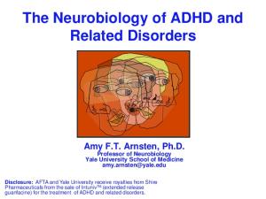
terns of brain damage. As discovered with the famous case of patient H.M., damage to the hippocampus and surrounding medial temporal lobe cortices led to a severe impairment in declarative memory – that is, an inability to form new memories for facts and events (Scoville & Milner, 1957). Notably, this devastating amnesia was selective: H.M. and other individuals with hippocampal damage remained able to learn new procedures and habits that were acquired gradually, such as playing the piano or developing diagnostic skills as a radiologist. This discovery spurred a wealth of subsequent human and animal research, which provided a detailed understanding of the cognitive and neural mechanisms by which the hippocampus and surrounding cortices contribute to memory function. As suggested by the terminology itself, early theories emphasized the declarative nature of memories that depend on the medial temporal lobes: they are explicit, verbalizable and available to conscious awareness (for review see Squire, 2004). Subsequent research has advanced our understanding by uncovering the mechanisms by which the medial temporal lobes support memory for events or episodes (also referred to as episodic memory), how subregions of the medial temporal lobes work in concert to support the formation of new memories, the representational characteristics of the memories that are built, and the contexts in which this system is used (Bakker, Kirwan, Miller, & Stark, 2008; Cohen & Eichenbaum, 1993; Daselaar, Fleck, & Cabeza, 2006; Davachi, 2006; Hannula, Tranel, & Cohen, 2006; Paller & Wagner, 2002; Uncapher & Rugg, 2008; Wirth et al., 2003). 2.1. Parkinson’s disease: a model of the role of the basal ganglia in learning and memory Relatively less is known about the cognitive and neural substrates of non-declarative memory. Habit learning, one form of non-declarative memory, has received much attention in the past decade. Habit learning is typically characterized in opposition to declarative memory: it is gradual, implicit, and can take place without conscious awareness. Converging evidence suggests that habit learning depends on the basal ganglia. Much of this evidence comes from studies of patients with Parkinson’s disease, a progressive neurological disorder that involves a loss of dopaminergic input to the striatum due to a reduction in dopamine neurons in the substantia nigra compacta (Kish, Shannak, & Hornykiewicz, 1988; Peran et al., 2010). Patients with Parkinson’s disease, who famously suffer from motor impairments, also have cognitive and mnemonic impairments, even in the earliest stages of the disease when damage is relatively selective to the striatum. Thus, individuals with Parkinson’s disease provide a useful model of basal ganglia dysfunction in humans and can be compared with individuals with medial temporal lobe dysfunction to ask questions about the unique contribution of each brain region to different kinds of memory. Such comparisons, detailed below, have revealed that the pattern of memory impairments found in patients with Parkinson’s disease is opposite to that found in patients with medial temporal lobe damage. In particular, patients with Parkinson’s disease are impaired on tasks that involve gradual learning of stimulus–response associations but are spared on tests of declarative memory. 2.2. Probabilistic category learning in Parkinson’s disease Probabilistic category learning paradigms have been central to understanding the contributions of the basal ganglia to learning and memory. One widely-used measure, the ‘‘weather prediction’’ task (see Fig. 1), requires that participants use trial-and-error feedback to learn to predict categorical outcomes (sun or rain) based on
625
Fig. 1. The ‘‘weather prediction’’ task is a popular probabilistic category learning task. (A) Each of 4 visual cues – cards with shapes – is independently and probabilistically associated with either ‘‘rain’’ or ‘‘sun’’. (B) On each trial, a combination of one to three cards is shown. Subjects respond based on their prediction of the weather for that trial, and receive response-contingent feedback.
four different visual cues (simple shapes). On each trial one, two, or three of the four cues can be present, yielding 14 possible stimuli (all possible combinations of the cues without displaying all four or none at all). The relationship between cues and outcomes is probabilistic, such that across all trials each cue predicts an outcome only some of the time. The complex cue structure and probabilistic nature of the associations, with no consistent one-to-one mapping between stimuli and outcomes, was originally thought to hamper attempts at explicitly encoding cue-outcome associations, making improved performance dependent on gradual, implicit learning (Knowlton et al., 1996; but see Gluck, Shohamy, & Myers, 2002). Indeed, an important study by Knowlton and colleagues demonstrated that amnesics were capable of incremental learning (measured as increased accuracy in performance) despite having no explicit memory for the testing episode (revealed by multiple choice questions about details of the testing events). By contrast, Parkinson’s disease patients were impaired at incremental learning of the task, but had intact explicit memory (Knowlton, Squire, & Gluck, 1994; Knowlton et al., 1996). This double dissociation was central in advancing the notion that the basal ganglia and medial temporal lobes support two dissociable memory systems. In particular, the findings provided evidence that the basal ganglia are necessary for non-motor, incremental learning of stimulus–response associations (Ashby, Noble, Filoteo, Waldron, & Ell, 2003; Knowlton et al., 1996; Owen et al., 1993; Reber, 1989; Robbins, 1996; SaintCyr, Taylor, & Lang, 1988; Shohamy, Myers, Geghman, Sage, & Gluck, 2006; Shohamy, Myers, Grossman, Sage, & Gluck, 2005; Shohamy et al., 2004; Swainson et al., 2000). These findings in humans converge with a large body of animal lesion work also demonstrating a key role for the basal ganglia in incremental, stimulus–response learning (Divac, Rosvold, & Szwarcbart, 1967; Kesner, Bolland, & Dakis, 1993; McDonald & White, 1993; Mishkin, Malamut, & Bachevalier, 1984; Packard, 1999; Packard, Hirsh, & White, 1989; Packard & McGaugh, 1996; White, 1997; Yin, Knowlton, & Balleine, 2004; Yin, Knowlton, & Balleine, 2006). 2.3. Multiple systems for non-declarative memory The double dissociation between declarative and non-declarative memory functions in patients with Parkinson’s disease and amnesics provided important evidence that the basal ganglia play a role in non-declarative learning. However, subsequent work has demonstrated that Parkinson’s disease patients are impaired at some but not all types of non-declarative learning tasks, as summarized in Table 1, leaving open questions about the specific role of the basal ganglia in learning and the possible contribution of
626
K. Foerde, D. Shohamy / Neurobiology of Learning and Memory 96 (2011) 624–636
Table 1 Learning paradigms where Parkinson’s disease (PD) patients are spared vs. impaired, sorted by their implicitness vs. the demand for feedback to drive learning. Task
Implicit/explicit
Feedback dependent
PD effect on performance
Weather prediction (trial-and-error) Weather prediction (observation) Artificial grammar Artificial grammar (trial-and-error) Motor sequence learning Motor sequence learning (trial-and-error) S-R simple task S-R complex (multi-trial integration needed) Information integration perceptual categorization Rule based perceptual categorization Rotor pursuit Mirror reading
other brain systems. Studies with other patient populations demonstrated that a variety of learning functions found under the umbrella of non-declarative memory do not depend on the basal ganglia, and instead depend on the cerebellum or visual cortex (Doyon, Penhune, & Ungerleider, 2003; Gabrieli, Stebbins, Singh, Willingham, & Goetz, 1997; Laforce & Doyon, 2001). FMRI studies have further corroborated the distinct contributions of the striatum and the cerebellum to learning (E.g. Diedrichsen, Hashambhoy, Rane, & Shadmehr, 2005; Jueptner, Frith, Brooks, Frackowiak, & Passingham, 1997; Jueptner & Weiller, 1998). Interestingly, many of the tasks used to study neuropsychological populations differ from the kinds of tasks used to investigate learning and memory functions of the basal ganglia in animals in that the tasks used in human studies rely heavily on motor and perceptual skills. For example, the mirror-drawing test, a prototypical ‘‘implicit’’ learning task, requires the participant to trace a complex figure, such as a star, while viewing his or her movements in a mirror. With practice, tracing becomes considerably easier and appears to be unrelated to verbalizable insight into how the skill is acquired. Medial temporal lobe damage renders such learning intact, but cerebellar damage can hamper learning of the mirrordrawing skill (Sanes, Dimitrov, & Hallett, 1990). Such tasks and others like it are likely implicit, but independent of the basal ganglia; instead, the skill relies on continuous visual feedback and a remapping between visual input and motor output. Results from studies using non-motor tasks to assess nondeclarative memory support this hypothesis; indeed, evidence from two category learning tasks have provided strong evidence that learning and memory can function independently of both the medial temporal lobes and the basal ganglia. For example, in artificial grammar learning, participants simply observe multiple nonsense letter strings that are generated according to the same underlying set of rules. If a string starts with the letter X, for example, the next ‘grammatically correct’ letter may be Y, which may be followed by T, S, or X with equal likelihood. Thus, a set of letter strings can be created that are either grammatical or not. After observation of multiple grammatical strings, participants are tested on their ability to identify grammatical vs. non-grammatical strings. Amnesics can succeed on this hallmark test of implicit learning, suggesting acquisition of relatively high level learning without accompanying declarative memory (Knowlton, Ramus, & Squire, 1992). However, learning is also spared in patients with Parkinson’s disease (Reber & Squire, 1999). A similar pattern of results emerges using a dot pattern categorization measure of prototype learning. This task involves observation of multiple stimuli that are generated based on a single prototype. For example, nine white dots on a black background may form a prototype. Task stimuli are then generated by moving the locations of the prototype dots 15% in a random direction. Thus, while none of the stimuli look exactly like the prototype, participants can reliably identify patterns of the prototype category vs.
random patterns. Studies assessing prototype learning have produced a similar pattern of results as those examining artificial grammar learning: performance is intact in amnesics, but it is also spared in patients with Parkinson’s disease (Reber & Squire, 1999; Smith, Siegert, McDowall, & Abernethy, 2001). Thus, while the weather prediction task provides evidence for a role for the basal ganglia in one form of habit learning, other forms of implicit learning are acquired independent of the basal ganglia. Together, these studies demonstrate that many questions remain about the specific role of the basal ganglia in learning and memory.
2.4. The basal ganglia and habit learning: summary and open questions Findings in both animals and humans indicated that the basal ganglia contribute to incremental learning of stimulus–response associations. These findings fit nicely with a selective role for the basal ganglia in procedural learning of habits or skills and suggested that the basal ganglia could provide a neural substrate for a nondeclarative memory system. However, this perspective left open many questions about the role of the basal ganglia in different forms of learning and how memory systems are organized in the brain. First, it remained unclear why Parkinson’s disease patients were impaired on some types of implicit learning, but not others. Second, questions were raised about whether participants completing the ‘‘weather prediction’’ task were necessarily learning the contingencies implicitly; subsequent studies demonstrated that healthy subjects could use simple explicit strategies to support learning. Additionally, these studies found that the hippocampus is activated during learning of the ‘‘weather prediction task’’, at least at an early stage (Poldrack et al., 2001; Shohamy, Myers, Onlaor, & Gluck, 2004). Indeed, as subsequent studies demonstrated, both healthy people and Parkinson’s disease patients might in fact rely on the hippocampus when learning in this task (Foerde, Knowlton, & Poldrack, 2006; Moody, Bookheimer, Vanek, & Knowlton, 2004). Thus, even in what is considered to be a prototypical implicit learning task, multiple memory systems contribute to learning. FMRI studies have highlighted the dynamics of memory systems over the course of learning and provide further evidence for joint contributions from multiple memory systems (Dickerson, Li, & Delgado, 2010; Poldrack et al., 2001; Seger, Peterson, Cincotta, LopezPaniagua, & Anderson, 2010). Third, both the medial temporal lobes and basal ganglia are innervated by dopamine neurons originating in the midbrain (Gasbarri, Packard, Campana, & Pacitti, 1994; Otmakhova & Lisman, 1996; Samson, Wu, Friedman, & Davis, 1990). These dopaminergic inputs are known to be crucial for plasticity in both the basal ganglia and the medial temporal lobes, but whether these dopaminergic inputs act in similar or different ways in the two systems is not yet understood (for review see Shohamy & Adcock, 2010). These
K. Foerde, D. Shohamy / Neurobiology of Learning and Memory 96 (2011) 624–636
recent discoveries have necessitated a move to think about memory systems in a more integrated manner. 3. The basal ganglia, dopamine, and reward learning Significant advances have been made in recent years into the functional neurophysiological, neurochemical, and neurocomputational characteristics of the basal ganglia and their dopaminergic inputs. Collectively, these studies suggest that dopamine neurons projecting to the basal ganglia are critical for learning to predict rewarding outcomes and acting to obtain them. This idea arose from a series of pivotal studies that reported on recordings from dopamine neurons in monkeys and demonstrated a role for midbrain dopamine in reward-related learning. First, midbrain dopamine neurons respond to unexpected rewards with an immediate, phasic, burst of activity. Second, if a cue consistently predicts a reward, the dopamine response shifts to occur immediately following the reward-predicting cue instead of the reward. Third, if an expected reward fails to arrive, there is a decrease in firing at the time of the expected reward (Fiorillo, Tobler, & Schultz, 2003; Schultz, Apicella, & Ljungberg, 1993; Waelti, Dickinson, & Schultz, 2001). However, cues often inconsistently predict rewards; that is, a cue will predict an outcome probabilistically. In this case, phasic responses, both at cue and outcome, vary monotonically with the probability of reward, such that even high levels of predictability lead to some phasic activity at the presentation of a rewarding outcome. Similarly, decreases in firing rate for reward omission also appear to vary in proportion to reward probability (Fiorillo et al., 2003). Taken together, these findings provide evidence for a phasic learning signal from midbrain dopamine neurons consistent with existing computational models of reward prediction (Tremblay, Hollerman, & Schultz, 1998; Waelti et al., 2001). In parallel, there has been a surge of evidence obtained using BOLD fMRI in humans that similarly links activity in midbrain dopamine regions to decision making and learning across a range of rewarding stimuli and experimental paradigms (Aron et al., 2004; D’Ardenne, McClure, Nystrom, & Cohen, 2008; Delgado, Nystrom, Fissell, Noll, & Fiez, 2000; O’Doherty, Dayan, Friston, Critchley, & Dolan, 2003; O’Doherty et al., 2004; Pagnoni, Zink, Montague, & Berns, 2002; Schonberg, Daw, Joel, & O’Doherty, 2007; Schonberg et al., 2010). Thus, emerging data demonstrate that the midbrain dopamine system supports feedback-dependent learning processes essential for predicting both certain and uncertain outcomes. Collectively, these models and data suggest a more nuanced view of the basal ganglia than the one indicated by the multiple memory systems framework. Indeed, the physiological data suggest that rather than supporting implicit learning, the basal ganglia are critical for supporting learning that is driven by feedback and is motivated by rewards. Furthermore, the anatomical connectivity between the basal ganglia, prefrontal cortex, and the medial temporal lobes indicate possible interactions between these brain regions, rather than strict dissociations between them (for review see Poldrack & Rodriguez, 2004). Notably, parallel advances have taken place in understanding the role of the hippocampus in memory. Studies have shown that, although memories supported by the hippocampus are often subjectively experienced as accessible to conscious awareness, this is not a necessary feature of such memories (Chun & Phelps, 1999; Greene, Spellman, Dusek, Eichenbaum, & Levy, 2001; Hannula, Ryan, Tranel, & Cohen, 2007; Schendan, Searl, Melrose, & Stern, 2003). Instead, much recent work has emphasized the specific role of the hippocampus in forming memories that encode relations among multiple stimuli, emphasizing the computational characteristics of the hippocampus rather than the verbalizability of the mnemonic contents.
627
4. An integrated view of the basal ganglia in learning Theories regarding the role of dopamine in reward prediction have had a substantial impact on understanding the role of the basal ganglia in learning. They shed light on the basis for previous inconsistencies and highlight the neural mechanisms that bridge seemingly disparate findings. Integrating the domains of electrophysiology, neuroimaging, computational modeling, and neuropsychology has led to some general principles regarding the role of the basal ganglia in learning, as detailed below.
4.1. Learning from response-contingent feedback One prediction that follows from existing physiological data and computational models is that the basal ganglia play a critical role in learning from response-contingent feedback, regardless of whether learning is implicit or explicit. This idea was tested in a functional magnetic resonance imaging (fMRI) study that compared learning of the standard ‘‘weather prediction’’ task (with response-contingent feedback) with a modified ‘‘observational’’ (nofeedback) version (Poldrack et al., 2001). In the observational version, instead of seeing a set of cues, predicting sun or rain, and then receiving feedback, on each trial the set of cues was shown together with a sun or rain outcome label. A test session without feedback assessed performance to determine how much learning had taken place based on observation. Healthy controls performed with similar levels of accuracy on both feedback-based and observational versions of the task. However, the basal ganglia were activated only when learning was feedback-dependent, not when learning was observational. Instead, observational learning engaged the medial temporal lobes. A parallel study found that Parkinson’s disease patients were impaired at ‘‘weather prediction’’ when learning was based on error-correcting feedback but not when it was based on observation (Shohamy et al., 2004). Feedback and observational learning have also been compared using perceptual categorization tasks. In one fMRI study, Cincotta and Seger (2007) compared feedback and observational learning of a perceptual categorization task and found that the head of the caudate was particularly modulated for feedback compared to observational learning. Thus, in this type of categorization task, the basal ganglia also appear to make unique contributions when error-correcting feedback is involved. The idea that the basal ganglia play a critical role in processing response-contingent feedback has also been tested in a nonlearning task. In a clever design, Tricomi and colleagues (2004) compared basal ganglia responses to monetary rewards and punishments that were either un-signaled, preceded by an anticipatory cue, or followed a cue, with the latter condition requiring participants either to make a pre-determined response or to make a self-selected response that participants perceived to affect outcomes. Critically, BOLD activation was observed in the caudate only when outcomes were perceived to be contingent on an action, thereby demonstrating the central role for the basal ganglia in providing response-contingent feedback. Together, these findings indicate that the basal ganglia are selectively involved in, and are necessary for, feedback-based learning. It is important to highlight that for most of the tasks reviewed here the structure of the acquired knowledge was identical. This point contradicts the prior assumption that the complexity of the information learned in the ‘‘weather prediction’’ task is a critical factor that determines whether learning depends on declarative or nondeclarative systems. Although these findings do not illuminate the role of implicit or explicit knowledge, they show that the role of feedback presents a useful way of describing the operating char-
628
K. Foerde, D. Shohamy / Neurobiology of Learning and Memory 96 (2011) 624–636
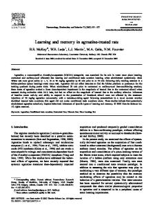
acteristics of learning systems that bridge multiple levels of analysis, from animal physiology to human neuropsychology. Importantly, subsequent studies have revisited other nondeclarative learning tasks, such as the artificial grammar learning (described earlier) and sequence learning, that had been shown to be intact in Parkinson’s disease patients. When these tasks were changed such that learning in these classic implicit tasks was dependent on trial-by-trial feedback, performance was indeed impaired in Parkinson’s disease patients (Seo, Beigi, Jahanshahi, & Averbeck, 2010; Smith & McDowall, 2006; Wilkinson, Khan, & Jahanshahi, 2009; see also Vriezen & Moscovitch, 1990). Taken together, these findings provide strong evidence for the fact that it is the ability to learn from feedback, not the ability to learn implicitly, that depends on the basal ganglia. 4.2. Learning from immediate feedback In a recent study, we tested another prediction about learning in Parkinson’s disease that emerges from examining electrophysiological data and computational models: that, in addition to the presence of feedback, the timing of this feedback is critical (Foerde & Shohamy, 2011). Dopamine is thought to enhance learning in the basal ganglia by facilitating cortico-striatal plasticity, presumably by reinforcing a representation of the rewarded response or stimulus (Reynolds & Wickens, 2002). Importantly for our hypotheses, dopamine responses to feedback occur approximately 100 ms following the reward (Redgrave & Gurney, 2006). Similarly, in prior studies of feedback-based learning, reinforcement was delivered almost immediately following a response (Knowlton et al., 1996; Poldrack et al., 2001; Shohamy et al., 2004). Such rapid feedback results in tight temporal proximity between the response and reward signal, which may facilitate the feedback-based strengthening of the appropriate action representation in the striatum. For example, a behavioral study in young, healthy participants found that learning on a feedback-dependent perceptual categorization task was impaired when feedback was not immediate (Maddox, Ashby, & Bohil, 2003). A recent study in animals demonstrated that the temporal precision of reward expectation in midbrain dopamine neurons decreased as the interval between cue and reward increased. In fact, responses to the longest delayed rewards (16 s) were similar to responses to unpredicted rewards (Fiorillo, Newsome, & Schultz, 2008; see also Roesch, Calu, & Schoenbaum, 2007), consistent with the hypothesis that the dopamine error signal is best suited for learning under short interval conditions. We tested this idea in Parkinson’s disease patients and controls by manipulating the time interval between choice and feedback in a probabilistic learning task (Foerde & Shohamy, 2011; Fig. 2). Participants were asked to categorize stimuli as belonging to one of two categories. As in the ‘‘weather prediction’’ task, the association between stimuli and outcomes was probabilistic and the relationship between cues and outcomes was learned based on trial-bytrial feedback. Learning in this study took place under two different conditions. In one condition, responses were followed by feedback immediately; in the other, feedback followed responses after a 6 s delay. Parkinson’s disease patients were severely impaired at learning when feedback was immediate, but not when feedback was delayed by 6 s (Foerde & Shohamy, 2011). Interestingly, healthy controls were able to learn equally well under both conditions. Together with the selective impairment in patients, this suggests that, despite similar performance among controls in both conditions, the neural systems supporting learning may differ. To address this question, we used fMRI to examine brain activation among young participants learning this task under immediate vs. delayed feedback conditions. We found that where learning from immediate feedback engaged mainly the striatum,
learning from delayed feedback engaged the hippocampus. Thus, feedback timing – and not just feedback presence – plays an important role in modulating the engagement of memory systems and in determining the pattern of impaired vs. spared learning in Parkinson’s disease. 4.3. Learning from rewards vs. punishments The valence of feedback is another obvious yet critical aspect to consider, and indeed one that has been receiving increased attention. It makes intuitive sense that one can learn both by trying to gain positive feedback and by avoiding negative feedback. The basal ganglia consist broadly of parallel functional paths that can be characterized in a number of ways, including the prevalence of D1or D2-type dopamine receptors. The type of receptor is thought to determine how dopamine affects each path, which have been termed Go and NoGo paths. Frank (2005) proposed that activity in the Go path facilitates responding while activity in the NoGo path facilitates the inhibition of responses. Consistent with this idea, dissociations in learning from rewards vs. punishments have been neatly demonstrated by Frank and colleagues in Parkinson’s disease patients (Frank, Seeberger, & O’Reilly, 2004; Moustafa, Cohen, Sherman, & Frank, 2008). In probabilistic learning paradigms, similar to the ones described above, Parkinson’s disease patients who were not on dopaminergic medication could learn from negative feedback, but not positive feedback; in patients on dopaminergic medication, the pattern was reversed. Similar results have been obtained from Parkinson’s disease patients on different types of tasks (Cools, Altamirano, & D’Esposito, 2006) as well as in genetic (Frank, Moustafa, Haughey, Curran, & Hutchison, 2007) and pharmacological studies (Cools et al., 2009). Taken together, these studies show that ability to learn is differentially affected by feedback valence depending on the levels of dopamine in the system, which can be influenced by disease, pharmacological intervention, or individual genetic variation. Nonetheless, there is still a ways to go in understanding how rewards and punishments are processed in the basal ganglia. As outlined previously, midbrain dopamine neurons that project to the basal ganglia respond with increased activity to cues that predict rewards and to outcomes that are better than expected. Outcomes that are worse than expected (or omitted) lead to decreases in responding (Schultz, 1997). However, recent studies have demonstrated that aversive stimuli can lead to increased activity in a subset of midbrain dopamine neurons (Brischoux, Chakraborty, Brierley, & Ungless, 2009). Similarly, fMRI studies have reported increases in BOLD activity in the striatum in response to punishments or negative prediction errors (Delgado, 2007; Delgado, Locke, Stenger, & Fiez, 2003; Delgado et al., 2000; Rodriguez, Aron, & Poldrack, 2006). One factor that may play a role in explaining these differing effects is the context in which rewards and punishments occur (Seymour, Daw, Dayan, Singer, & Dolan, 2007); that is, whether rewards are presented intermixed with punishments or only with the absence of reward. Importantly, neurons that respond with increased activity to rewards vs. punishments appear to be anatomically segregated (Brischoux et al., 2009; Seymour et al., 2007), which suggests that both types of signals can make distinct and meaningful contributions to learning. However, as will be discussed more below, Parkinson’s disease differentially affects distinct pathways in the midbrain and basal ganglia as the disease progresses. This suggests that there could be asymmetric consequences for learning from reward and punishment over the course of the disease. Overall, the valence of feedback plays a central role in how the basal ganglia are engaged to support learning and memory, but a deeper understanding of the interplay of individual differences
K. Foerde, D. Shohamy / Neurobiology of Learning and Memory 96 (2011) 624–636
629
Fig. 2. Learning from immediate vs. delayed feedback (A). Examples of trials where feedback is provided immediately or after a 6-s delay. (B). Performance of Parkinson’s disease patients and age-matched controls in a post-learning test phase where participants make the same choices as during learning but without receiving feedback. These results show how well each group has learned after training with feedback of different delays.
(across healthy and impaired populations), context, and the basic physiology of the relevant circuits will be critical in order to make further progress. 4.4. Building inflexible stimulus–response associations The multiple memory systems framework has emphasized a role for the striatum in habit learning. Indeed, evidence points to a role for the basal ganglia in incremental stimulus–response learning, which fits with our intuitive notion of what habits are. In animals, habits are defined as behaviors that persist even when the resulting outcomes have become undesirable and are contrasted with goal-directed behaviors that are sensitive to changes in the value of outcomes (Adams & Dickinson, 1981; Balleine & O’Doherty, 2010). This idea has been tested in rats using devaluation procedures. In a typical study, a rat may learn to press one lever for one reward (a food pellet, for example) and another lever for an alternative food reward. One of these food rewards is then devalued by feeding the rat the food until satiated or by pairing the food with a drug that makes the rat ill. In either case, the specific food is no longer desirable to the rat. If the rat’s behavior is goal-directed, behavior leading to the food reward that has become undesirable should decrease in frequency; in contrast, if behavior is habitual, it may continue even when it leads to the newlyundesirable outcome. A series of experiments demonstrated that damage to the dorsolateral striatum impairs habit learning and renders behavior goal-directed, whereas dorsomedial striatal lesions lead to the opposite pattern of behavior (Yin & Knowlton, 2006; Yin, Knowlton, & Balleine, 2005; Yin et al., 2004; Yin et al., 2006). In humans, two general approaches have been used to capture the habitual nature of learning. One approach has been to literally replicate the rodent conditioning and devaluation paradigms in humans (Tricomi, Balleine, & O’Doherty, 2009; Valentin, Dickinson, & O’Doherty, 2007). In an fMRI study, for example, healthy subjects learned to make responses to gain M&M’s or Fritos to consume upon leaving the scanner. Half of the participants trained for 1 day, and half trained for 3 days. Subsequently, one food was devalued by feeding to satiation. Behavior was then tested to see if participants’ responding would continue as before, even once the food outcome was no longer desirable. Training for 3 days led to continued responding to earn the devalued food, indicating that responding had become habitual, but training for 1 day led to a marked decrease in participants’ responding for that food. As behavior became habitual, neural activity increased in posterior dorsolateral striatum (Tricomi et al.). These behavioral and fMRI
results are consistent with those in the rodent lesion studies described above in implicating the dorsolateral striatum in habit learning. Although the application of the behavioral assays outlined above have been key in developing our understanding of the type of learning that is controlling behavior, this view of flexibility is somewhat limited, and does not perfectly overlap with how flexibility of mnemonic representations (or behavior) is defined and used in cognitive psychology or neuropsychology. In many animal behavior studies, the focus has primarily been on trying to understand whether stimulus–response or action-outcome associations are controlling behavior. Another approach to determine the mnemonic flexibility of learned information has been to measure how easily it can be used in a novel context. This approach is also inspired by studies of animal behavior, but is centered on studies characterizing the unique contributions of the hippocampus (Cohen & Eichenbaum, 1993). Here, the critical factor is representational flexibility of stimulus– stimulus associations; that is, whether a representation retains information about the relationship among the components of a memory. Typically, for an episodic memory, the particular relationship and combination of items is critical. For example, in the ‘‘weather prediction’’ task, post-learning tests ask people to make judgments about task elements presented in novel configurations or to assess the strength of relationships between cues and outcomes instead of making the categorical responses used during learning (Foerde et al., 2006; Reber, Knowlton, & Squire, 1996). Alternatively, to break away from the use of explicit test questions, other studies have used two-phase tasks that consist of a learning phase, followed by a probe phase to determine the flexibility of the learned representations. In such tasks, participants first learn repeated stimulus–response associations, then use these associations in a second phase where they must flexibly integrate across learned associations to respond to novel items. This approach has demonstrated that the striatum supports inflexible stimulus–response associations whereas the hippocampus is necessary for flexible generalization to novel contexts (Myers et al., 2003; Shohamy & Wagner, 2008; Shohamy et al., 2006). A final type of flexibility that is very important (and, in fact, commonly studied in Parkinson’s disease but is beyond the scope of this review), is higher-level flexibility, critical for cognitive control and task-switching behaviors. Such flexibility includes the ability to switch from responding to one stimulus to another, as well as the ability to pivot from one set of rules for responding to another set. This goes beyond simply remembering which rule is currently operating, because it also depends on the ability to re-
630
K. Foerde, D. Shohamy / Neurobiology of Learning and Memory 96 (2011) 624–636
frain from applying a previous rule. Extensive research has demonstrated the key role of the prefrontal cortex in such task-switching and cognitive control behavior (for a review, see Cools, Altamirano, & D’Esposito, 2006). The striatum’s role in habit learning through repetition is closely related to its role in the chunking of action sequences; through repeated practice, behaviors that are composed of multiple actions can become unitized into a single action (Barnes et al., 2011; Graybiel, 1998; Jog, Kubota, Connolly, Hillegaart, & Graybiel, 1999). The chunking process is consistent with our intuition of procedural learning and also comports with the idea that, once unitized, the individual components of behavior lose flexibility and are no longer accessible. The development towards such welllearned sequences is associated with a change in reliance on the anterior striatum to the posterior striatum (Hikosaka, Miyashita, Miyachi, Sakai, & Lu, 1998). The general notion of chunking is also related to sequence learning tasks, which have frequently been used to study implicit vs. explicit learning in humans (Aizenstein et al., 2004; Curran & Keele, 1993; Doyon et al., 1997; Doyon et al., 1998; Nissen & Bullemer, 1987; Reber & Squire, 1994; Reber & Squire, 1998; Seidler et al., 2005; Willingham, Salidis, & Gabrieli; 2002). Findings across studies of sequence learning in Parkinson’s disease are mixed, but there are several reports of impairments in this type of learning in Parkinson’s disease (see Doyon, 2008 for review and discussion of possible factors underlying variable findings). In such tasks, learning is generally measured in terms of reaction times to stimuli that occur in predictable sequences. Critically, learning of the predictable sequence is commonly unrelated to explicit knowledge of the sequence, which may or may not develop in parallel. In fact, procedural and declarative knowledge could exist without influencing each other (Reber & Squire, 1998). Just as in probabilistic category learning, different types of knowledge may be acquired but are used in different ways. Sequence learning is unique, however, in that explicit trial-by-trial feedback is not commonly provided. Altogether, then, despite mixed results in patients with Parkinson’s disease, animal studies and fMRI studies in humans generally do implicate the striatum in sequence learning. This raises the question of whether the roles of the striatum in supporting learning from feedback and in sequencing behavior are related or independent of each other. One intriguing resolution may lie in thinking about feedback more broadly. A relevant example comes from the study of birdsong, where the auditory feedback provided by producing the song is itself essential in maintaining vocal performance (Brainard & Doupe, 2000). This finding suggests that feedback need not be provided in the explicit manner commonly seen in studies in animals and humans. Future studies applying such a broader view of feedback could expand our understanding of the role of the striatum and help reconcile inconsistencies in patient studies across multiple tasks. It is interesting that the striatum appears to play a general role in supporting inflexible representations across these different classes of representations that have been assessed in terms of their flexibility. Despite the shared term, these different forms of flexibility are quite distinct – they appear to be mediated by distinct neural systems (anterior striatum, hippocampus, prefrontal cortex) that are differentially affected by disease (Parkinson’s, for example). Perhaps the striatum is able to play a role across multiple forms of flexibility by virtue of its characteristic connectivity to other brain regions, as discussed in the next section. 4.5. Summary Findings from Parkinson’s disease, the most influential model of basal ganglia dysfunction, demonstrate that the basal ganglia support trial-by-trial learning, driven by error correcting feedback as
well as sequencing behavior. This appears to be independent of whether learning is implicit or explicit (see also Henke, 2010; Seger, Dennison, Lopez-Paniagua, Peterson, & Roark, 2011). The system seems to be biased towards learning from reward, evidenced by the selective pattern of impairments in medicated vs. non-medicated patients on learning from rewards vs. punishments. Finally, data from Parkinson’s disease support the idea that the basal ganglia are specialized for forming specific, inflexible representations that do not easily generalize to new choices, which contrasts with the role of the hippocampus in building flexible, relational representations that are well suited to guide behavior in novel contexts. However, an increasing body of evidence from fMRI and animal work also suggests that the basal ganglia as a whole play a broader role in learning, supporting not just habitual learning, but also more goal-directed motivated behaviors. Therefore, greater attention is being paid to the role of subregions within the basal ganglia and their interactions with other learning systems.
5. Interactions between the basal ganglia and other brain systems for learning Neuropsychological and animal data, together with reinforcement learning models, have provided a detailed understanding of the circumstances under which the basal ganglia support learning and insights into the mechanisms by which they do so. Armed with this new understanding, there has been a renewed interest in understanding how this learning system is complemented by, and interacts with, other learning systems. These efforts have taken place in three main areas:
5.1. Interactions between basal ganglia and medial temporal lobes: competitive vs. cooperative Studies in neuropsychological populations such as Parkinson’s disease patients have been critical in understanding memory systems, but there are limitations to what they can tell us about how the healthy brain functions. The use of functional imaging (e.g. fMRI) to explore how memory systems are engaged in the healthy brain has revealed that, although one memory system or another may be absolutely necessary to support a function, multiple systems are commonly engaged to support behavior. This has prompted questions about whether these systems might influence each other, and if so, whether they act oppositionally, independently, or synergistically. Initial evidence for oppositional interactions between striatal and medial temporal lobe systems came from lesion studies in animals (Chang & Gold, 2003a; Chang & Gold, 2003b; Eichenbaum, Fagan, Mathews, & Cohen, 1988; McDonald & White, 1995; Mitchell & Hall, 1988; Packard et al., 1989; Rabe & Haddad, 1969; Schroeder, Wingard, & Packard, 2002). These studies showed that damage to one system could lead to improved performance by the other system. This remarkable finding provided strong evidence for the idea that, under normal conditions, there is a competitive interaction between these systems, such that removing this interference could enhance learning. Other experiments found that, on some tasks where multiple learning strategies were available and dependent on separate systems, lesions to one system could lead to use of the alternative strategy of the other system, although the lesioned system might be naturally preferred (Chang & Gold, 2003a; Chang & Gold, 2003b; Packard & Knowlton, 2002; Schroeder et al., 2002). These results suggest that multiple systems learn viable response strategies and that competition could occur at the time of action selection or responding. Note that this is very different from a form of competition that interferes with learning
K. Foerde, D. Shohamy / Neurobiology of Learning and Memory 96 (2011) 624–636
itself; this kind of competition occurs between learned response strategies. There is also evidence from fMRI studies in humans to support the idea that multiple memory systems may support performance on a single task (Doeller, King, & Burgess, 2008; Foerde et al., 2006; Hartley & Burgess, 2005; Voermans et al., 2004). Mirroring findings from animal work, these studies found that different systems support information with distinct qualities, in concordance with the type of representation that characterizes each system. For example, Foerde et al. (2006), used fMRI to examine subjects learning ‘‘weather prediction’’ under divided attention (the subject had to perform another task while simultaneously learning to predict the weather) or full attention (the participant only performed the ‘‘weather prediction’’ task). When full attention was afforded to the task, subjects engaged the medial temporal lobe more, and had more flexible knowledge of what they learned. However, when learning with divided attention, subjects engaged the basal ganglia more and had less flexible representations. One question raised by such studies is why Parkinson’s disease patients do not simply switch to strategies that depend on functional neural systems. In some cases this can indeed happen (Moody et al., 2004), and understanding how to capitalize on such possibilities should be informed by future research aimed at understanding how we can manipulate and modulate distinct memory systems. The evidence for competition between systems received renewed attention when fMRI studies found evidence for ‘‘neural competition’’ between systems: negative correlations between BOLD activity in striatal regions and medial temporal lobe regions, such that when one system was ‘on’ the other was ‘off’ (Lieberman, Chang, Chiao, Bookheimer, & Knowlton, 2004; Poldrack et al., 2001; Seger & Cincotta, 2005). One limitation with such results is that it is not known whether this effect reflects direct communication between systems, or whether it is mediated by other regions. Further analysis has suggested that, in some cases, the prefrontal cortex may play a role in mediating between multiple systems (Poldrack & Rodriguez, 2004). Another limitation is that ‘‘neural competition’’ does not reveal whether interactions might reflect interference with learning per se, or only with the ability to express what is being learned (that is, the behavioral output of learning). Finally, these interactions found in imaging studies have not been linked to behavioral evidence for competition as they have in the animal work. Moreover, evidence for this type of competition has been scant in neuropsychological populations (but see Cavaco, Anderson, Allen, Castro-Caldas, & Damasio, 2004). Thus, in humans, these results are still in need of further explication. FMRI has also provided evidence for synergistic relationships between systems. For example, evidence supports such a relationship between the dopaminergic midbrain and medial temporal lobes, as correlations between these regions predict how well individuals perform behaviorally on tests of memory (Adcock, Thangavel, Whitfield-Gabrieli, Knutson, & Gabrieli, 2006; Duzel et al., 2010; Shohamy & Wagner, 2008; Wittmann et al., 2005). Consistent with these results, a number of theories and findings have proposed that such midbrain–hippocampus interactions are critical for long-term episodic memory (Lisman & Grace, 2005; Redondo & Morris, 2011; Shohamy & Adcock, 2010). Given the interconnectivity of these systems, one would expect to observe consequences for Parkinson’s disease patients beyond those behaviors that depend on the basal ganglia. However, as outlined in the introduction, the general research approach has emphasized dissociation rather than interaction: impairment in one task and intact performance on another task. As neuropsychological research adopts paradigms that have been refined in other domains, this emphasis on dissociation may start to change (see Schonberg et al., 2010 for an example). As we become better at targeting specific computations through task design and apply a more
631
integrated view of memory systems to our experimental logic, we may begin to discover the effects of Parkinson’s disease on memory system interactions. However, as we discuss in the next section, caution is warranted when making overarching claims about ‘distinct’ systems and how they interact with other systems; these systems are heterogeneous and patterns of interaction may vary across different sub-regions. 5.2. Subregions of the basal ganglia and distinct cortical loops One critical advance in our understanding of the role of the basal ganglia in behavior has come from seminal anatomical work showing that the basal ganglia are a node in a system of corticostriatal loops (Alexander, DeLong, & Strick, 1986; Lehericy et al., 2004; Middleton & Strick, 2000). Cortical regions project to distinct regions of the striatum, from the striatum neurons project to the globus pallidus, from the globus pallidus to the thalamus, and from the thalamus back to cortical regions. Initially, at least five distinct loops were described (Alexander et al., 1986) but three loops, labeled the limbic, associative, and motor circuits, capture the essential segregation (Joel & Weiner, 2000). The motor loop involves motor regions of the frontal lobe (primary, supplementary, premotor) and dorsolateral putamen and caudate within the striatum (mainly posterior regions); the associative loop involves regions such as dorsolateral prefrontal cortex and the caudate and anterior putamen regions; the limbic loop involves orbitofrontal and cingulate cortex of the frontal lobes, but also amygdala and hippocampus inputs, and targets the ventral striatum including nucleus accumbens and ventral aspects of the caudate and putamen (Haber, Fudge, & McFarland, 2000; Joel & Weiner, 2000). Views on the connections amongst the cortico-striatal loops continue to develop, but there appears to have been a shift from a view of loops as fundamentally segregated to one of a mixture of discrete and interactive loops. Haber and colleagues (2000) describe such connections as spiraling in a ventromedial to dorsolateral gradient in the striatum, from the limbic through the associative to the motor loop via the dopaminergic midbrain. Given this architecture, it is not surprising that a variety of motor and cognitive tasks have been associated with the basal ganglia, and the architecture comports well with the observation that various loops appear to be preferentially involved in different types of tasks and that engagement of specific loops may change as practice unfolds (Jueptner et al., 1997). It also underscores the fact that the basal ganglia are heterogeneous structures and a simple overarching labeling of their functions is challenging. However, it has been suggested that these heterogeneous loops can account for basal ganglia involvement in various psychiatric and neurological disorders, with dysfunction in specific sub-loops accounting for distinct neuropsychiatric symptoms (Mega & Cummings, 1994; Middleton & Strick, 2000). Furthermore, the various targets in the striatum differ neurochemically and various pharmacological treatments affect sub-regions within the basal ganglia differently; positive effects on some cognitive functions and detrimental effects for others are seen as a result of medications targeting the basal ganglia (E.g. Cools, Barker, Sahakian, & Robbins, 2001; Deutch & Cameron, 1992). This becomes important in Parkinson’s disease, which entails a progression of denervation from dorsal towards ventral striatum (Bernheimer, Birkmayer, Hornykiewicz, Jellinger, & Seitelberger, 1973; Kish et al., 1988). As noted above, different striatal sub-regions have highly divergent connectivity with other brain regions and this should result in consequences for learning and memory functions beyond the striatum that change as the disease progresses. Therefore, continued study of learning and memory functions of the basal ganglia and how these are affected over time in both medicated and unmedicated patients with basal ganglia dysfunction is of great interest.
632
K. Foerde, D. Shohamy / Neurobiology of Learning and Memory 96 (2011) 624–636
More recent techniques have allowed assessments of functional connectivity, which have revealed intriguing differences in the relationships between striatal sub-regions and the rest of the brain. Di Martino and colleagues (2008) found that caudate and putamen regions showed opposite relationships with a number of other regions. For example, the caudate showed a positive relationship with the parahippocampal gyrus, with the relationship more pronounced in the inferior ventral striatum than the dorsal caudate. In contrast, the putamen showed a negative relationship with this region. Thus, tasks that rely on the putamen could rely on one form of interaction with other brain regions, such as the parahippocampal gyrus, while the caudate and ventral striatum could exhibit an entirely different pattern. The consequences of such differential interactions for behavior are beginning to be investigated (E.g. Poldrack et al., 2001; Shohamy & Wagner, 2008), but the implications for neurological and psychiatric disorders remain unknown. Furthermore, recent work has suggested that anatomical connectivity between the striatum, limbic, and prefrontal regions is associated with individual differences in traits such as novelty seeking and reward dependence (Cohen, Schoene-Bake, Elger, & Weber, 2009). Pairing such variability with changes that occur as Parkinson’s disease progresses presents new complexities, but at the same time presents an opportunity to understand variation across time and across individuals. 5.3. Dopamine modulation of multiple forms of learning In learning and memory research, Parkinson’s disease is often viewed simply as a model of disrupted striatal function. Notably, Parkinson’s disease patients are predominantly treated with medications that increase dopamine levels in the brain. The most common treatment, L-dopa, is a dopamine precursor that is converted to dopamine in the brain and alleviates many of the motor symptoms of the disease. In addition to L-dopa, many Parkinson’s disease patients are treated with a variety of dopamine agonists with differential affinity for different dopamine receptor subtypes. Medication is thought to cause a global increase in baseline dopamine levels (Agid, Javoy-Agid, & Ruberg, 1987). This provides an opportunity to examine how changes in dopamine levels impact cognitive function, by comparing performance in Parkinson’s disease patients tested ‘on’ vs. ‘off’ medication. Importantly, electrophysiological and computational models specify that the timing and relative changes in phasic dopamine release are critical for learning. Thus, this suggests that medication may worsen learning, putatively by masking stimulus-specific phasic effects (while benefiting, or not affecting, non-learning functions). Indeed, dopaminergic medication can worsen learning performance in Parkinson’s disease patients (Cools, Lewis, Clark, Barker, & Robbins, 2007; Cools et al., 2001; Frank, 2005; Frank, Seeberger, & O’Reilly, 2004; Shohamy et al., 2006), despite improving or sparing other forms of cognitive function. Several complementary theories have been advanced to account for these results. One proposal relates to the separable frontostriatal circuits and the progression in Parkinson’s disease from affecting one to several of these circuits. Treatment with dopaminergic medication may rectify dopamine hypo-function within one circuit and alleviate symptoms associated with that circuit, while ‘‘overdosing’’ the other, not yet affected, circuits (Cools et al., 2001; Cools et al., 2007). Even as disease progresses and more circuits are affected by dopamine loss, balancing the dopamine needs of distinct circuits would remain a challenge. Another proposal, as noted earlier, is based on the idea that a basic feature of basal ganglia loop architecture is the presence of both Go and NoGo paths; that is, learning both to execute and inhibit responses is important, and the valence of feedback during learning drives these paths differentially. Critically, positive and
negative feedback are differentially able to function in an environment of tonic increases in dopamine (Frank, 2005). Experimental work has confirmed that learning from positive feedback is intact in Parkinson’s disease patients on medication but impaired in those off medication, whereas learning from negative feedback is impaired in Parkinson’s disease patients on medication but improved in those off medication. Understanding the role of the striatum in learning is thus closely linked to understanding dopamine in learning; Parkinson’s disease has played a key role in advancing how we understand this link in humans. Far less is known so far about the role of dopamine in modulating the hippocampus in humans. Midbrain dopamine regions in the ventral tegmental area project to the hippocampus, where dopamine plays an essential role in hippocampal plasticity (Frey, Schroeder, & Matthies, 1990; Redondo & Morris, 2011). This suggests that information about expectations, reward and learning is projected to both the basal ganglia and the hippocampus modulating learning in both targets. Indeed, recent findings and theories suggest that dopaminergic modulation of the hippocampus may be critical for declarative memory (Lisman & Grace, 2005; Shohamy & Adcock, 2010). For example, fMRI studies have shown enhancements in medial temporal lobe dependent declarative memory when midbrain dopaminergic regions are also engaged (Adcock et al., 2006; Shohamy & Wagner, 2008; Wittmann, Bunzeck, Dolan, & Duzel, 2007; Wittmann et al., 2005). More direct evidence for a role for dopamine comes from a recent PET study that showed associations between memory and D2, but not D1, receptor binding in the hippocampus (Takahashi et al., 2008). Declarative memory deficits associated with dopamine loss are documented in healthy aging (Bäckman et al., 2000) and there has been some attempt to unify observations about aging and midbrain-medial temporal lobe interactions into a coherent framework (Duzel, Bunzeck, Guitart-Masip, & Duzel, 2010). Pharmacological studies have found long-lasting enhancements in word learning as a result of dopaminergic manipulations. Interestingly, enhancements were found as a consequence of L-dopa (Knecht et al., 2004), thought to enhance phasic responses, but impairments were found in response to pergolide, a tonic D1 and D2 receptor agonist (Breitenstein et al., 2006). Thus, in healthy young adults manipulation of some dopamine mechanisms leads to an ‘‘overdose’’ like effect where raising dopamine levels above what is ‘‘normal’’ has detrimental effects (as described for some types of learning in Parkinson’s disease patients on dopaminergic medication), but manipulating transient effects can lead to benefits even in an otherwise healthy brain. In schizophrenia, dysregulation of dopamine in the hippocampus does seem linked to mnemonic dysfunction, suggesting that restoring ‘‘normal’’ levels is advantageous (Shohamy et al., 2010). These results suggest similar dynamics for dopamine in the medial temporal lobes and the basal ganglia, yet many questions remain regarding the mechanisms of dopamine action in the medial temporal lobes; findings of distinct dopamine receptor distribution in the basal ganglia vs. the medial temporal lobes suggest possibly distinct modes of action in these regions (see Shohamy & Adcock, 2010 for detail). The importance of dopamine in circuits that are thought to be spared by Parkinson’s disease (medial temporal lobes) raises questions about how the disease affects functions that rely on these neural systems. It should be noted that there have been some investigations of declarative memory deficits in Parkinson’s disease (Cohn, Moscovitch, & Davidson, 2010; Davidson, Anaki, Saint-Cyr, Chow, & Moscovitch, 2006). However, it remains unclear whether such deficits are present in non-demented Parkinson’s disease patients or only when dementia is present. Thus far, it is difficult to know whether declarative memory deficits are related to executive dysfunction and strategic problems associated with
K. Foerde, D. Shohamy / Neurobiology of Learning and Memory 96 (2011) 624–636
the prefrontal cortex or whether the hippocampus and medial temporal lobes are compromised as well (Hay, Moscovitch, & Levine, 2002; Whittington, Podd, & Kan, 2000; Whittington, Podd, & Stewart-Williams, 2006). Increasingly, studies of declarative memory in animals and healthy young participants are guided by what has been learned from computational models to target the critical operations more specifically, rather than relying on features like awareness of and ability to report on learning and memory. Hopefully, the advances that have been made in theory and in imaging research design will enable increasingly fruitful investigations of these questions. 6. Conclusions and future directions Volumes of research have highlighted the unique contributions of the basal ganglia and medial temporal lobes to learning and memory. However, some of the resulting characterizations, such as assigning implicit and explicit learning and memory to these systems, have turned out to be oversimplified. Moreover, a focus on assigning unique attributes to these two systems has overlooked the many ways in which these brain regions jointly contribute to behavior. Converging evidence from electrophysiology, neuroimaging, computational modeling, and neuropsychology, and, moreover, communication between these fields, have advanced a more integrated view of basal ganglia function. Paradigms that translate across varied approaches have been key in developing the emerging view of basal ganglia function, and the study of learning in Parkinson’s disease patients has been integral to this progress. This integrative approach will help provide the next critical steps towards advancing our view of how the basal ganglia interact with the medial temporal lobes and other brain systems, as well as promote an understanding of the role of dopamine and other neuromodulators in multiple brain systems for learning and memory. Acknowledgments We thank Suzanne Wood for comments on an earlier draft and members of the Learning Lab at Columbia University for helpful conversations about the research reviewed here. K.F. is supported by an NINDS NRSA training grant (F32 NS063632); D.S. is supported by a NARSAD Young Investigator Award, the Michael J Fox Foundation, and a National Science Foundation Early Career Award. References Adams, C. D., & Dickinson, A. (1981). Instrumental responding following reinforcer devaluation. Quarterly Journal of Experimental Psychology, 33, 109–122. Adcock, R. A., Thangavel, A., Whitfield-Gabrieli, S., Knutson, B., & Gabrieli, J. D. (2006). Reward-motivated learning: Mesolimbic activation precedes memory formation. Neuron, 50(3), 507–517. Agid, Y., Javoy-Agid, M., & Ruberg, M. (Eds.). (1987). Biochemistry of neurotransmitters in Parkinson’s disease. (Vol. 2). London: Butterworth’s and Co. Aizenstein, H. J., Stenger, V. A., Cochran, J., Clark, K., Johnson, M., Nebes, R. D., et al. (2004). Regional brain activation during concurrent implicit and explicit sequence learning. Cerebral Cortex, 14(2), 199–208. Alexander, G. E., DeLong, M. R., & Strick, P. L. (1986). Parallel organization of functionally segregated circuits linking basal ganglia and cortex. Annual Review of Neuroscience, 9, 357–381. Aron, A. R., Shohamy, D., Clark, J., Myers, C., Gluck, M. A., & Poldrack, R. A. (2004). Human midbrain sensitivity to cognitive feedback and uncertainty during classification learning. Journal of Neurophysiology, 92(2), 1144–1152. Ashby, F. G., Noble, S., Filoteo, J. V., Waldron, E. M., & Ell, S. W. (2003). Category learning deficits in Parkinson’s disease. Neuropsychology, 17(1), 115–124. Bäckman, L., Ginovart, N., Dixon, R. A., Wahlin, T. B., Wahlin, A., Halldin, C., et al. (2000). Age-related cognitive deficits mediated by changes in the striatal dopamine system. American Journal of Psychiatry, 157(4), 635–637. Bakker, A., Kirwan, C. B., Miller, M., & Stark, C. E. (2008). Pattern separation in the human hippocampal CA3 and dentate gyrus. Science, 319(5870), 1640–1642. Balleine, B. W., & O’Doherty, J. P. (2010). Human and rodent homologies in action control: Corticostriatal determinants of goal-directed and habitual action. Neuropsychopharmacology, 35(1), 48–69.
633
Barnes, T. D., Mao, J. B., Hu, D., Kubota, Y., Dreyer, A. A., Stamoulis, C., et al. (2011). Advance-cueing produces enhanced action-boundary patterns of spike activity in the sensorimotor striatum. Journal of Neurophysiology. Bernheimer, H., Birkmayer, W., Hornykiewicz, O., Jellinger, K., & Seitelberger, F. (1973). Brain dopamine and the syndromes of Parkinson and Huntington. Clinical, morphological and neurochemical correlations. Journal of the Neurological Sciences, 20(4), 415–455. Brainard, M. S., & Doupe, A. J. (2000). Interruption of a basal ganglia-forebrain circuit prevents plasticity of learned vocalizations. Nature, 404(6779), 762–766. Breitenstein, C., Korsukewitz, C., Floel, A., Kretzschmar, T., Diederich, K., & Knecht, S. (2006). Tonic dopaminergic stimulation impairs associative learning in healthy subjects. Neuropsychopharmacology, 31(11), 2552–2564. Brischoux, F., Chakraborty, S., Brierley, D. I., & Ungless, M. A. (2009). Phasic excitation of dopamine neurons in ventral VTA by noxious stimuli. Proceedings of the National Academy of Sciences of the United States of America, 106(12), 4894–4899. Cavaco, S., Anderson, S. W., Allen, J. S., Castro-Caldas, A., & Damasio, H. (2004). The scope of preserved procedural memory in amnesia. Brain, 127(Pt 8), 1853–1867. Chang, Q., & Gold, P. E. (2003a). Intra-hippocampal lidocaine injections impair acquisition of a place task and facilitate acquisition of a response task in rats. Behavioral Brain Research, 144(1–2), 19–24. Chang, Q., & Gold, P. E. (2003b). Switching memory systems during learning: Changes in patterns of brain acetylcholine release in the hippocampus and striatum in rats. Journal of Neuroscience, 23(7), 3001–3005. Chun, M. M., & Phelps, E. A. (1999). Memory deficits for implicit contextual information in amnesic subjects with hippocampal damage. Nature Neuroscience, 2(9), 844–847. Cincotta, C. M., & Seger, C. A. (2007). Dissociation between striatal regions while learning to categorize via feedback and via observation. Journal of Cognitive Neuroscience, 19(2), 249–265. Cohen, M. X., Schoene-Bake, J. C., Elger, C. E., & Weber, B. (2009). Connectivity-based segregation of the human striatum predicts personality characteristics. Nature Neuroscience, 12(1), 32–34. Cohen, N. J., & Eichenbaum, H. (1993). Memory. Cambridge, MA, US: The MIT Press amnesia, and the hippocampal system. Cohn, M., Moscovitch, M., & Davidson, P. S. (2010). Double dissociation between familiarity and recollection in Parkinson’s disease as a function of encoding tasks. Neuropsychologia, 48(14), 4142–4147. Cools, R., Altamirano, L., & D’Esposito, M. (2006). Reversal learning in Parkinson’s disease depends on medication status and outcome valence. Neuropsychologia, 44(10), 1663–1673. Cools, R., Barker, R. A., Sahakian, B. J., & Robbins, T. W. (2001). Enhanced or impaired cognitive function in Parkinson’s disease as a function of dopaminergic medication and task demands. Cerebral Cortex, 11(12), 1136–1143. Cools, R., Frank, M. J., Gibbs, S. E., Miyakawa, A., Jagust, W., & D’Esposito, M. (2009). Striatal dopamine predicts outcome-specific reversal learning and its sensitivity to dopaminergic drug administration. Journal of Neuroscience, 29(5), 1538–1543. Cools, R., Lewis, S. J., Clark, L., Barker, R. A., & Robbins, T. W. (2007). L-dopa disrupts activity in the nucleus accumbens during reversal learning in Parkinson’s disease. Neuropsychopharmacology, 32(1), 180–189. Curran, T., & Keele, S. W. (1993). Attentional and nonattentional forms of sequence learning. Journal of Experimental Psychology: Learning, Memory, & Cognition., 19(1), 189–202. D’Ardenne, K., McClure, S. M., Nystrom, L. E., & Cohen, J. D. (2008). BOLD responses reflecting dopaminergic signals in the human ventral tegmental area. Science, 319(5867), 1264–1267. Daselaar, S. M., Fleck, M. S., & Cabeza, R. (2006). Triple dissociation in the medial temporal lobes: Recollection, familiarity, and novelty. Journal of Neurophysiology, 96(4), 1902–1911. Davachi, L. (2006). Item, context and relational episodic encoding in humans. Current Opinion in Neurobiology, 16(6), 693–700. Davidson, P. S., Anaki, D., Saint-Cyr, J. A., Chow, T. W., & Moscovitch, M. (2006). Exploring the recognition memory deficit in Parkinson’s disease: Estimates of recollection versus familiarity. Brain, 129(Pt 7), 1768–1779. Delgado, M. R. (2007). Reward-related responses in the human striatum. Annals of the New York Academy of Sciences, 1104, 70–88. Delgado, M. R., Locke, H. M., Stenger, V. A., & Fiez, J. A. (2003). Dorsal striatum responses to reward and punishment: Effects of valence and magnitude manipulations. Cognitive Affect Behavioral Neuroscience, 3(1), 27–38. Delgado, M. R., Nystrom, L. E., Fissell, C., Noll, D. C., & Fiez, J. A. (2000). Tracking the hemodynamic responses to reward and punishment in the striatum. Journal of Neurophysiology, 84(6), 3072–3077. Deutch, A. Y., & Cameron, D. S. (1992). Pharmacological characterization of dopamine systems in the nucleus accumbens core and shell. Neuroscience, 46(1), 49–56. Di Martino, A., Scheres, A., Margulies, D. S., Kelly, A. M., Uddin, L. Q., Shehzad, Z., et al. (2008). Functional connectivity of human striatum: A resting state FMRI study. Cerebral Cortex, 18(12), 2735–2747. Dickerson, K. C., Li, J., & Delgado, M. R. (2010). Parallel contributions of distinct human memory systems during probabilistic learning. Neuroimage. Diedrichsen, J., Hashambhoy, Y., Rane, T., & Shadmehr, R. (2005). Neural correlates of reach errors. Journal of Neuroscience, 25(43), 9919–9931. Divac, I., Rosvold, H. E., & Szwarcbart, M. K. (1967). Behavioral effects of selective ablation of the caudate nucleus. Journal of Comparative and Physiological Psychology, 63(2), 184–190.
634
K. Foerde, D. Shohamy / Neurobiology of Learning and Memory 96 (2011) 624–636
Doeller, C. F., King, J. A., & Burgess, N. (2008). Parallel striatal and hippocampal systems for landmarks and boundaries in spatial memory. Proceedings of the National Academy of Sciences of the United States of America, 105(15), 5915–5920. Doyon, J. (2008). Motor sequence learning and movement disorders. Current Opinion in Neurology, 21(4), 478–483. Doyon, J., Penhune, V., & Ungerleider, L. G. (2003). Distinct contribution of the cortico-striatal and cortico-cerebellar systems to motor skill learning. Neuropsychologia, 41(3), 252–262. Doyon, J., Gaudreau, D., Laforce, R., Jr., Castonguay, M., Bedard, P. J., Bedard, F., et al. (1997). Role of the striatum, cerebellum, and frontal lobes in the learning of a visuomotor sequence. Brain and Cognition, 34(2), 218–245. Doyon, J., Laforce, R., Jr., Bouchard, G., Gaudreau, D., Roy, J., Poirier, M., et al. (1998). Role of the striatum, cerebellum and frontal lobes in the automatization of a repeated visuomotor sequence of movements. Neuropsychologia, 36(7), 625–641. Duzel, E., Bunzeck, N., Guitart-Masip, M., & Duzel, S. (2010). NOvelty-related motivation of anticipation and exploration by dopamine (NOMAD): Implications for healthy aging. Neuroscience and Biobehavioral Reviews, 34(5), 660–669. Duzel, E., Bunzeck, N., Guitart-Masip, M., Wittmann, B., Schott, B. H., & Tobler, P. N. (2009). Functional imaging of the human dopaminergic midbrain. Trends in Neurosciences, 32(6), 321–328. Eichenbaum, H., Fagan, A., Mathews, P., & Cohen, N. J. (1988). Hippocampal system dysfunction and odor discrimination learning in rats: Impairment or facilitation depending on representational demands. Behavioral Neuroscience, 102(3), 331–339. Fiorillo, C. D., Newsome, W. T., & Schultz, W. (2008). The temporal precision of reward prediction in dopamine neurons. Nature Neuroscience, 11(8), 966–973. Fiorillo, C. D., Tobler, P. N., & Schultz, W. (2003). Discrete coding of reward probability and uncertainty by dopamine neurons. Science, 299(5614), 1898–1902. Foerde, K., Knowlton, B. J., & Poldrack, R. A. (2006). Modulation of competing memory systems by distraction. Proceedings of the National Academy of Sciences of the United States of America, 103(31), 11778–11783. Foerde, K., & Shohamy, D. (2011). Feedback timing modulates brain systems for learning in humans. Journal of Neuroscience, 31(37), 13157–13167. Frank, M. J. (2005). Dynamic dopamine modulation in the basal ganglia: A neurocomputational account of cognitive deficits in medicated and nonmedicated Parkinsonism. Journal of Cognitive Neuroscience, 17(1), 51–72. Frank, M. J., Moustafa, A. A., Haughey, H. M., Curran, T., & Hutchison, K. E. (2007). Genetic triple dissociation reveals multiple roles for dopamine in reinforcement learning. Proceedings of the National Academy of Sciences of the United States of America, 104(41), 16311–16316. Frank, M. J., Seeberger, L. C., & O’Reilly, R. C. (2004). By carrot or by stick: Cognitive reinforcement learning in parkinsonism. Science, 306(5703), 1940–1943. Frey, U., Schroeder, H., & Matthies, H. (1990). Dopaminergic antagonists prevent long-term maintenance of posttetanic LTP in the CA1 region of rat hippocampal slices. Brain Research, 522(1), 69–75. Gabrieli, J. D. (1998). Cognitive neuroscience of human memory. Annual Review of Psychology, 49, 87–115. Gabrieli, J. D., Stebbins, G. T., Singh, J., Willingham, D. B., & Goetz, C. G. (1997). Intact mirror-tracing and impaired rotary-pursuit skill learning in patients with Huntington’s disease: Evidence for dissociable memory systems in skill learning. Neuropsychology, 11(2), 272–281. Gasbarri, A., Packard, M. G., Campana, E., & Pacitti, C. (1994). Anterograde and retrograde tracing of projections from the ventral tegmental area to the hippocampal formation in the rat. Brain Research Bulletin, 33(4), 445–452. Gluck, M. A., Shohamy, D., & Myers, C. (2002). How do people solve the ‘‘weather prediction’’ task?: Individual variability in strategies for probabilistic category learning. Learning and Memory, 9(6), 408–418. Graybiel, A. M. (1998). The basal ganglia and chunking of action repertoires. Neurobiology of Learning and Memory, 70(1/2), 119–136. Greene, A. J., Spellman, B. A., Dusek, J. A., Eichenbaum, H. B., & Levy, W. B. (2001). Relational learning with and without awareness: Transitive inference using nonverbal stimuli in humans. Memory and Cognition, 29(6), 893–902. Haber, S. N., Fudge, J. L., & McFarland, N. R. (2000). Striatonigrostriatal pathways in primates form an ascending spiral from the shell to the dorsolateral striatum. Journal of Neuroscience, 20(6), 2369–2382. Hannula, D. E., Ryan, J. D., Tranel, D., & Cohen, N. J. (2007). Rapid onset relational memory effects are evident in eye movement behavior, but not in hippocampal amnesia. Journal of Cognitive Neuroscience, 19(10), 1690–1705. Hannula, D. E., Tranel, D., & Cohen, N. J. (2006). The long and the short of it: Relational memory impairments in amnesia, even at short lags. Journal of Neuroscience, 26(32), 8352–8359. Hartley, T., & Burgess, N. (2005). Complementary memory systems: Competition, cooperation and compensation. Trends in Neurosciences, 28(4), 169–170. Hay, J. F., Moscovitch, M., & Levine, B. (2002). Dissociating habit and recollection: Evidence from Parkinson’s disease, amnesia and focal lesion patients. Neuropsychologia, 40(8), 1324–1334. Henke, K. (2010). A model for memory systems based on processing modes rather than consciousness. Nature Reviews Neuroscience, 11(7), 523–532. Hikosaka, O., Miyashita, K., Miyachi, S., Sakai, K., & Lu, X. (1998). Differential roles of the frontal cortex, basal ganglia, and cerebellum in visuomotor sequence learning. Neurobiology of Learning and Memory, 70(1–2), 137–149. Joel, D., & Weiner, I. (2000). The connections of the dopaminergic system with the striatum in rats and primates: An analysis with respect to the functional
and compartmental organization of the striatum. Neuroscience, 96(3), 451–474. Jog, M. S., Kubota, Y., Connolly, C. I., Hillegaart, V., & Graybiel, A. M. (1999). Building neural representations of habits. Science, 286(5445), 1745–1749. Jueptner, M., Frith, C. D., Brooks, D. J., Frackowiak, R. S., & Passingham, R. E. (1997). Anatomy of motor learning II. Subcortical structures and learning by trial and error. Journal of Neurophysiology, 77(3), 1325–1337. Jueptner, M., & Weiller, C. (1998). A review of differences between basal ganglia and cerebellar control of movements as revealed by functional imaging studies. Brain, 121(Pt 8), 1437–1449. Kesner, R. P., Bolland, B. L., & Dakis, M. (1993). Memory for spatial locations, motor responses, and objects: Triple dissociation among the hippocampus, caudate nucleus, and extrastriate visual cortex. Experimental Brain Research, 93(3), 462–470. Kish, S. J., Shannak, K., & Hornykiewicz, O. (1988). Uneven pattern of dopamine loss in the striatum of patients with idiopathic Parkinson’s disease. Pathophysiologic and clinical implications. New England Journal of Medicine, 318(14), 876–880. Knecht, S., Breitenstein, C., Bushuven, S., Wailke, S., Kamping, S., Floel, A., et al. (2004). Levodopa: Faster and better word learning in normal humans. Annals of Neurology, 56(1), 20–26. Knowlton, B. J., Mangels, J. A., & Squire, L. R. (1996). A neostriatal habit learning system in humans. Science, 273(5280), 1399–1402. Knowlton, B. J., Ramus, S. J., & Squire, L. R. (1992). Intact artificial grammar learning in amnesia: Dissociation of classification learning and explicit memory for specific instances. Psychological Science, 3(3), 172–179. Knowlton, B. J., Squire, L. R., & Gluck, M. A. (1994). Probabilistic classification learning in amnesia. Learning and Memory, 1(2), 106–120. Laforce, R., Jr., & Doyon, J. (2001). Distinct contribution of the striatum and cerebellum to motor learning. Brain and Cognition, 45(2), 189–211. Lehericy, S., Ducros, M., Van de Moortele, P. F., Francois, C., Thivard, L., Poupon, C., et al. (2004). Diffusion tensor fiber tracking shows distinct corticostriatal circuits in humans. Annals of Neurology, 55(4), 522–529. Lieberman, M. D., Chang, G. Y., Chiao, J., Bookheimer, S. Y., & Knowlton, B. J. (2004). An event-related fMRI study of artificial grammar learning in a balanced chunk strength design. Journal of Cognitive Neuroscience, 16(3), 427–438. Lisman, J. E., & Grace, A. A. (2005). The hippocampal-VTA loop: Controlling the entry of information into long-term memory. Neuron, 46(5), 703–713. Maddox, W. T., Ashby, F. G., & Bohil, C. J. (2003). Delayed feedback effects on rulebased and information-integration category learning. Journal of Experimental Psychology. Learning, Memory and Cognition, 29(4), 650–662. McDonald, R. J., & White, N. M. (1993). A triple dissociation of memory systems: Hippocampus, amygdala, and dorsal striatum. Behavioral Neuroscience, 107(1), 3–22. McDonald, R. J., & White, N. M. (1995). Hippocampal and nonhippocampal contributions to place learning in rats. Behavioral Neuroscience, 109(4), 579–593. Mega, M. S., & Cummings, J. L. (1994). Frontal-subcortical circuits and neuropsychiatric disorders. Journal of Neuropsychiatry and Clinical Neurosciences, 6(4), 358–370. Middleton, F. A., & Strick, P. L. (2000). Basal ganglia output and cognition: Evidence from anatomical, behavioral, and clinical studies. Brain and Cognition, 42(2), 183–200. Mishkin, M., Malamut, B., & Bachevalier, J. (1984). Memories and habits: Two neural systems. In G. Lynch, J. L. McGaugh, & N. Weinberger (Eds.), Neurobiology of learning and memory (pp. 65–77). New York: Guilford Press. Mitchell, J. A., & Hall, G. (1988). Caudate-putamen lesions in the rat may impair or potentiate maze learning depending upon availability of stimulus cues and relevance of response cues. Quarterly Journal of Experimental Psychology. B Comparative and Physiological Psychology, 40(3), 243–258. Moody, T. D., Bookheimer, S. Y., Vanek, Z., & Knowlton, B. J. (2004). An implicit learning task activates medial temporal lobe in patients with Parkinson’s disease. Behavioral Neuroscience, 118(2), 438–442. Moustafa, A. A., Cohen, M. X., Sherman, S. J., & Frank, M. J. (2008). A role for dopamine in temporal decision making and reward maximization in parkinsonism. Journal of Neuroscience, 28(47), 12294–12304. Myers, C. E., Shohamy, D., Gluck, M. A., Grossman, S., Kluger, A., Ferris, S., et al. (2003). Dissociating hippocampal versus basal ganglia contributions to learning and transfer. Journal of Cognitive Neuroscience, 15(2), 185–193. Nissen, M. J., & Bullemer, P. (1987). Attentional requirements of learning: Evidence from performance measures. Cognitive Psychology, 19(1), 1–32. O’Doherty, J. P., Dayan, P., Friston, K., Critchley, H., & Dolan, R. J. (2003). Temporal difference models and reward-related learning in the human brain. Neuron, 38(2), 329–337. O’Doherty, J. P., Dayan, P., Schultz, J., Deichmann, R., Friston, K., & Dolan, R. J. (2004). Dissociable roles of ventral and dorsal striatum in instrumental conditioning. Science, 304(5669), 452–454. Otmakhova, N. A., & Lisman, J. E. (1996). D1/D5 dopamine receptor activation increases the magnitude of early long-term potentiation at CA1 hippocampal synapses. Journal of Neuroscience, 16(23), 7478–7486. Owen, A. M., Beksinska, M., James, M., Leigh, P. N., Summers, B. A., Marsden, C. D., et al. (1993). Visuospatial memory deficits at different stages of Parkinson’s disease. Neuropsychologia, 31(7), 627–644. Packard, M. G. (1999). Glutamate infused posttraining into the hippocampus or caudate-putamen differentially strengthens place and response learning. Proceedings of the National Academy of Sciences of the United States of America, 96(22), 12881–12886.
K. Foerde, D. Shohamy / Neurobiology of Learning and Memory 96 (2011) 624–636 Packard, M. G., Hirsh, R., & White, N. M. (1989). Differential effects of fornix and caudate nucleus lesions on two radial maze tasks: Evidence for multiple memory systems. Journal of Neuroscience, 9(5), 1465–1472. Packard, M. G., & Knowlton, B. J. (2002). Learning and memory functions of the basal ganglia. Annual Review of Neuroscience, 25, 563–593. Packard, M. G., & McGaugh, J. L. (1996). Inactivation of hippocampus or caudate nucleus with lidocaine differentially affects expression of place and response learning. Neurobiology of Learning and Memory, 65(1), 65–72. Pagnoni, G., Zink, C. F., Montague, P. R., & Berns, G. S. (2002). Activity in human ventral striatum locked to errors of reward prediction. Nature Neuroscience, 5(2), 97–98. Paller, K. A., & Wagner, A. D. (2002). Observing the transformation of experience into memory. Trends Cognitive Science, 6(2), 93–102. Peran, P., Cherubini, A., Assogna, F., Piras, F., Quattrocchi, C., Peppe, A., et al. (2010). Magnetic resonance imaging markers of Parkinson’s disease nigrostriatal signature. Brain, 133(11), 3423–3433. Poldrack, R. A., Clark, J., Pare-Blagoev, E. J., Shohamy, D., Moyano, J. C., Myers, C., et al. (2001). Interactive memory systems in the human brain. Nature, 414(6863), 546–550. Poldrack, R. A., & Rodriguez, P. (2004). How do memory systems interact? Evidence from human classification learning. Neurobiology of Learning and Memory, 82(3), 324–332. Rabe, A., & Haddad, R. K. (1969). Acquisition of two-way shuttle-box avoidance after selective hippocampal lesions. Physiology and Behavior, 4(3), 319–323. Reber, A. S. (1989). Implicit learning and tacit knowledge. Journal of Experimental Psychology: General, 118(3), 219–235. Reber, P. J., Knowlton, B. J., & Squire, L. R. (1996). Dissociable properties of memory systems: Differences in the flexibility of declarative and nondeclarative knowledge. Behavioral Neuroscience, 110(5), 861–871. Reber, P. J., & Squire, L. R. (1994). Parallel brain systems for learning with and without awareness. Learning and Memory, 1(4), 217–229. Reber, P. J., & Squire, L. R. (1998). Encapsulation of implicit and explicit memory in sequence learning. Journal of Cognitive Neuroscience, 10(2), 248–263. Reber, P. J., & Squire, L. R. (1999). Intact learning of artificial grammars and intact category learning by patients with Parkinson’s disease. Behavioral Neuroscience, 113(2), 235–242. Redgrave, P., & Gurney, K. (2006). The short-latency dopamine signal: A role in discovering novel actions? Nature Review Neuroscience, 7(12), 967–975. Redondo, R. L., & Morris, R. G. (2011). Making memories last: The synaptic tagging and capture hypothesis. Nature Review Neuroscience, 12(1), 17–30. Reynolds, J. N., & Wickens, J. R. (2002). Dopamine-dependent plasticity of corticostriatal synapses. Neural Network, 15(4–6), 507–521. Robbins, T. W. (1996). Dissociating executive functions of the prefrontal cortex. Philosophical Transactions of the Royal Society of London, Series B: Biological Sciences, 351(1346), 1463–1470. Rodriguez, P., Aron, A., & Poldrack, R. (2006). Ventral-striatal/nucleus-accumbens sensitivity to prediction errors during classification learning. Human Brain Mapping, 27(4), 306–313. Roesch, M. R., Calu, D. J., & Schoenbaum, G. (2007). Dopamine neurons encode the better option in rats deciding between differently delayed or sized rewards. Nature Neuroscience, 10(12), 1615–1624. Saint-Cyr, J. A., Taylor, A. E., & Lang, A. E. (1988). Procedural learning and neostriatal dysfunction in man. Brain, 111(Pt 4), 941–959. Samson, Y., Wu, J. J., Friedman, A. H., & Davis, J. N. (1990). Catecholaminergic innervation of the hippocampus in the cynomolgus monkey. Journal of Comparative Neurology, 298(2), 250–263. Sanes, J. N., Dimitrov, B., & Hallett, M. (1990). Motor learning in patients with cerebellar dysfunction. Brain, 113(Pt 1), 103–120. Schendan, H. E., Searl, M. M., Melrose, R. J., & Stern, C. E. (2003). An FMRI study of the role of the medial temporal lobe in implicit and explicit sequence learning. Neuron, 37(6), 1013–1025. Schonberg, T., Daw, N. D., Joel, D., & O’Doherty, J. P. (2007). Reinforcement learning signals in the human striatum distinguish learners from nonlearners during reward-based decision making. Journal of Neuroscience, 27(47), 12860–12867. Schonberg, T., O’Doherty, J. P., Joel, D., Inzelberg, R., Segev, Y., & Daw, N. D. (2010). Selective impairment of prediction error signaling in human dorsolateral but not ventral striatum in Parkinson’s disease patients: Evidence from a modelbased fMRI study. Neuroimage, 49(1), 772–781. Schroeder, J. P., Wingard, J. C., & Packard, M. G. (2002). Post-training reversible inactivation of hippocampus reveals interference between memory systems. Hippocampus, 12(2), 280–284. Schultz, W. (1997). Dopamine neurons and their role in reward mechanisms. Current Opinion in Neurobiology, 7(2), 191–197. Schultz, W., Apicella, P., & Ljungberg, T. (1993). Responses of monkey dopamine neurons to reward and conditioned stimuli during successive steps of learning a delayed response task. Journal of Neuroscience, 13(3), 900–913. Scoville, W. B., & Milner, B. (1957). Loss of recent memory after bilateral hippocampal lesions. Journal of Neurology, Neurosurgery and Psychiatry, 20(1), 11–21. Seger, C. A., & Cincotta, C. M. (2005). The roles of the caudate nucleus in human classification learning. Journal of Neuroscience, 25(11), 2941–2951. Seger, C. A., Dennison, C. S., Lopez-Paniagua, D., Peterson, E. J., & Roark, A. A. (2011). Dissociating hippocampal and basal ganglia contributions to category learning using stimulus novelty and subjective judgments. Neuroimage. Seger, C. A., Peterson, E. J., Cincotta, C. M., Lopez-Paniagua, D., & Anderson, C. W. (2010). Dissociating the contributions of independent corticostriatal systems to
635
visual categorization learning through the use of reinforcement learning modeling and Granger causality modeling. Neuroimage, 50(2), 644–656. Seidler, R. D., Purushotham, A., Kim, S. G., Ugurbil, K., Willingham, D., & Ashe, J. (2005). Neural correlates of encoding and expression in implicit sequence learning. Experimental Brain Research, 165(1), 114–124. Seo, M., Beigi, M., Jahanshahi, M., & Averbeck, B. B. (2010). Effects of dopamine medication on sequence learning with stochastic feedback in Parkinson’s disease. Frontiers in Systems Neuroscience, 4. Seymour, B., Daw, N., Dayan, P., Singer, T., & Dolan, R. (2007). Differential encoding of losses and gains in the human striatum. Journal of Neuroscience, 27(18), 4826–4831. Shohamy, D., & Adcock, R. A. (2010). Dopamine and adaptive memory. Trends in Cognitive Science, 14(10), 464–472. Shohamy, D., Mihalakos, P., Chin, R., Thomas, B., Wagner, A. D., & Tamminga, C. (2010). Learning and generalization in schizophrenia: Effects of disease and antipsychotic drug treatment. Biological Psychiatry, 67(10), 926–932. Shohamy, D., Myers, C. E., Geghman, K. D., Sage, J., & Gluck, M. A. (2006). L-dopa impairs learning, but spares generalization, in Parkinson’s disease. Neuropsychologia, 44(5), 774–784. Shohamy, D., Myers, C. E., Grossman, S., Sage, J., & Gluck, M. A. (2005). The role of dopamine in cognitive sequence learning: Evidence from Parkinson’s disease. Behavioural Brain Research, 156(2), 191–199. Shohamy, D., Myers, C. E., Grossman, S., Sage, J., Gluck, M. A., & Poldrack, R. A. (2004). Cortico-striatal contributions to feedback-based learning: Converging data from neuroimaging and neuropsychology. Brain, 127(Pt 4), 851–859. Shohamy, D., Myers, C. E., Onlaor, S., & Gluck, M. A. (2004). Role of the basal ganglia in category learning: How do patients with Parkinson’s disease learn? Behavioral Neuroscience, 118(4), 676–686. Shohamy, D., & Wagner, A. D. (2008). Integrating memories in the human brain: Hippocampal–midbrain encoding of overlapping events. Neuron, 60(2), 378–389. Smith, J., Siegert, R. J., McDowall, J., & Abernethy, D. (2001). Preserved implicit learning on both the serial reaction time task and artificial grammar in patients with Parkinson’s disease. Brain and Cognition, 45(3), 378–391. Smith, J. G., & McDowall, J. (2006). When artificial grammar acquisition in Parkinson’s disease is impaired: The case of learning via trial-by-trial feedback. Brain Research, 1067(1), 216–228. Squire, L. R. (2004). Memory systems of the brain: A brief history and current perspective. Neurobiology of Learning and Memory, 82(3), 171–177. Squire, L. R., & Zola, S. M. (1996). Structure and function of declarative and nondeclarative memory systems. Proceedings of the National Academy of Sciences of the United States of America, 93(24), 13515–13522. Swainson, R., Rogers, R. D., Sahakian, B. J., Summers, B. A., Polkey, C. E., & Robbins, T. W. (2000). Probabilistic learning and reversal deficits in patients with Parkinson’s disease or frontal or temporal lobe lesions: Possible adverse effects of dopaminergic medication. Neuropsychologia, 38(5), 596–612. Takahashi, H., Kato, M., Takano, H., Arakawa, R., Okumura, M., Otsuka, T., et al. (2008). Differential contributions of prefrontal and hippocampal dopamine D(1) and D(2) receptors in human cognitive functions. Journal of Neuroscience, 28(46), 12032–12038. Tremblay, L., Hollerman, J. R., & Schultz, W. (1998). Modifications of reward expectation-related neuronal activity during learning in primate striatum. Journal of Neurophysiology, 80(2), 964–977. Tricomi, E., Balleine, B. W., & O’Doherty, J. P. (2009). A specific role for posterior dorsolateral striatum in human habit learning. European Journal of Neuroscience, 29(11), 2225–2232. Tricomi, E., Delgado, M., & Fiez, J. (2004). Modulation of caudate activity by action contingency. Neuron, 41(2), 281–292. Uncapher, M. R., & Rugg, M. D. (2008). Fractionation of the component processes underlying successful episodic encoding: A combined fMRI and dividedattention study. Journal of Cognitive Neuroscience, 20(2), 240–254. Valentin, V. V., Dickinson, A., & O’Doherty, J. P. (2007). Determining the neural substrates of goal-directed learning in the human brain. Journal of Neuroscience, 27(15), 4019–4026. Voermans, N. C., Petersson, K. M., Daudey, L., Weber, B., Van Spaendonck, K. P., Kremer, H. P., et al. (2004). Interaction between the human hippocampus and the caudate nucleus during route recognition. Neuron, 43(3), 427–435. Vriezen, E. R., & Moscovitch, M. (1990). Memory for temporal order and conditional associative-learning in patients with Parkinson’s disease. Neuropsychologia, 28(12), 1283–1293. Waelti, P., Dickinson, A., & Schultz, W. (2001). Dopamine responses comply with basic assumptions of formal learning theory. Nature, 412(6842), 43–48. White, N. M. (1997). Mnemonic functions of the basal ganglia. Current Opinion in Neurobiology, 7(2), 164–169. White, N. M., & McDonald, R. J. (2002). Multiple parallel memory systems in the brain of the rat. Neurobiology of Learning and Memory, 77(2), 125–184. Whittington, C. J., Podd, J., & Kan, M. M. (2000). Recognition memory impairment in Parkinson’s disease: Power and meta-analyses. Neuropsychology, 14(2), 233–246. Whittington, C. J., Podd, J., & Stewart-Williams, S. (2006). Memory deficits in Parkinson’s disease. Journal of Clinical and Experimental Neuropsychology, 28(5), 738–754. Wilkinson, L., Khan, Z., & Jahanshahi, M. (2009). The role of the basal ganglia and its cortical connections in sequence learning: Evidence from implicit and explicit sequence learning in Parkinson’s disease. Neuropsychologia, 47(12), 2564–2573.
636
K. Foerde, D. Shohamy / Neurobiology of Learning and Memory 96 (2011) 624–636
Willingham, D. B., Salidis, J., & Gabrieli, J. D. (2002). Direct comparison of neural systems mediating conscious and unconscious skill learning. Journal of Neurophysiology, 88(3), 1451–1460. Wirth, S., Yanike, M., Frank, L. M., Smith, A. C., Brown, E. N., & Suzuki, W. A. (2003). Single neurons in the monkey hippocampus and learning of new associations. Science, 300(5625), 1578–1581. Wittmann, B. C., Bunzeck, N., Dolan, R. J., & Duzel, E. (2007). Anticipation of novelty recruits reward system and hippocampus while promoting recollection. Neuroimage, 38(1), 194–202. Wittmann, B. C., Schott, B. H., Guderian, S., Frey, J. U., Heinze, H. J., & Duzel, E. (2005). Reward-related FMRI activation of dopaminergic midbrain is associated with enhanced hippocampus-dependent long-term memory formation. Neuron, 45(3), 459–467.
Yin, H. H., & Knowlton, B. J. (2006). The role of the basal ganglia in habit formation. Nature Reviews Neuroscience, 7, 464–476. Yin, H. H., Knowlton, B. J., & Balleine, B. W. (2004). Lesions of dorsolateral striatum preserve outcome expectancy but disrupt habit formation in instrumental learning. European Journal of Neuroscience, 19(1), 181–189. Yin, H. H., Knowlton, B. J., & Balleine, B. W. (2005). Blockade of NMDA receptors in the dorsomedial striatum prevents action-outcome learning in instrumental conditioning. European Journal of Neuroscience, 22(2), 505–512. Yin, H. H., Knowlton, B. J., & Balleine, B. W. (2006). Inactivation of dorsolateral striatum enhances sensitivity to changes in the action-outcome contingency in instrumental conditioning. Behavioral Brain Research, 166(2), 189–196.