Indian Journal of Medical Case Reports ISSN: 2319–3832(Online) An Open Access, Online International Journal Available at http://www. cibtech.org/jcr.htm 2016 Vol. 5 (1) January-March, pp. 35-41/Rafi
Case Report
HERLYN-WERNER-WUNDERLICH SYNDROME- ONE OF THE RARE CAUSES OF LOWER ABDOMEN PAIN IN YOUNG FEMALE *
Mohamed Rafi Department of Radio diagnosis, Sri Manakula Vinayagar Medical College and Hospital, Kalitheerthalkuppam, Pondicherry-605107, India. *Author for Correspondence ABSTRACT Herlyn-Werner-Wunderlich syndrome (HWWS), characterized by uterus didelphys, obstructed hemivagina, and associated with ipsilateral renal agenesis, is an uncommon combined Mullerian and mesonephric duct anomaly. We are reporting a case of 12 year old Female presented with lower abdominal pain, clinically as suspected to have lower abdominal mass. On subsequent radiological investigation patient was diagnosed to have uterus didelphys, obstructed left hemivagina, and ipsilateral renal agenesis. Patient was referred for further surgical management. Keywords: Uterus Didelphys, Hematometra Hematosalphinx
INTRODUCTION Herlyn-Werner-Wunderlich syndrome (HWWS) is often a misdiagnosed condition and need a high index of suspicion is required in patients with Mullerian and mesonephric duct anomalies. It usually presents at puberty with pelvic pain, the form of pyometra, ischiorectal swelling, urinary obstruction, and primary infertility. Since this condition can be treated by vaginal septum excision, and delay in diagnosis may worsen the associated endometriosis, early diagnosis is beneficial (1). This entity is also known as obstructed hemivagina and ipsilateral renal anomaly (OHVIRA). It is not to be confused with the Wunderlich syndrome.Wunderlich syndrome is a rare condition, in which spontaneous nontraumatic renal haemorrhage occurs into the subcapsular and perirenal spaces. Wunderlich syndrome is clinically characterized by the Lenk's triad viz 1.acute flank pain, 2. flank mass 3. hypovolemic shock. Magnetic resonance imaging (MRI) is the modality of choice for the diagnosis of HWWS and other such anomalies because of better anatomic delineation of pelvic structures and higher sensitivity for blood products. CASES A young adolescent girl aged about 12 years presented with lower abdominal pain for the past one month. Pain was more towards the left iliac fossa. It was more of a dragging pain and radiating to the back. Patient noticed a swelling in the lower abdomen towards the left side. Pain was not relieved with antiinflammatory drugs. The patient had no history of fever, no vaginal discharge. She had attained menarche 2 months ago and has regular cycles of 28 days with flow for 3-5 days. Past and family history was not of significance. No other significant history was noted. On clinical evaluation it was found that patient was moderately built and nourished and had normal vital signs. There was firm tender mass of size 4 x 4 cms with palpable in the left iliac fossa. Per vaginal examination was not done as patient was unmarried. Per rectal examination was done and Uterus could not be felt separately and external genital organs were normal. Patient was then referred for USG. USG of the liver, pancreas, GB, right kidney and spleen were normal Left kidney is not seen in left renal fossa, LIF and in RIF. USG of bladder was normal USG of the pelvis shows well defined cystic lesion with mobile internal echoes noted within the left ovary and two separate uterine corpus and fundus noted. Left uterus horn shows distended endometrial © Copyright 2014 | Centre for Info Bio Technology (CIBTech)
35
Indian Journal of Medical Case Reports ISSN: 2319–3832(Online) An Open Access, Online International Journal Available at http://www. cibtech.org/jcr.htm 2016 Vol. 5 (1) January-March, pp. 35-41/Rafi
Case Report cavity filled with fluid containing mobile internal echoes. Left adnexa show well-defined saccular elongated lobulated hypo echoic structure with incomplete septation and mobile internal echoes within it. Minimal free fluid in POD noted. Diagnosis was made as two separate uterine corpus and fundus – uterus didelphyus with obstructed left uterine horn causing left hematometra and left hematosalphinx with left ovarian endometrioma/ hemorrhagic cyst with absent left kidney.
Figure1
Figure 2
Figure 1and 2: USG image showing normal liver, GB and Pancreas.
Figure 3
Figure 4
Figure 3 and 4: Normal right kidney in the right renal fossa, with non-visualization of left kidney into the left renal fossa, LIF, RIF and thorax. © Copyright 2014 | Centre for Info Bio Technology (CIBTech)
36
Indian Journal of Medical Case Reports ISSN: 2319–3832(Online) An Open Access, Online International Journal Available at http://www. cibtech.org/jcr.htm 2016 Vol. 5 (1) January-March, pp. 35-41/Rafi
Case Report
Figure 5
Figure 6
Figure 5 and 6: Normal right ovary in the right adnexa. Left ovary shows well defined anechoic cystic lesion of size measuring 5.1x4.4x5.0 cm with fine mobile internal echoes within s/o left endometrioma/ hemorrhagic cyst.
Figure 7
Figure 8
Figure 7 and 8: Shows two separate uterine horn with left endometrial cavity is distended with filled with fluid collection with fine mobile internal echoes.
The patient underwent MRI after this to confirm the diagnosis. MRI shows two separate uterine horn with hemorrhagic fluid collection in the left uterine horn, left follapian tube suggestive of left hydrometra and left hematosalpinx and endometriotic cyst in the left ovary.
© Copyright 2014 | Centre for Info Bio Technology (CIBTech)
37
Indian Journal of Medical Case Reports ISSN: 2319–3832(Online) An Open Access, Online International Journal Available at http://www. cibtech.org/jcr.htm 2016 Vol. 5 (1) January-March, pp. 35-41/Rafi
Case Report
Figure 9: T2 Coronal
Figure 10: T1 Coronal
Figure 9 and 10: T2 and T1 images shows right kidney in the right renal fossa with absent left kidney.
Figure 11: T2 Axial
Figure 12: T2 Coronal
Figure 11 and 12: T2 Axial and Coronal images shows bicornuate uterus with fluid collection in the left uterine horn and in the left fallopian tube.
© Copyright 2014 | Centre for Info Bio Technology (CIBTech)
38
Indian Journal of Medical Case Reports ISSN: 2319–3832(Online) An Open Access, Online International Journal Available at http://www. cibtech.org/jcr.htm 2016 Vol. 5 (1) January-March, pp. 35-41/Rafi
Case Report
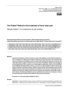
Figure 13: T2 Coronal
Figure 14: T2 Coronal
Figure 9 and 10: T2 Coronal images shows bicornuate uterus with fluid collection in the left uterine horn and in the left hematosalpinx and left hemorrhagic ovarian cyst Hence, a diagnosis of uterus didelphys with left hematometra, hematosalpinx, and left hemivaginal obstruction with left ovarian endometriosis was made. Considering that she also had absent left kidney, a final diagnosis of HWWS or obstructed hemivagina ipsilateral renal anomaly (OHVIRA) syndrome was made. DISCUSSION Anomalies of the internal female reproductive tract referred to as the Müllerian duct anomalies, account for about 7% of female population. Even though the occurrence is sporadic, there is a strong association of congenital renal and ano-rectal malformations indicating common early developmental etiology. Mullerian duct anomalies are classified according to the American fertility society criteria as follows: Class I or uterine hypoplasia or agenesis. Class IIor unicornuate uterus: A banana - shaped uterus with a single fallopian tube. A rudimentary horn (communicating or non-communicating) may be present. Class IIIor uterus didelphys. Two complete uteruses, each with its own cervix. A sagittal vaginal septum is seen in the majority of cases. Class IV or bicornuate uterus. Two uterine cavities with one cervix. MRI shows widely separated uterine horns with an intercornual distance of >4 cm and concavity of the fundal contour or an external fundal cleft of >1 cm in depth. Class V or septate uterus: A fibrous septum is seen that appear hypointense on T2W images. While the muscular septum appears intermediate in intensity. MRI criteria includes a convex or flat external fundal contour or external fundal cleft of