ONCOLOGY REPORTS 38: 3297-3308, 2017
Formyl peptide receptor 2 expression predicts poor prognosis and promotes invasion and metastasis in epithelial ovarian cancer Xiaohui Xie, Mengyuan Yang, Yiling Ding, Ling Yu and Jianlin Chen Department of Obstetrics and Gynecology, Second Xiangya Hospital of Central South University, Changsha, Hunan 410011, P.R. China Received March 31, 2017; Accepted August 31, 2017 DOI: 10.3892/or.2017.6034 Abstract. Formyl peptide receptor 2 (FPR2) has been identified as a member of the G protein-coupled chemoattractant receptor (GPCR) family and has been implicated as playing a role in both inflammation and cancer development. Epithelial ovarian cancer (EOC) has been suggested to be correlated with both infectious and non-infectious inflammation. To date, the role of FPR2 in EOC remains poorly understood and controversial. In the present study, we aimed to investigate the potential of FPR2 in regulating EOC. We performed immunohistochemistry and RT-qPCR to analyzed expression of FPR2 in EOC tissues and the correlation between FPR2 and EOC clinicopathological characteristics as well as prognosis were also analyzed. To test the role of FPR2 in EOC cell migration, we established FPR2-knockdown SKOV3 cells and performed wound-healing, Transwell and angiogenesis assays to detect the metastatic potential of these EOC cells. Our studies found that FPR2 was overexpressed in EOC tissues and was positively correlated with EOC clinicopathological characteristics including the International Federation of Gynecology and Obstetrics (FIGO) stage, histological grade and ovarian cancer type. Survival analyses suggested that FPR2 overexpression indicated the poorer prognosis of EOC patients and FPR2 may act as an independent risk factor for EOC prognosis. FPR2 knockdown decreased the migration potential of the ovarian cancer cells. Moreover, serum amyloid A (SAA) may stimulate the migration of SKOV3 cells through FPR2. The present study suggested that FPR2 promoted the invasion and metastasis of EOC and it could be a prognostic marker for EOC.
Correspondence to: Dr Mengyuan Yang, Department of Obstetrics and Gynecology, Second Xiangya Hospital of Central South University, Changsha, Hunan 410011, P.R. China E-mail:
[email protected]
Key words: epithelial ovarian cancer, FPR2, metastasis, prognosis, SAA
Introduction Ovarian cancer is one of the deadliest gynecological cancers worldwide. The most recent update from the Cancer Statistics of America stated that ovarian cancer is the fifth most common cause of cancer-related deaths. In 2017, the number of newly diagnosed cases of ovarian cancer was projected to be ~22,440, and the number of deaths was predicted to be 14,080 (1). In 2015, the number of new cases of ovarian cancer in China was ~52,100, whereas the number of deaths was ~22,500; this places ovarian cancer as the seventh leading cause of cancer-related death among Chinese women (2). Among the diverse histological subtypes of ovarian cancer, epithelial ovarian cancer (EOC) accounts for ~85-90% of all types of ovarian carcinoma, and up to 75% of patients with EOC were diagnosed with advanced stage disease and were characterized by rapid progression, poor prognosis and a high frequency of TP53 mutations (3-5). The cause of EOC is still unclear, but in recent years, accumulating studies have demonstrated that inflammation plays an important role in ovarian cancer tumorigenesis and progression (6-9). Formyl peptide receptor 2 (FPR2) has been identified as a member of the G protein-coupled chemoattractant receptor (GPCR) family. It is a seven-transmembrane receptor with 351 amino acids and is located on human chromosome 19q13.3-q13.4 (10). It was first detected on the cell membrane of macrophages and neutrophils and is activated by bacterial formylated chemotactic peptides. FPR2 was later reported to be expressed in a wide range of cells, tissues and organs, including inflammatory, microglial and astrocytoma and neuroblastoma cells, hepatocytes, microvascular endothelial cells, fibroblasts, spleen, lung, testes, ovaries, placenta, brain and bone marrow (11,12). FPR2 is a multi-functional receptor that is associated with diverse pathophysiologic processes, such as inflammation, cancer, amyloidosis and neurodegenerative diseases, wound healing, diabetes and obesity and AIDS (13). There is also a close relationship between FPR2 and cancer, as FPR2 has been detected via flow cytometry in seven ovarian cancer cell lines and the FPR2 agonist-antimicrobial peptide LL-37 was suggested to stimulate the invasiveness of ovarian cancer cells by interacting with FPR2 (14). FPR2 has been proposed as a bridge between inflammation and cancer. Serum amyloid A (SAA) is an acute phase protein that can be stimulated by inflammation, infection, trauma or
3298
xie et al: FPR2 is a novel prognostic marker for EOC
tumorigenesis. In a study of Urieli-Shoval et al, SAA was found to be strongly expressed in ovarian cancer tissues by IHC, ISH and RT-PCR (15). SAA is one of the primary agonists of FPR2, and previous studies have suggested that SAA-induced activation of FPR2 stimulates inflammation, cell migration, adhesion and infiltration (16). The SAA-FPR2 interaction also contributes to inflammation-mediated neovascularization in the cornea (17). Thus, we hypothesized that SAA may be expressed by ovarian cancer cells and interacts with FPR2 to stimulate ovarian cancer invasion and migration. In the present study, we explored the effects of FPR2 in the presence or absence of SAA on EOC progression. Materials and methods Quantitative real-time PCR. Total RNA was extracted from either tissues or cells using TRIzol reagents (Pufei Biotechnology, Shanghai, China). Reverse transcription was performed using M-MLV reverse transcriptase following the manufacturer's instructions (Promega, Madison, WI, USA). We used 2 µg of total RNA in a sterile RNase-free microcentrifuge tube, and then added 0.5 µg of the primer or per microgram of the total RNA sample in a total volume of 15 µl in water. The tube was heated to 70̊C for 5 min to melt secondary structure within the template. The tube was cooled immediately on ice to prevent secondary structure from reforming, and then spinned briefly to collect the solution at the bottom of the tube. Then, the following components were added to the annealed primer/template: M-MLV 5X reaction buffer 5 µl; dATP, 10 mM 1.25 µl; dCTP, 10 mM 1.25 µl; dGTP, 10 mM 1.25 µl; dTTP, 10 mM 1.25 µl; recombinant RNasin ribonuclease inhibitor 25 units; M-MLV RT 200 units; nuclease-free water to a final volume of 25 µl. Gentle mixing was performed by flicking the tube, and incubation was carried out for 60 min at 42̊C for FPR2 primers. The extension temperature was 37̊C. Quantitative PCR was performed using SYBR-Green Real‑Time PCR Master Mix (Toyobo, Osaka, Japan) according to the manufacturer's protocol. The primer sequences were as follows: FPR2 forward, 5'-AGTCTGCTG GCTACACTGTTC-3' and reverse, 5'-TGGTAATGTGGCCG TGAAAGA-3'; STAT3 forward, 5'-AGAAGGACATCAGCG GTAAG-3' and reverse, 5'-CCTTGGGAATGTCAGGATA GAG-3'; NF-κ B forward, 5'-AGGATTTCGTTTCCGTTAT GT-3' and reverse, 5'-CCTGAGGGTAAGACTTCTTGT TC-3'; MAPK1 forward, 5'-GTCGCCATCAAGAAAATCA GC-3' and reverse, 5'-GGAAGGTTTGAGGTCACGGT-3'; Notch3 forward, 5'-TGTGGACGAGTGCTCTATCG-3' and reverse, 5'-AATGTCCACTCGCAATAGG-3'; GAPDH forward, 5'-TGACTTCAACAGCGACACCCA-3' and reverse, 5'-CACCCTGTTGCTGTAGCCAAA-3'. Immunohistochemistry (IHC). The ovarian cancer tissues were fixed in 10% formalin and paraffin-embedded to form tissue blocks from which sections (4-µm thickness) could be sliced for IHC studies. The tissue sections were deparaffinized in xylene and rehydrated in graded concentrations of alcohol, and then were placed in a microwave for 30 min for antigen retrieval. After the slides were dewaxed, 3% H2O2 was added for 10 min to inhibit any endogenous peroxidase activity. The sections were incubated with the FPR2 antibody (13448-1-AP; 1:50
dilution; Proteintech, San Diego, CA, USA) at 4̊C overnight and subsequently incubated with a streptavidin‑peroxidase system (Zhongshan Goldenbridge Biotechnology, Beijing, China) according to the manufacturer's instructions. Cell culture. The human ovarian cancer cell lines SKOV3, OVCAR3 and the HUVECs were obtained from the America Type Culture Collection (ATCC; Manassas, VA, USA), and the HO-8910 and HO-8910PM cell lines were obtained from the China Center for Type Culture Collection (CCTCC). The cell lines were cultured in either Dulbecco's modified Eagle's medium (DMEM) or RPMI-1640 medium supplemented with 10% fetal bovine serum (FBS) and 100 UI/ml of penicillin and 100 mg/ml of streptromycin (all from Biological Industries, Cromwell, CT, USA) in a 37̊C incubator containing 5% CO2. Vector construction and transfection. The FPR2 knockdown and control lentiviruses were purchased from GeneChem (Shanghai, China). The shRNA sequences for FPR2 knockdown were as follows: shRNA-1, ccggAATCATTGACAT CCTGGTTAActcgagTTAACCAGGATGTCAATGATTtttttg; shRNA-2, ccggGGCCAAGACTTCCGAGAGAGActcgagTC TCTCTCGGAAGTCTTGGCCtttttg; shRNA‑3, ccggGTCC TATGAGTCTGCTGGCTActcgagTAGCCAGCAGACTCAT AGGACtttttg. The shRNA-2 sequence exhibited the best interference efficiency. Wound-healing assay. Cells (3x10 4/well) were seeded on 96-well plates and grown to 90% confluence, after which a scratch was made in the monolayer using a 10-µl pipette tip. Then, the cells were incubated at 37̊C in 5% CO2 for another 4 h according to the result of the pre-experiment, and the images were obtained at the time points of 0, 2 and 4 h. Each experiment was performed three times. Transwell assay. The assay was performed using a pre-coated cell invasion kit (pore size, 8.0 µm; Corning Inc., Corning, NY, USA) and Matrigel (BD Biosciences, Bedford, MA, USA) was inserted in the upper chambers. Approximately 1x10⁵ cells in 100 µl serum-free medium were placed into the upper chambers, the cells were cultured in 5% CO2 at 37̊C for 16 h (according to the pre-experiment). The lower chambers contained 30% FBS, thus, the cells migrated to the lower chambers. The cells remaining in the upper chambers were removed with a cotton swab and the cells that migrated through the membrane to the lower surface were stained with Giemsa's staining for 3-5 min at room temperature. The number of cells that migrated through the lower membrane of the inserts was counted under a light microscope. Each experiment was performed three times. Tube formation assay. HUVECs (4x10 4 cells/well) were seeded on Matrigel-coated 96-well plates and incubated at 37̊C. Serum-free supernatant from SKOV3, shFPR2 SKOV3, SAA+shFPR2 SKOV3 and SAA+SKOV3 cells were collected and centrifuged. The supernatants were then incubated with the HUVECs at 37̊C for 24 h. Afterwards, the cells were fixed with 4% paraformaldehyde at 37̊C for 15 min and the capillary-like structures were obtained using Cellomics (CCX7C1115; Thermo Fisher Scientific, Waltham, MA, USA). Branching points and lengths were measured using ImageJ
ONCOLOGY REPORTS 38: 3297-3308, 2017
3299
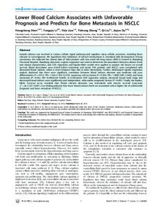
Figure 1. Expression of FPR2 mRNA in epithelial ovarian cancer (EOC) and normal ovarian tissues. (A) The FPR2 mRNA expression level in EOC was significantly higher than that in the normal ovarian tissues (*P