Int J Clin Exp Pathol 2014;7(9):5940-5949 www.ijcep.com /ISSN:1936-2625/IJCEP0001451
Original Article REDD1 and p-AKT over-expression may predict poor prognosis in ovarian cancer Wei Jia1, Bin Chang2, Lili Sun1,3, Huimin Zhu1,4, Lijuan Pang1, Lin Tao1, Hong Zou1, Jinze Du1, Yuling Dong1, Yan Qi1, Jinfang Jiang1, Weihua Liang1, Feng Li1, Xia Zhao5 Department of Pathology, Shihezi University School of Medicine, Shihezi, China; 2Department of Pathology, Shanghai Cancer Hospital of Fudan University, Shanghai, China; 3Department of Pathology, The People’s Hospital of Puyang, Puyang City, Henan, China; 4Department of Gynecology & Obstetrics, The Third Affiliated Hospital of Luohe Higher Medical School, Luohe, China; 5Department of Gynecology & Obstetrics, The First Affiliated Hospital of Shihezi University School of Medicine, Shihezi, China 1
Received July 18, 2014; Accepted August 23, 2014; Epub August 15, 2014; Published September 1, 2014 Abstract: We investigated the clinical significance of regulated in development and DNA damage response (REDD1) and p-AKT expression in human ovarian cancer (OC), explored the correlation of KRAS mutations with REDD1 expression, and assessed the therapeutic relevance of REDD1 in OC. We collected and immunohistochemically analyzed 118 formalin-fixed paraffin-embedded tumor tissue samples (100 primary OC and 18 borderline tumors) and 14 normal fallopian tubes, for REDD1 and p-AKT expression. Direct DNA sequencing for KRAS mutations and quantitative real-time polymerase chain reaction for detecting REDD1 mRNA expression were performed. REDD1 and p-AKT expressions were significantly higher in serous adenocarcinoma than other histological types, and this increase positively correlated with late-stage disease. REDD1 expression correlated with ascites formation, while p-AKT expression correlated with higher histological grade and chemoresistance. Kaplan Meier survival analysis showed significantly reduced disease-free survival (DFS) and overall survival (OS) in OC patients with both REDD1 and p-AKT overexpression. Patients with KRAS mutations had a longer DFS and OS. However, KRAS mutation and REDD1 over-expression was not correlated. Together, REDD1 and p-AKT over-expression may serve as a prognostic biomarker in OC, but KRAS mutations and REDD1 protein over-expression were not correlated in OC. We believe that with increasing knowledge of the role of REDD1 in cell migration, invasion, and proliferation pathways, the potential of REDD1 as a therapeutic target in OC may be uncovered. Keywords: REDD1, p-AKT, KRAS mutation, ovarian cancer, prognosis, biomarker
Introduction Ovarian cancer (OC) is the most lethal gynecological malignancy. The majority of patients are diagnosed with OC at advanced stages, with an overall survival rate as low as 30% [1][Lalwani, 2011 #248]. OC is remarkably heterogeneous at the clinical, cellular, and molecular levels. Histologically, OC is classified into serous (S-OC), mucinous (M-OC), endometrioid (E-OC), clear cell (CC-OC), transitional (Brenner cell), and undifferentiated types. S-OC, which is the most common histological subtype, accounts for about two-thirds of OC [2]. Patients with this malignancy have an extremely poor prognosis due to late clinical presentation, subtle symptomatology, and rapid disease progression. In
order to develop better preventive and diagnostic approaches, as well as more effective treatment modalities, a deep understanding is required of the molecular mechanisms implicated in the complex process of ovarian carcinogenesis. Despite numerous diagnostic and therapeutic advances in the management of OC in recent years, there remains an urgent need to identify novel biomarkers to aid in prognostication, and for use as possible therapeutic options. Regulated in development and DNA damage response 1 (REDD1) (also known as RTP801/ Dig1/DDIT4), first identified in 2002, was initially identified as a stress response gene induced following DNA damage and other cellu-
REDD1 & p-AKT over-expression and poor prognosis in ovarian cancer Table 1. Associations of REDD1 and p-AKT expression with clinicopathological characteristics in 100 ovarian cancer samples Characteristics Age ≤ 50 > 50 FIGO stage Stage I Stage II Stage III Stage IV Ascites Yes No Unknown Histology type S-OC M-OC E-OC CC-OC Mixed-type Chemotherapy response Responders Partial-responders Unknown response Histological grade Grade 1 Grade 2 Grade 3
N 100 45 55 100 20 40 28 12 65 48 17 35 100 42 17 17 17 7 39 26 13 61 100 28 32 40
REDD1 Expression Over-expression (%) Low expression (%) 43 57
χ2
P
p-AKT Expression Over-expression (%) Low expression (%) 53 47
χ2
P
21 (46.7) 22 (40.0)
24 (53.3) 33 (60.0)
0.449
0.503
25 (55.6) 24 (43.6)
20 (44.4) 31 (56.4)
1.407
0.236
3 (15.0) 15 (37.5) 18 (64.3) 7 (58.3)
17 (85.0) 25 (62.5) 10 (35.7) 5 (41.7)
13.218
0.004
4 (20.0) 22 (55.0) 18 (64.3) 9 (75.0)
16 (80.0) 18 (45.0) 10 (35.7) 3 (25.0)
5.982
0.014
30 (62.5) 5 (29.4)
18 (37.5) 12 (70.6)
5.530
0.019
31 (64.6) 9 (52.9)
17 (35.4) 8 (47.1)
0.719
0.397
27 (64.3) 3 (17.6) 5 (29.4) 6 (35.3) 2 (28.6)
15 (35.7) 14 (82.4) 12 (70.6) 11 (64.7) 5 (71.4)
14.509
0.006
33 (78.6) 8 (47.1) 5 (29.4) 4 (23.5) 3 (42.9)
9 (21.4) 9 (52.9) 12 (70.6) 13 (76.5) 4 (57.1)
21.280
0.001
14 (53.8) 6 (46.2)
12 (46.2) 7 (53.8)
0.205
0.651
3 (11.5) 8 (61.5)
23 (88.5) 5 (38.5)
10.700 0.002
12 (42.9) 12 (37.5) 19 (47.5)
16 (57.1) 20 (62.5) 21 (52.5)
0.726
0.696
11 (39.3) 14 (43.7) 28 (70.0)
17 (60.7) 18 (56.3) 12 (30.0)
7.854
0.020
S-OC, serous ovarian cancer; M-OC, mucinous ovarian cancer; E-OC, endometrioid ovarian cancer; C-OC, clear-cell ovarian cancer.
5941
Int J Clin Exp Pathol 2014;7(9):5940-5949
REDD1 & p-AKT over-expression and poor prognosis in ovarian cancer lar stress stimuli [3, 4]. A low-level of REDD1 protein is ubiquitously expressed and is found in most mature tissues [4]. REDD1 is expressed in response to diverse stress conditions, and is also an important regulator of response to many transcription factors, including p53, p63, ATF4, Sp1, and HIF1 [4-7]. Abnormalities in REDD1-mediated signaling may disrupt energy homeostasis and the regulation of tumorigenesis and can act as a pro-apoptotic or anti-apoptotic factor depending on different cellular microenvironments [6, 8]. Recent studies have demonstrated that AKT is activated in response to DNA damage via the action of the PI3K pathways [9]. Evidence suggests that REDD1 phosphorylates downstream AKT through the upstream tuberous sclerosis complex (TSC), to inhibit mTORC1 signaling [10, 11].
cally, 42 tumors were considered as the S-OC type; 17 each were M-OC, E-OC, and CC-OC types; and seven were of the mixed histology type. Tumors were staged according to the FIGO classification: 60 Stage I/II tumors and 40 Stage III/IV tumors. The majority of patients had undergone chemotherapy followed by surgical debulking of the tumor mass, as summarized in Table 1. A total of 73 cases were followed up by interview in the clinic or by telephone; follow-up ranged 3-192 months. The last follow-up time-point was December 17, 2013. At this final follow-up point, 25 patients had died. Among the 48 patients still alive, 18 had experienced tumor recurrence. The median age of patients was 58 years old (range 20-77 years old).
In a previous study, we demonstrated that REDD1 expression is required for RAS-mediated transformation of ovarian epithelial cells, and it serves as a transforming oncogene in ovarian cancer carcinogenesis [12]. However, to the best of our knowledge, the role of REDD1 in human OC has not been reported in the literature. We hypothesized that over-expression of REDD1 may be induced by KRAS mutations and synergize to activate AKT in OC.
Tissue microarray construction and immunohistochemistry
The aims of the present study were to (1) determine whether REDD1 expression synergizes with phosphorylated AKT (p-AKT) and to investigate its clinical significance, (2) to examine the correlation of KRAS mutations with REDD1 expression, and (3) to assess the therapeutic relevance of REDD1 in OC patients. Materials and methods Patients and tissue specimens One hundred and eighteen formalin-fixed paraffin-embedded tumor tissue samples (100 primary ovarian cancer and 18 borderline tumors) and 14 normal fallopian tubes, obtained from 132 patients were collected from the Department of Pathology, the First Affiliated Hospital of Shihezi University School of Medicine. The collection of specimens was approved and supervised by the Research Ethics Committee of first Affiliated Hospital of Shihezi University School of Medicine. Clinical, demographic and pathological data were obtained from the patients’ medical records. Histopathological evaluation was carried out independently by two pathologists. Histologi-
5942
We constructed tissue microarray (TMA) blocks by selecting one representative paraffinembedded tissue block from every patient, and taking two cores from morphologically representative areas of the block, according to established methods [13]. Each core was reviewed on a hematoxylin and eosin (H & E)-stained section to ensure that tumor cells were present in at least 70% of the core. Immunohistochemistry (IHC) staining was performed on TMA slides using the EnVision system (DAKO, Carpinteria, CA, USA), as previously described [14]. Primary antibodies used for IHC were as follows: monoclonal rabbit anti-phospho-Akt (#3787, Cell Signaling Technology, Danvers, MA, USA; dilution, 1:100) and polyclonal rabbit anti-REDD1 (ab63059, Abcam, Cambridge, UK; dilution, 1:100). The reaction product was visualized with 3, 3’-diaminobenzidine peroxidase (DAB) substrate kit (DAKO, Carpinteria, CA, USA) for 5 min at room temperature, and the sections were counterstained with hematoxylin. Negative control sections were incubated with phosphate-buffered saline instead of primary antibody. IHC staining for REDD1 and p-AKT was analyzed by two gynecological pathologists, without prior knowledge of the clinical information. The IHC scoring system for REDD1 and p-AKT used in this work, has been widely established [15]; the percentage of positive cells (range, 0-100%) was multiplied by the staining intensity score (1, buff; 2, yellow; and 3, brown). Scores ≥ 150 were scored as Int J Clin Exp Pathol 2014;7(9):5940-5949
REDD1 & p-AKT over-expression and poor prognosis in ovarian cancer
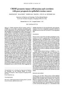
Figure 1. Immunohistochemical (IHC) staining of REDD1 and p-AKT in ovarian cancer (OC) tissue and non-cancerous tissues. Paraffin-embedded sections of OC, borderline ovary tumor, and normal fallopian tube were stained using anti-REDD1 polyclonal antibody and p-AKT monoclonal antibody, as described in the Methods section. A-F. Represent REDD1 IHC staining in serous-OC, mucinous-OC, endometrioid-OC, clear cell-OC, borderline tumor, and normal fallopian tube, respectively. G-L. Represent p-AKT IHC staining in serous-OC, mucinous-OC, endometrioidOC, clear cell-OC, borderline tumor, and normal fallopian tube, respectively. M. REDD1 and p-AKT protein levels are correlated (P < 0.05).
5943
Int J Clin Exp Pathol 2014;7(9):5940-5949
REDD1 & p-AKT over-expression and poor prognosis in ovarian cancer over-expression, while those < 150 were scored as low expression. DNA extraction and KRAS mutation analysis Genomic DNA was extracted from tumor areas of FFPE tissues according to the manufacturer’s instructions using a QIAamp DNA FFPE Tissue kit (Qiagen Inc., Hilden, Germany). Exon 1 of the KRAS gene was selected for mutation analysis by direct sequencing following PCR amplification. PCR amplifications were performed using a PCR thermocycler (Bio-Rad, Hercules, CA, USA), with denaturation at 94°C for 3 min, followed by 33 cycles of 94°C for 1 min, 58.5°C for 1 min, and 72°C for 1 min, followed by a final extension at 72°C for 10 min. After DNA purification, direct sequencing was carried out by Biotechnologies, Sangon, China. The primers used were as follows: Ras exon 1, forward primer: 5’ GGC CTG CTG AAA ATG ACT GA 3’ and reverse primer: 5’ GTC CTG CAC CAG TAA TAT GC 3’. Statistical analysis All statistical analyses were performed using the Statistical Package for the Social Science (SPSS) (SPSS 13.0, Chicago, IL, USA). Pearson’s Chi-square test was performed to analyze the statistical significance of REDD1 and p-AKT expression in OC and control groups, as well as the associations between p-AKT expression and clinicopathological factors. Spearman’s rank correlation coefficient was used to analyze the correlation between KRAS mutations and the expression of REDD1 and p-AKT. The Kaplan-Meier method was used carry out survival analysis. A P-value of < 0.05 was considered to be significant. Results REDD1 and p-AKT expression by immunohis tochemistry in ovarian cancer and control groups IHC staining for REDD1 and p-AKT expression was performed on 100 primary ovarian tumors, 18 borderline tumors, and 14 normal fallopian tube tissue samples (Figure 1A-L). REDD1 and p-AKT were mainly expressed in OC tissue. The expression of the two proteins was markedly different between cancerous and non-cancerous tissue; positive staining for REDD1 and
5944
p-AKT in OC tissue was 43% and 53%, respectively, whereas staining was much lower in borderline tissue (2% and 3%, respectively) and normal fallopian tubes (3% and 2%, respectively) (p=0.001) (Tables S1, S2). A positive correlation between REDD1 and p-AKT was found in OC samples. In the OC group, there were 29 cas es of REDD1-over-expression/p-AKT-over-expression, 33 cases of REDD1-low expression/pAKT-low expression, 14 cases of REDD1-overexpression/p-AKT-low expression, and 24 cases of REDD1-low expression/p-AKT-overexpression. Pearson’s Chi-square test indicated that p-AKT over-expression was significantly associated with REDD1 over-expression (P= 0.012, r=0.251) (Figure 1M). Associations between expression levels of REDD1 and p-AKT and clinicopathological features Associations between REDD1 and p-AKT expression and clinicopathological characteristics were analyzed in 100 OC patients. REDD1 and p-AKT expressions in OC samples were significantly higher in patients with serous adenocarcinoma (P=0.006 and P=0.001, respectively), and late FIGO stage (P=0.004 and p=0.014, respectively). REDD1 expression correlated with ascites formation (P=0.019), but no correlation was found with chemotherapy response or histological grade (P=0.651 and P=0.696, respectively). p-AKT expression was higher in patients with higher histological grade (P= 0.020), and partial or non-response to chemotherapy (P=0.002), but did not correlate with ascites formation (P=0.397) (Table 1). Correlation of REDD1 and p-AKT expression with disease-free survival (DFS) and overall survival (OS) In our study, survival analysis showed that patients with low expression of REDD1 had a longer disease-free survival (DFS) and overall survival (OS) than those with REDD1 overexpression (P=0.020 and P=0.023, respectively, Figure 2A and 2B). Similarly, patients with p-AKT positive expression had shorter DFS and OS (P=0.005 and P=0.017, respectively, Figure 2C and 2D). When both REDD1 and p-AKT were taken into account, patients in the REDD1negative/p-AKT-negative group had the longest DFS and OS (P=0.001 and P=0.011, respectively, Figure 2E and 2F).
Int J Clin Exp Pathol 2014;7(9):5940-5949
REDD1 & p-AKT over-expression and poor prognosis in ovarian cancer
Figure 2. Kaplan-Meier disease-free survival (DFS) and overall survival (OS) analysis of ovarian cancer patients. A, B. Patients with low expression of REDD1 had a longer DFS and OS than those with REDD1 over-expression (P=0.020 and P=0.023, respectively). C, D. Patients with p-AKT over-expression had shorter DFS and OS than those with low p-AKT expression (P=0.005 and P=0.017, respectively). E, F. Patients in the REDD1-low expression/p-AKTlow expression group had the longest DFS and OS (P=0.001 and P=0.011, respectively). (-): low expression, (+): over-expression.
5945
Int J Clin Exp Pathol 2014;7(9):5940-5949
REDD1 & p-AKT over-expression and poor prognosis in ovarian cancer Table 2. KRAS mutation profile in different ovarian tissues Histology type n Mutated (n) Mutated (%) S-OC 35 3 8.6 M-OC 17 4 23.5 E-OC 13 1 7.7 CC-OC 14 3 21.4 Mixed-type 3 0 0.0 Borderline tumors 18 4 22.2 Normal fallopian tubes 9 1 11.1 Total 109 16 14.7 S-OC, serous ovarian cancer; M-OC, mucinous ovarian cancer; E-OC, endometrioid ovarian cancer; C-OC, clear-cell ovarian cancer.
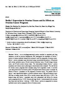
Analysis of KRAS mutations in ovarian cancer samples and control groups The KRAS mutation status was assessed in 82 cases of OC, 18 borderline tumors, and 9 normal fallopian tube tissues. Mutations were more common in borderline lesions 4/18 (22.2%) than in the tumor tissue 11/82 (13.4%). A single KRAS mutation was found in a benignappearing fallopian tube. Mutations were more common in mucinous lesions than in serous lesions (23.5% vs. 8.6%), but no statistical significance was observed in this regard (Table 2). Of 82 cases of OC, 11 (13.4%) displayed mutations in the KRAS gene, 9 (10.9%) of which were found in codon 12 and 2 (2.4%) in codon 13; the most commonly affected amino acid was G12V (gly12→val12) (Table S3). No mutation in codon 61 was found in any specimen. In this study, KRAS mutations were significantly associated with lower grade (P=0.002) and Stage I tumors (P=0.024). No association was found between KRAS mutation status and age, or clinical stage. Kaplan-Meier analysis of 57 OC patients revealed that KRAS mutations were significantly associated with improved DFS and OS (P=0.031 and 0.014, respectively) compared to KRAS wild-type patients (Figure 3A and 3B). Discussion Previously, we have found that REDD1 is a downstream target of the RAS oncogene, as shown by an increased expression of the REDD1 protein in RAS-transformed cells, indicating that REDD1 may be a transforming oncogene in ovarian cancer cells [12]. In the present study, IHC analyses demonstrated that REDD1
5946
over-expression was common in high-grade S-OC and was significantly correlated with late clinical stage and ascites formation. However, correlations between REDD1 over-expression and RAS mutations had not been investigated in human ovarian cancer. In this study, we examined the frequency of mutations in the KRAS gene and found that 13.4% of OC cases displayed KRAS mutations, corresponding to mutation frequencies previously reported [16, 17]. However, KRAS mutations did not correlate with REDD1 over-expression in OC, suggesting that there might be other oncogenic molecular alterations involved in the activation of REDD1. REDD1 can inhibit mTORC1 via TSC1/2, which acts as a negative regulator of mTORC1 activity [8]. Jin et al. [11] found that constitutive expression of REDD1 suppressed mTORC1 activity in a manner mediated by the TSC1/2 complex, and induced AKT S473 phosphorylation in H1299 cell lines. In the present study, IHC analyses indicated that AKT S473 activation was associated with over-expression of REDD1 in OC tissues. This suggests that over-expression of REDD1 may synergize with p-AKT expression, leading to ovarian carcinogenesis via the PI3K/ AKT/mTOR signaling pathway. While the exact mechanism underlying REDD1-induced AKT activation in OC is unclear at present, Maren Cam et al. [9] indicated that DNA damage can induce REDD1 to disrupt TSC2/14-3-3 binding, providing a means to rapidly extinguish mTORC1 activation, which is known to activate AKT. In the present study, patients had undergone chemotherapy followed by surgical debulking of the tumor mass, such as cisplatin and paclitaxel agents. Initially, the majority of patients responded well to chemotherapy, but following recurrence, ovarian tumors are more aggressive and resistant to chemotherapy. Our findings suggest that some of the patients with p-AKT over-expression had chemoresistance. Kim et al. [18] and Craig et al. [19] also reported that over-expression of p-AKT was involved in chemoresistance in human ovarian cancer cell lines. The PI3K/AKT pathway could be a key oncogenic pathway regulating survival, tumorigenesis, and chemoresistance in OC. Our IHC analyses demonstrated that p-AKT over-expression is a common event in OC, in line with published findings [20-23]. We also analyzed correlations between the expression status of p-AKT and clinicopathological characteristics and survival, to investigate the potential of
Int J Clin Exp Pathol 2014;7(9):5940-5949
REDD1 & p-AKT over-expression and poor prognosis in ovarian cancer
Figure 3. Kaplan-Meier disease-free survival (DFS) and overall survival (OS) analyses of ovarian cancer patients. A, B. OS and DFS analyses were performed according to the mutation status of KRAS. Patients with KRAS mutations were significantly associated with a longer DFS and OS (P=0.031 and 0.014, respectively) than patients with KRAS wild-type patients.
p-AKT expression as a novel prognostic factor. The results showed that patients with p-AKT over-expression had a poorer prognosis, and this over-expression was commonly associated with high-grade, late-stage OC. Furthermore, patients with over-expression of both p-AKT and REDD1 tended to have a more unfavorable prognosis. Thus, we can conclude that overexpression of p-AKT and REDD1 may predict poor survival in ovarian cancer. Chang et al. [12] showed that transfection of REDD1 into non-tumorigenic immortalized ovarian epithelial cell lines generated neoplasms that mimic high-grade papillary serous carcinoma in the peritoneal cavity in nude mice. In the present study, we demonstrated that REDD1 protein expression was enhanced in human OC tissue, which indicates that REDD1 may act as an oncogene in human OC tissue. Chang et al. [12] also revealed the functional role of REDD1 in RAS-mediated transformation of ovarian epithelial cells. However, we did not find a similar relationship between REDD1 over-expression and KRAS mutation in OC tissue. The OC tissue samples examined in this study included four histological types: S-OC, M-OC, E-OC, CC-OC, and we postulate that REDD1/RAS may only be involved in the carcinogenesis of S-OC histological type. In 2004, Shih and Kurman proposed a 2-tier system for classification of OC [24]. In recent years, this system has become more widely
5947
accepted. Type I (low malignant potential) tends to be associated with KRAS, BRAF, and PTEN mutations, whereas type II (high-grade carcinoma) accounts for 90% of all serous carcinoma, which are characterized by TP53 mutations and genetic instability. In the present study, KRAS mutations are more frequent in type I tumors than in type II. KRAS mutations have been shown to characterize type I tumors and are generally associated with a more favorable clinical course [25-27]. Our findings also indicate that patients with KRAS mutations had more favorable prognosis than patients with wild-type KRAS. In conclusion, REDD1 and p-AKT over-expression may serve as biomarkers of adverse prognosis in OC, but KRAS mutations and REDD1 protein over-expression in OC may not be correlated. To the best of our knowledge, the effects of REDD1 over-expression on ovarian tissue have not been reported in the literature. We believe that with increasing knowledge of the role of REDD1 in cell migration, invasion, and proliferation pathways, the potential of REDD1 as a therapeutic target in OC may be uncovered in future studies. Acknowledgements This work was supported by grants from the National Natural Science Foundation of China (No. 81160316) and the Preeminent Youth
Int J Clin Exp Pathol 2014;7(9):5940-5949
REDD1 & p-AKT over-expression and poor prognosis in ovarian cancer Foundation of Shihezi University School of Medicine (No. 2013ZRKXYQ-YD18). The funding agencies had no role in design, execution, and analysis of the study, or the decision to submit the paper for publication.
[8]
[9]
Disclosure of conflict of interest None. Address correspondence to: Dr. Xia Zhao, Department of Gynecology & Obstetrics, The First Affiliated Hospital of Shihezi University School of Medicine, Shihezi, Xinjiang 832002, China. Tel: +86-993205-7136; E-mail:
[email protected]
References [1]
[2]
[3]
[4]
[5]
[6]
[7]
Lalwani N, Prasad SR, Vikram R, Shanbhogue AK, Huettner PC and Fasih N. Histologic, molecular, and cytogenetic features of ovarian cancers: implications for diagnosis and treatment. Radiographics 2011; 31: 625-646. Barda G, Menczer J, Chetrit A, Lubin F, Beck D, Piura B, Glezerman M, Modan B and Sadetzki S. Comparison between primary peritoneal and epithelial ovarian carcinoma: a population-based study. Am J Obstet Gynecol 2004; 190: 1039-1045. Corradetti MN, Inoki K and Guan KL. The stress-inducted proteins RTP801 and RTP801L are negative regulators of the mammalian target of rapamycin pathway. J Biol Chem 2005; 280: 9769-9772. Ellisen LW, Ramsayer KD, Johannessen CM, Yang A, Beppu H, Minda K, Oliner JD, McKeon F and Haber DA. REDD1, a developmentally regulated transcriptional target of p63 and p53, links p63 to regulation of reactive oxygen species. Mol Cell 2002; 10: 995-1005. Lee M, Bikram M, Oh S, Bull DA and Kim SW. Sp1-dependent regulation of the RTP801 promoter and its application to hypoxia-inducible VEGF plasmid for ischemic disease. Pharm Res 2004; 21: 736-741. Shoshani T, Faerman A, Mett I, Zelin E, Tenne T, Gorodin S, Moshel Y, Elbaz S, Budanov A, Chajut A, Kalinski H, Kamer I, Rozen A, Mor O, Keshet E, Leshkowitz D, Einat P, Skaliter R, Feinstein E. Identification of a novel hypoxiainducible factor 1-responsive gene, RTP801, involved in apoptosis. Mol Cell Biol 2002; 22: 2283-2293. Whitney ML, Jefferson LS and Kimball SR. ATF4 is necessary and sufficient for ER stressinduced upregulation of REDD1 expression. Biochem Biophys Res Commun 2009; 379: 451-455.
5948
[10]
[11]
[12]
[13]
[14]
[15]
[16]
[17]
[18]
[19]
DeYoung MP, Horak P, Sofer A, Sgroi D and Ellisen LW. Hypoxia regulates TSC1/2-mTOR signaling and tumor suppression through REDD1-mediated 14-3-3 shuttling. Genes Dev 2008; 22: 239-251. Cam M, Bid HK, Xiao L, Zambetti GP, Houghton PJ and Cam H. p53/TAp63 and AKT regulate mammalian target of rapamycin complex 1 (mTORC1) signaling through two independent parallel pathways in the presence of DNA damage. J Biol Chem 2014; 289: 4083-4094. Dennis MD, McGhee NK, Jefferson LS and Kimball SR. Regulated in DNA damage and development 1 (REDD1) promotes cell survival during serum deprivation by sustaining repression of signaling through the mechanistic target of rapamycin in complex 1 (mTORC1). Cell Signal 2013; 25: 2709-2716. Jin HO, Hong SE, Kim JH, Choi HN, Kim K, An S, Choe TB, Hwang CS, Lee JH and Kim JI, Kim HA, Kim EK, Noh WC, Hong YJ, Hong SI, Lee JK, Park IC. Sustained overexpression of Redd1 leads to Akt activation involved in cell survival. Cancer Lett 2013; 336: 319-324. Chang B, Liu G, Yang G, Mercado-Uribe I, Huang M and Liu J. REDD1 is required for RASmediated transformation of human ovarian epithelial cells. Cell cycle 2009; 8: 780-786. Rimm DL, Camp RL, Charette LA, Costa J, Olsen DA and Reiss M. Tissue microarray: a new technology for amplification of tissue resources. Cancer J 2000; 7: 24-31. Qi Y, Wang CC, He YL, Zou H, Liu CX, Pang LJ, Hu JM, Jiang JF, Zhang WJ and Li F. The correlation between morphology and the expression of TGF-beta signaling pathway proteins and epithelial-mesenchymal transition-related proteins in synovial sarcomas. Int J Clin Exp Pathol 2013; 6: 2787-2799. Carvalho S, Milanezi F, Costa JL, Amendoeira I and Schmitt F. PIKing the right isoform: the emergent role of the p110β subunit in breast cancer. Virchows Arch 2010; 456: 235-243. Auner V, Kriegshäuser G, Tong D, Horvat R, Reinthaller A, Mustea A and Zeillinger R. KRAS mutation analysis in ovarian samples using a high sensitivity biochip assay. BMC cancer 2009; 9: 111. Vang R, Shih IM and Kurman RJ. Ovarian lowgrade and high-grade serous carcinoma: pathogenesis, clinicopathologic and molecular biologic features, and diagnostic problems. Adv Anat Pathol 2009; 16: 267. Kim SH, Juhnn YS and Song YS. Akt involvement in paclitaxel chemoresistance of human ovarian cancer cells. Ann N Y Acad Sci 2007; 1095: 82-89. Carden CP, Stewart A, Thavasu P, Kipps E, Pope L, Crespo M, Miranda S, Attard G, Garrett
Int J Clin Exp Pathol 2014;7(9):5940-5949
REDD1 & p-AKT over-expression and poor prognosis in ovarian cancer MD Clarke PA, Workman P, de Bono JS, Gore M, Kaye SB, Banerji U. The association of PI3 kinase signaling and chemoresistance in advanced ovarian cancer. Mol Cancer Ther 2012; 11: 1609-1617. [20] Altomare DA, Wang HQ, Skele KL, De Rienzo A, Klein-Szanto AJ, Godwin AK and Testa JR. AKT and mTOR phosphorylation is frequently detected in ovarian cancer and can be targeted to disrupt ovarian tumor cell growth. Oncogene 2004; 23: 5853-5857. [21] Huang J, Zhang L, Greshock J, Colligon TA, Wang Y, Ward R, Katsaros D, Lassus H, Butzow R Godwin AK, Testa JR, Nathanson KL, Gimotty PA, Coukos G, Weber BL, Degenhardt Y. Frequent genetic abnormalities of the PI3K/ AKT pathway in primary ovarian cancer predict patient outcome. Genes Chromosomes Cancer 2011; 50: 606-618. [22] Kurose K, Zhou XP, Araki T, Cannistra SA, Maher ER and Eng C. Frequent loss of PTEN expression is linked to elevated phosphorylated Akt levels, but not associated with p27 and cyclin D1 expression, in primary epithelial ovarian carcinomas. Am J Pathol 2001; 158: 2097-2106.
5949
[23] Yuan ZQ, Sun M, Feldman RI, Wang G, Ma X-l, Jiang C, Coppola D, Nicosia SV and Cheng JQ. Frequent activation of AKT2 and induction of apoptosis by inhibition of phosphoinositide3-OH kinase/Akt pathway in human ovarian cancer. Oncogene 2000; 19: 2324-2330. [24] Shih I-M and Kurman RJ. Ovarian tumorigenesis: a proposed model based on morphological and molecular genetic analysis. Am J Pathol 2004; 164: 1511-1518. [25] Garrett AP, Lee KR, Colitti CR, Muto MG, Berkowitz RS and Mok SC. K-ras mutation may be an early event in mucinous ovarian tumorigenesis. Int J Gynecol Pathol 2001; 20: 244251. [26] Ichikawa Y, Nishida M, Suzuki H, Yoshida S, Tsunoda H, Kubo T, Uchida K and Miwa M. Mutation of K-ras protooncogene is associated with histological subtypes in human mucinous ovarian tumors. Cancer Res 1994; 54: 33-35. [27] Palayekar M and Herzog T. The emerging role of epidermal growth factor receptor inhibitors in ovarian cancer. Int J Gynecol Cancer 2008; 18: 879-890.
Int J Clin Exp Pathol 2014;7(9):5940-5949
REDD1 & p-AKT over-expression and poor prognosis in ovarian cancer Table S1. Expression of REDD1 in different ovarian tissues Specimens Cancer Borderline Normal
REDD1 expression Over (%) Low (%) 43 (43.0) 57 (57.0) 2 (11.1) 16 (88.9) 3 (21.4) 11 (78.6)
N
P
100 18 14
0.016
Table S2. Expression of p-AKT in different ovarian tissues Specimens Cancer Borderline Normal
p-AKT expression Over (%) Low (%) 53 (53.0) 47 (47.0) 3 (16.7) 15 (83.3) 2 (14.3) 12 (85.7)
N
P
100 18 14
0.001
Table S3. KAS mutation types in codons 12 and 13 Mutation 12ASP (G12D) 12ARG (G12R) 12CYS (G12C) 12VAL (G12V) 13ASP (G13D) Total
Base change GGT > GAT GGT > CGT GGT > TGT GGT > GTT GGC > GAC
Amino acid gly12→asp12 gly12→arg12 gly12→cys12 gly12→val12 gly13→asp13
N (%) 2 (12.5) 1 (6.3) 2 (12.5) 8 (50.0) 3 (18.8) 16
Gly, glycine; asp, aspartic acid, arg, arginine; cys, cysteine; val, valine.
1