Journal of Orthopaedic & Sports Physical Therapy Official Publication of the Orthopaedic and Sports Physical Therapy Sections of the American Physical Therapy Association
Electromyographic Analysis of the Rotator Cuff and Deltoid Musculature During Common Shoulder External Rotation Exercises
Coordinator of Rehabilitative Research and Clinical Education, Healthsouth Rehabilitation, American Sports Medicine Institute, Birmingham, AL. 2 National Director of Research and Education, Healthsouth Rehabilitation, American Sports Medicine Institute, Birmingham, AL. 3 Smith and Nephew Chair of Research, American Sports Medicine Institute, Birmingham, AL. 4 Coordinator of Joint Biomechanics Research, American Sports Medicine Institute, Birmingham, AL. 5 Biomechanist, American Sports Medicine Institute, Birmingham, AL. 6 Assistant Research Professor, Department of Physical Therapy, University of Florida, Gainesville, FL. 7 Assistant Professor, Department of Orthopedics and Rehabilitation, University of Vermont, Burlington, VT. 8 Medical Director, American Sports Medicine Institute, Alabama Sports Medicine and Orthopedic Center, Birmingham, AL. This study was approved by the American Sports Medicine Institute Institutional Review Board, Birmingham, AL. Address correspondence to Michael M. Reinold, Coordinator of Rehabilitative Research and Clinical Education, Healthsouth Sports Medicine and Rehabilitation Center, American Sports Medicine Institute, 1201 11th Avenue South, Suite 100, Birmingham, AL 35205. E-mail:
[email protected] 1
Journal of Orthopaedic & Sports Physical Therapy
was compared among exercises using a 1-way repeated-measures analysis of variance (P!.05). Results: EMG activity varied significantly among the 7 exercises. Sidelying ER produced the greatest amount of EMG activity for the infraspinatus (62% MVIC) and teres minor (67% MVIC). The greatest amount of activity of the supraspinatus (82% MVIC), middle deltoid (87% MVIC), and posterior deltoid (88% MVIC) was observed during prone horizontal abduction at 100° with full ER. Conclusions: Results from this study provide initial information to develop rehabilitation programs. It also provides information helpful for the design and conduct of future studies. J Orthop Sports Phys Ther 2004;34:385-394.
Key Words: dynamic stabilization, infraspinatus, supraspinatus, teres minor
T
he glenohumeral joint exhibits the greatest amount of motion of any articulation in the human body, consequently little inherent stability is provided by its osseous configuration.42 Functional stability of the 385
REPORT
Study Design: Prospective single-group repeated-measures design. Objectives: To quantify electromyographic (EMG) muscle activity of the infraspinatus, teres minor, supraspinatus, posterior deltoid, and middle deltoid during exercises commonly used to strengthen the shoulder external rotators. Background: Exercises to strengthen the external rotators are commonly prescribed in rehabilitation, but the amount of EMG activity of the infraspinatus, teres minor, supraspinatus, and deltoid during these exercises has not been thoroughly studied to determine which exercises would be most effective to achieve strength gains. Methods and Measures: EMG measured using intramuscular electrodes were analyzed in 10 healthy subjects during 7 shoulder exercises: prone horizontal abduction at 100° of abduction and full external rotation (ER), prone ER at 90° of abduction, standing ER at 90° of abduction, standing ER in the scapular plane (45° abduction, 30° horizontal adduction), standing ER at 0° of abduction, standing ER at 0° of abduction with a towel roll, and sidelying ER at 0° of abduction. The peak percentage of maximal voluntary isometric contraction (MVIC) for each muscle
RESEARCH
Michael M. Reinold, DPT, ATC 1 Kevin E. Wilk, PT 2 Glenn S. Fleisig, PhD 3 Nigel Zheng, PhD 4 Steven W. Barrentine, M S 5 Terri Chmielewski, PT, PhD 6 Rayden C. Cody, M D 7 Gene G. Jameson, MA 5 James R. Andrews, M D 8
shoulder is accomplished through the integrated functions of the joint capsule, ligaments, and glenoid labrum, as well as the dynamic stabilization of the surrounding musculature, particularly the rotator cuff muscles.2,8,15,37 The rotator cuff musculature maintains stability by compressing the humeral head into the concave glenoid fossa during upper extremity motion.42 Thus, the rotator cuff muscles play a vital role in normal arthrokinematics and asymptomatic shoulder function. The overhead athlete requires the rotator cuff to maintain an adequate amount of glenohumeral joint congruency for asymptomatic function.41 Sufficient strength of the external rotators (infraspinatus and teres minor), in particular, is integral during the overhead throwing motion to develop an approximation force on the upper arm at the shoulder equal to body weight to prevent joint distraction.11 Andrews and Angelo1 found that overhead throwers most often present with rotator cuff tears located from the midsupraspinatus and extending posteriorly to the midinfraspinatus area, which they believe to be a result of the force produced to resist distraction, horizontal adduction, and internal rotation at the shoulder during arm deceleration. Furthermore, Walch et al39 documented the undersurface fraying of the infraspinatus with internal impingement. Thus, the external rotators are muscles that appear to be involved in the pathomechanics leading to, or resulting from, different shoulder pathologies such as internal impingement,17,39 joint laxity,8,11,17,36,37 labral lesions,11,17,36 and rotator cuff lesions,11,29,36 particularly in overhead athletes.40,44 Consequently, many authors5,10,38,40,41,44-46 have advocated emphasis on shoulder external rotation strengthening during rehabilitation or athletic conditioning programs to enhance muscular strength and endurance in overhead athletes. Rehabilitation programs for specific pathologies, such as rotator cuff impingement or repair surgery, also emphasize strengthening of the shoulder external rotators. Furthermore, rotator cuff strengthening exercises are often incorporated in athletic conditioning programs for injury prevention and performance enhancement. The balance between external and internal rotation strength is important to normal glenohumeral joint function, especially during athletic activities.42 An adequate external-internal rotator muscle strength ratio has been emphasized in the literature.7,10,40,43 Several studies documented the electromyographic (EMG) activity of the glenohumeral musculature during specific shoulder exercises.3,5,6,13,22,25,26,28,38,47 Townsend et al38 determined that the best exercise for the infraspinatus muscle was to perform prone shoulder horizontal abduction with external rotation (ER), producing 88% of maximal voluntary isometric contraction (MVIC), while the most effective exercise 386
for the teres minor muscle was sidelying ER, producing 80% MVIC. In a different study comparing ER in the scapular and frontal planes during isokinetic testing, Greenfield et al13 suggested that the highest amount of muscle activity for the external rotators occur when the shoulder is in the scapular plane. Ballantyne et al3 compared sidelying ER with ER in the prone position at 90° of shoulder abduction. The authors report no significant difference between exercises with approximately 50% EMG activity (mean EMG was normalized as a percent of the highest mean value of repetitions) for both the infraspinatus and teres minor. Conversely, Blackburn et al5 similarly compared the sidelying ER and prone ER exercises and noted greater activity during prone ER for the infraspinatus (prone, 80% EMG; sidelying, 30% EMG) and teres minor (prone, 88% EMG; side, 45% EMG). EMG values were normalized in relation to the maximum amount of EMG activity produced by each muscle across the test series. Blackburn et al5 also reported high levels of EMG activity of the infraspinatus (80% EMG) and teres minor (70% EMG) when performing a prone shoulder horizontal abduction movement at 90° and 100° of abduction with full external rotation. Current trends in rehabilitation have focused more closely on functional rehabilitation through sportspecific exercises designed to strengthen the external rotators in a position that replicates the capsular strain and muscular length-tension relationships observed during athletic competition, such as external rotation in the 90° abducted position for overhead athletes.44-46 In addition, many clinicians have advocated the use of a towel roll placed between the arm and side of the body while performing external rotation strengthening to enhance stability and to increase posterior cuff muscular activation.46 Thus, it appears that there is controversy regarding the optimal exercises for the external rotators and that many exercises are being utilized and advocated without justification based on EMG analysis. Several authors have analyzed the EMG activity of the supraspinatus musculature and deltoid muscles to determine exercises that produce the most supraspinatus activity with the least deltoid involvement.20,25,35,38,47 This has been theorized to avoid potential deleterious superior humeral head migration associated with high deltoid activity.20,35,47 Anatomically, based on the fiber orientation posterior to the longitudinal axis of rotation, the supraspinatus and posterior deltoid may be active during the external rotation movement.21 However, there is a lack of data regarding the contribution of the supraspinatus and deltoid musculature during external rotation exercises. The purpose of this investigation was to measure the normalized EMG activity of the infraspinatus, teres minor, supraspinatus, and deltoid muscles durJ Orthop Sports Phys Ther • Volume 34 • Number 7 • July 2004
ing a variety of commonly prescribed rehabilitation exercises to strengthen the shoulder external rotators.
METHODS Subjects The participants for this investigation were 5 males and 5 females (mean age, 28.1 years; age range, 22-38 years). Each volunteered for the study and reported no history of shoulder injury or instability, nor did any subjects report a history of shoulder pain. Each subject signed an informed consent and the rights of each subject were protected. The research protocol was approved by the Institutional Review Board of the American Sports Medicine Institute.
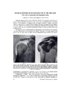
FIGURE 1. Prone horizontal abduction at 100° with full external rotation.
Electrode Insertion
REPORT
J Orthop Sports Phys Ther • Volume 34 • Number 7 • July 2004
RESEARCH
The dominant shoulder of each subject was utilized for testing. The skin near the electrode insertion sites was shaved and cleaned with alcohol for testing. Paired hook wires (44-gauge stainless steel × 100 mm) were inserted into the supraspinatus, infraspinatus, teres minor, posterior deltoid, and middle deltoid using a 27-gauge, 30-mm needle as a cannula (Nicolet Biomedical, Madison, WI). Ethyl Chloride was sprayed on the skin at each electrode insertion point to help minimize the discomfort associated with electrode insertion. One of the investigators (R.C.C.) experienced with the use of intramuscular electrodes and their placement inserted all electrodes. Technique and location of insertion for all muscles were performed according to Basmajian and DeLuca,4 and Perotto.32 Electrode placement was assisted by palpation and visual localization of the muscle. Electrodes were inserted into the supraspinatus 1.5 cm superior to the midpoint of the spine of the scapula.32 Electrodes were placed into the infraspinatus 2.5 cm inferior to the midpoint of the spine of the scapula.32 Electrodes for the teres minor were inserted at a point one third of the way between the acromion and inferior angle of the scapula along the lateral border.32 Electrode placement for the rotator cuff musculature involved needle insertion until contact was made with the base of the scapula to assure that adequate depth was achieved. Electrodes were inserted into the posterior deltoid 2.5 cm inferior to the posterior margin of the acromion.32 Electrodes for the middle deltoid were inserted at a point half way between the tip of the acromion and the deltoid tubercle.32 The inserted wires were then attached to the leads of the EMG system (Noraxon USA, Scottsdale, AZ). The subject was then passively moved through their available range of motion and instructed to actively move the shoulder joint in internal and external
FIGURE 2. Prone external rotation at 90° abduction.
rotation at 0°, 45°, and 90° of abduction. Resisted isometric contractions for each of the muscles were then performed as described below. Passive, active, and resisted movements were performed to determine subject comfort, quality of EMG data, and to fix the wire hooks within the muscle as recommended by Kelley et al.19
MVIC Data collection for each subject began with a series of isometric contractions selected to obtain the MVIC of each muscle tested. The position used for the infraspinatus and teres minor was with the shoulder at 0° abduction, neutral rotation, and elbow flexed to 90°, with resistance applied just above the wrist to create shoulder internal rotation.24 The position used for the supraspinatus was with the shoulder elevated 90° in the scapular plane, elbow extended, and shoulder externally rotated (thumb up).20 The testing position for the middle deltoid was with the arm 387
FIGURE 3. Standing external rotation at 90° abduction.
between the trunk and elbow (Figure 6), and sidelying ER at 0° of abduction (Figure 7). Each subject performed 10 repetitions of each exercise. The speed of the repetitions was regulated by a metronome set to 60 beats per minute, where each concentric and eccentric phase was performed during 1 beat. The subject performed each exercise holding a dumbbell selected by a physical therapist (T.C.). Dumbbell mass determination was made based on the maximum mass that the subject could use while maintaining proper form for 10 repetitions and maintaining a proper cadence (10-repetition maximum). This determination was performed in a testing session conducted prior to the EMG experiment. Mean (±SD) mass used for each exercise were 2.2 ± 0.8 kg for prone horizontal abduction at 100° with full ER, 1.9 ± 1.0 kg for prone ER at 90° of abduction, 3.6 ± 0.9 kg for standing ER at 90° of abduction, 3.6 ± 1.4 kg for standing ER in the scapular plane, 7.1 ± 2.7 kg for standing ER at 0° abduction (same weight was used with and without towel), and 4.0 ± 1.9 kg for sidelying ER at 0° abduction.
Data Collection and Processing For the MVIC trials, the subject was instructed to ‘‘ramp up’’ to maximum effort. The investigator verbally encouraged the subject to reach and maintain maximum effort while EMG data were collected for 5 seconds. Once data collection was completed the subject was instructed to relax. EMG data during the first and last second of each MVIC trial was
FIGURE 4. Standing external rotation in the scapular plane.
abducted to 90° with neutral rotation (palm down) with resistance applied just proximal to the elbow in an inferior direction.21 The testing position for the posterior deltoid was with the arm abducted 90° with neutral rotation (palm down) with resistance applied just proximal to the elbow in the anterior direction.21
Exercises The subjects were then tested for 7 shoulder rehabilitation exercises performed in random order. The exercises performed were: prone horizontal abduction at 100° with full ER (Figure 1), prone ER at 90° abduction (Figure 2), standing ER at 90° of abduction (Figure 3), standing ER in the scapular plane (ie, 45° abduction, 30° horizontal adduction)24 (Figure 4), standing ER at 0° abduction (Figure 5), standing ER at 0° abduction with a towel roll placed 388
FIGURE 5. Standing external rotation at 0° abduction. J Orthop Sports Phys Ther • Volume 34 • Number 7 • July 2004
Statistical Analysis Statistical analysis was performed using SPSS 10.0 statistical software (SPSS, Inc., Chicago, IL). An intraclass correlation coefficient (ICC3,1) was used to detect same-day test-retest reliability of EMG data using the values of the 4 trials for analysis. EMG differences among the exercises were tested for statistical significance using a separate 1-way repeatedmeasures analysis of variance for each muscle. Statistical significance was set at P!.05. The Tukey test was used for post hoc analysis to compare specific pairs of exercises, using P!.05 for the level of significance.
RESULTS FIGURE 6. Standing external rotation at 0° abduction with a towel placed between elbow and body.
DISCUSSION
FIGURE 7. Sidelying external rotation.
discarded and the remaining 3 seconds of data were used. The data were processed with a 25-Hz high-pass filter, then rectified, and finally integrated using a 100-millisecond moving average window. Peak value was then identified as MVIC.48 Each muscle was tested once. For the exercise trials, EMG data were collected at 1000 Hz for 10 seconds. Data collection was initiated after the third repetition. Because the metronome paced each 10-repetition exercise set to be about 20 seconds, the 10-second data collection captured the 4 middle repetitions (repetitions 4-7). The data were then normalized by expressing the peak EMG value for each muscle for each trial as a percentage of the MVIC of the corresponding muscle. Maximum EMG expressed in percent MVIC values for each muscle were averaged for the subject’s 4 repetitions. J Orthop Sports Phys Ther • Volume 34 • Number 7 • July 2004
TABLE 1. Mean (±SD) electromyographic (EMG) activation of the infraspinatus expressed as a percentage of maximum voluntary isometric contraction (MVIC) for 7 shoulder exercises. Intraclass correlation coefficients (ICC3,1) are also provided. Exercise* 1. 2. 3. 4. 5. 6. 7.
Sidelying external rotation at 0° of abduction Standing external rotation in the scapular plane (45° abduction, 30° horizontal adduction) Prone external rotation at 90° of abduction Standing external rotation at 90° of abduction Standing external rotation at 0° abduction with a towel roll Standing external rotation at 0° abduction without a towel roll Prone horizontal abduction at 100° with full external rotation
% MVIC
ICC
†
62 ± 13
0.81
53 ± 25
0.87
50 ± 23
0.92
50 ± 25
0.97
50 ± 14
0.76
40 ± 14
0.86
39 ± 17
0.73
* The 1-way repeated-measures ANOVA indicated a significant main effect across exercises (F = 3.288, P = .008). † Exercise 1 is significantly different than exercise 6 (P = .011) and exercise 7 (P = .008). 389
REPORT
For all 5 muscles, statistically significant differences were noted in the amount of EMG activity across the 7 exercises tested.
RESEARCH
The EMG activity of each muscle (percent MVIC) during each exercise, the corresponding ICCs, and the statistically significant findings are listed in Tables 1 through 5. The statistical analysis revealed a statistically significant difference between some of the exercises for each muscle tested. Most ICCs were high, ranging from 0.71 to 0.99, with a mean of 0.92. The exception was for prone ER at 90° of abduction for the middle deltoid, with an ICC of 0.45.
TABLE 2. Mean (±SD) electromyographic (EMG) activation of the teres minor expressed as a percentage of maximum voluntary isometric contraction (MVIC) for 7 shoulder exercises. Intraclass correlation coefficients (ICC3,1) are also provided. Exercise* 1. 2. 3. 4. 5. 6. 7.
Sidelying external rotation at 0° of abduction Standing external rotation in the scapular plane (45° abduction, 30° horizontal adduction) Prone external rotation at 90° of abduction Standing external rotation at 0° abduction with a towel roll Prone horizontal abduction at 100° with full external rotation Standing external rotation at 90° of abduction Standing external rotation at 0° abduction without a towel roll
% MVIC
ICC
†
67 ± 34
0.87
55 ± 30
0.79
48 ± 27
0.97
46 ± 21
0.90
44 ± 25
0.97
39 ± 13
0.97
34 ± 13
0.90
* The 1-way repeated-measures ANOVA indicated a significant main effect across exercises (F = 3.726, P = .004). † Exercise 1 is significantly different than exercise 6 (P = .014) and exercise 7 (P = .003). TABLE 3. Mean (±SD) electromyographic (EMG) activation of the supraspinatus expressed as a percentage of maximum voluntary isometric contraction (MVIC) for 7 shoulder exercises. Intraclass correlation coefficients (ICC3,1) are also provided. Exercise* 1. 2. 3. 4. 5. 6. 7.
Prone horizontal abduction at 100° with full external rotation Prone external rotation at 90° of abduction Standing external rotation at 90° of abduction Sidelying external rotation at 0° of abduction Standing external rotation at 0° abduction with a towel roll Standing external rotation at 0° abduction without a towel roll Standing external rotation in the scapular plane (45° abduction, 30° horizontal adduction)
% MVIC
ICC
†
82 ± 37
0.97
68 ± 33‡
0.97
57 ± 32
0.94
51 ± 47
0.89
41 ± 37
0.71
41 ± 38
0.94
32 ± 24
0.93
* The 1-way repeated-measures ANOVA indicated a significant main effect across exercises (F = 8.802, P!.001). † Exercise 1 is significantly different than exercises 4 (P = .008), 5, 6, and 7 (P !.001). ‡ Exercise 2 is significantly different than exercises 5, 6 (P = .005), and 7 (P = .002).
Infraspinatus and Teres Minor EMG activity across the 7 exercises varied from 62% MVIC to 39% MVIC for the infraspinatus and from 67% MVIC to 34% MVIC for the teres minor. Based on the statistical analysis, a greater percent MVIC was generated in the infraspinatus while performing sidelying ER at 0° abduction, as compared to standing ER at 0° abduction without a towel roll (P = .011) and prone horizontal abduction at 100° with 390
full ER (P = .008). Similarly, a statistically greater percent MVIC was generated for the teres minor while performing sidelying ER at 0° abduction, as compared to standing ER at 90° abduction (P = .014) and standing ER at 0° without a towel roll (P = .003). Therefore, these data would support the hypothesis that to specifically strengthen both muscles, sidelying ER at 0° abduction should be favored over the other 3 exercises. Despite the differences in EMG values measured for the first 5 exercises in Tables 1 and 2, varying from 62% to 50% for the infraspinatus and 67% to 44% for the teres minor, the statistical analysis showed no significant differences among these exercises, which suggests that all 5 exercises may have similar strengthening effects. However, this lack of statistical significance needs to be interpreted with caution because it is likely due to this study’s limited number of subjects. Consequently, further studies are needed to establish if statistically or clinically significant differences exist across these exercises. Activity of the infraspinatus (39%) and teres minor (44%) were low to moderate during prone horizontal abduction with external rotation. This was different than the results of Townsend et al,38 which showed high activity of the infraspinatus (88%) during this exercise. The difference between studies is most likely due to methodological differences. Although Townsend et al38 used methods similar to those of the current study with indwelling EMG, they did not report the positioning or procedure for obtaining their MVIC, making comparison between the studies difficult.
Supraspinatus and Deltoid The greatest activity for the supraspinatus (82%) and deltoid muscles (posterior, 88% MVIC; middle, 82% MVIC) were observed during prone horizontal abduction at 100° of abduction and full ER. This was consistent with the data from Worrell et al,47 Malanga et al,25 and Blackburn et al,5 who also showed high activity level of these muscles during this exercise. The supraspinatus and deltoid muscles showed activity throughout each external rotation exercise. High activity of the supraspinatus, middle, and posterior deltoid were observed during prone and standing external rotation at 90° of abduction. The high activity during the exercises at 90° abduction may be in part due to the need to stabilize the upper extremity in the 90° abduction position while performing the external rotation movement. The finding of supraspinatus activity in our study is consistent with Kronberg et al,22 who noted approximately 50% activity of the supraspinatus during the external rotation movement. J Orthop Sports Phys Ther • Volume 34 • Number 7 • July 2004
Clinical Implications
1. 2. 3. 4. 5. 6. 7.
Prone horizontal abduction at 100° with full external rotation Standing external rotation at 90° of abduction Prone external rotation at 90° of abduction Standing external rotation in the scapular plane (45° abduction, 30° horizontal adduction) Sidelying external rotation at 0° of abduction Standing external rotation at 0° abduction with a towel roll Standing external rotation at 0° abduction without a towel roll
% MVIC
ICC
†
82 ± 32
0.95
55 ± 23‡
0.99
49 ± 15§
0.45
38 ± 19
0.81
36 ± 23
0.97
11 ± 6
0.96
11 ± 7
0.94
* The 1-way repeated-measures ANOVA indicated a significant main effect across exercises (F = 13.444, P!.001) † Exercise 1 is significantly different than exercises 2 (P = .046), 3 (P = .043), 4, 5, 6, and 7 (P!.001). ‡ Exercise 2 is significantly different than exercises 6 (P = .007) and 7 (P!.001). § Exercise 3 is significantly different than exercises 6 (P = .017) and 7 (P = .002).
TABLE 5. Mean (±SD) electromyographic (EMG) activation of the posterior deltoid expressed as a percentage of maximum voluntary isometric contraction (MVIC) for 7 shoulder exercises. Intraclass correlation coefficients (ICC3,1) are also provided. Exercise* 1. 2. 3. 4. 5. 6. 7.
Prone horizontal abduction at 100° with full external rotation Prone external rotation at 90° of abduction Standing external rotation at 90° of abduction Sidelying external rotation at 0° of abduction Standing external rotation in the scapular plane (45° abduction, 30° horizontal adduction) Standing external rotation at 0° abduction with a towel roll Standing external rotation at 0° abduction without a towel roll
% MVIC
ICC
†
88 ± 33
0.90
79 ± 31‡
0.96
59 ± 33§
0.95
52 ± 42!
0.84
43 ± 30
0.98
31 ± 27
0.92
27 ± 27
0.93
* The 1-way repeated-measures ANOVA indicated a significant main effect across exercises (F = 12.433, P!.001). † Exercise 1 is significantly different than exercises 3 (P = .046), 4 (P = .005), 5, 6, and 7 (P!.001). ‡ Exercise 2 is significantly different than exercises 6 and 7 (P!.001). § Exercise 3 is significantly different than exercises 6 (P = .007) and 7 (P = .005). ! Exercise 4 is significantly different than exercises 6 (P = .038) and 7 (P = .047).
Sidelying ER at 0° abduction may be a better choice than standing ER with the arm at the side; while both include ER with 0° of abduction, all 5 muscles demonstrated higher activity during sidelying 391
REPORT
J Orthop Sports Phys Ther • Volume 34 • Number 7 • July 2004
Exercise*
RESEARCH
Exercises designed to strengthen the rotator cuff with minimal deltoid involvement are often desired to minimize the amount of superior humeral head migration, thus reducing the chance of subacromial impingement.20,25,35,47 In this study, the high amount of middle (82%) and posterior deltoid (88%) EMG activity during prone horizontal abduction at 100° may make this exercise disadvantageous to patients who have pathology affecting the rotator cuff’s ability to provide dynamic stabilization or who suffer from or are at risk of subacromial impingement. Furthermore, in general, exercises in the scapular plane or at 0° abduction showed significantly less middle and posterior deltoid activity than those at 90° abduction. It is our hypothesis that during external rotation at 90° abduction, the high amount of middle deltoid activity may provide an assistive compressive force rather than superior humeral head migration due to the muscle tissue alignment and resultant force vectors.33 Poppen and Walker33 have shown that at 90° abduction, the resultant force vector of the deltoid and rotator cuff muscles produces a centralized compressive force of the humeral head within the glenoid fossa, rather than purely a superior-oriented vector. Similarly, we could postulate that the slightly higher EMG activity of the posterior deltoid during ER at 90° may function to provide a compressive force as well as assist in external rotation. The moderate activity of all muscles tested during ER at 90° of abduction (both prone and standing) supports the use of these exercises to simulate the position of athletic competition when a sport-specific position is desired in the rehabilitation of overhead athletes. Exercise performed in the 90°-abducted position, either standing or prone, replicates the shoulder position, capsular strain, and muscle fiber length-tension relationships observed in sporting activities, making strength gains in this position advantageous.11,12,41,44 In this position, the glenohumeral and scapulothoracic musculature function to provide joint movement and dynamic stabilization simultaneously. While standing external rotation at 90° abduction may have a functional advantage over 0° of abduction and in the scapular plane due to the close replication of this position in sporting activities, the combination of abduction and external rotation places strain on the shoulder’s capsule, particularly the anterior band of the inferior glenohumeral ligament.30,42 When the arm is not in an abducted position, ER places less strain on this portion of the joint capsule. Therefore, although muscle activity was low to moderate during ER at 0° of abduction, this rehabilitation exercise may be worthwhile when strain of the inferior glenohumeral ligament complex is of concern.
TABLE 4. Mean (±SD) electromyographic (EMG) activation of the middle deltoid expressed as a percentage of maximum voluntary isometric contraction (MVIC) for 7 shoulder exercises. Intraclass correlation coefficients (ICC3,1) are also provided.
ER. This could possibly be due to a greater effect of gravity observed when performing ER in the sidelying position. This was similar to the results of Townsend et al38 and Ballantyne et al,3 but differed from the results of Blackburn et al,5 who reported greater activity of the infraspinatus and teres minor during prone ER. No significant differences between sidelying ER and prone ER were observed in the present study. Initially, the sidelying position may be a better choice than the prone position when protection of the glenohumeral ligaments is warranted, progressing to incorporate prone and standing ER at 90° abduction when protection of the capsuloligamentous tissue is not warranted and strengthening and dynamic stabilization with the shoulder in a sport-specific position is desired. Theoretically, ER in the scapular plane at 45° of abduction and 30° of horizontal adduction may be a beneficial shoulder ER exercise, as it offers a compromise between the functional benefit of ER at 90° of abduction and the reduced capsular strain of ER at 0° of abduction.42 The scapular plane positions the muscles of the glenohumeral joint in their optimal alignment and length-tension relationship to provide dynamic stabilization. Furthermore, because the long axis of the humerus intersects near the center of the glenoid fossa of the scapula when the arm is in this position, humeral head translation during ER may be minimized.42 The infraspinatus (53% MVIC) and teres minor (55% MVIC) were particularly active during ER in the scapular plane, consistent with the results of Greenfield.13 Theoretically, ER at 0° of abduction with a towel roll provides both low capsular strain and low activity of the muscles that adduct the arm to hold the towel. Our clinical experience has shown that adding a towel roll to the external rotation exercise provides assistance to the patient by keeping the elbow to the side, ensuring that proper technique is observed without shoulder abduction and muscle substitution of the deltoid. In this study, while the use of a towel roll resulted in greater activation of the infraspinatus and teres minor, these differences were not statistically significant. Also, activity of the middle and posterior deltoid was similar between the 2 exercises. Despite the lack of significant findings, we have clinically incorporated the use of a towel roll during external rotation at 0° of abduction.
Limitations There are several limitations to EMG studies, including reliability and validity concerns. This study represents a common application of EMG during rehabilitation exercises and was as reliable and valid as possible using this mode of objective research. The results of our study may be influenced by improper electrode placement and submaximal effort by the patient. Our electrode placement was similar to that 392
of previous studies.3-5,19,20,25,32,38,47 During data collection each subject was working at an intensity level consistent with a 10-repetition maximum trial. While we found statistically significant differences across the 7 exercises for all 5 muscles, the lack of additional differences between exercises, in particular for the infraspinatus and teres minor, may have been due to the low number of subjects and large standard deviations. Accordingly, this lack of statistical difference between exercises should be interpreted with caution in light of the likely low statistical power. Therefore, for some of our comparisons, our findings of no statistical difference may be related to low power rather than an actual lack of difference between exercises.14,23 The descriptive results (mean and standard deviation) of this study may be a starting point when designing rehabilitation programs. Future research should consider using these data to estimate appropriate sampling size. Furthermore, the current study analyzed the EMG response of these exercises in young asymptomatic patients, using dumbbells. Future studies involving groups of asymptomatic and symptomatic subjects, with varied ages and pathologies such as instability or subacromial impingement, and with different types of resistance (ie, resistive elastic bands) should be performed.
CONCLUSION Muscle activity patterns from this study can help clinicians choose the exercises that best fit their objectives. Based on the results of this study, EMG activity varied based on the arm position during exercise. The exercise that produced the greatest amount of EMG activity of the infraspinatus and teres minor was sidelying ER. Prone horizontal abduction at 100° with full ER produced maximum muscle activity of the supraspinatus, middle deltoid, and posterior deltoid muscles. Considerations when selecting external rotation exercises may be made based on the amount of infraspinatus and teres minor activity as well as the amount of desired concomitant activity of the supraspinatus and deltoid musculature.
REFERENCES 1. Andrews JR, Angelo RL. Shoulder arthroscopy for the throwing athlete. Tech O rthop. 1988;3:75. 2. Apreleva M, Hasselman CT, Debski RE, Fu FH, Woo SL, Warner JJ. A dynamic analysis of glenohumeral motion after simulated capsulolabral injury. A cadaver model. J Bone Joint Surg Am. 1998;80:474-480. 3. Ballantyne BT, O’Hare SJ, Paschall JL, et al. Electromyographic activity of selected shoulder muscles in commonly used therapeutic exercises. Phys Ther. 1993;73:668-677; discussion 677-682. J Orthop Sports Phys Ther • Volume 34 • Number 7 • July 2004
393
REPORT
J Orthop Sports Phys Ther • Volume 34 • Number 7 • July 2004
24. Magee DJ. O rthopedic Physical Assessment. Philadelphia, PA: W.B. Saunders; 1997. 25. Malanga GA, Jenp YN, Growney ES, An KN. EMG analysis of shoulder positioning in testing and strengthening the supraspinatus. M ed S ci S ports Exerc. 1996;28:661-664. 26. McCann PD, Wootten ME, Kadaba MP, Bigliani LU. A kinematic and electromyographic study of shoulder rehabilitation exercises. Clin O rthop. 1993;179-188. 27. Moseley JB, Jr., Jobe FW, Pink M, Perry J, Tibone J. EMG analysis of the scapular muscles during a shoulder rehabilitation program. Am J Sports Med. 1992;20:128134. 28. Moynes DR, Perry J, Antonelli DJ, Jobe FW. Electromyography and motion analysis of the upper extremity in sports. Phys Ther. 1986;66:1905-1911. 29. Neer CS, 2nd, Craig EV, Fukuda H. Cuff-tear arthropathy. J Bone Joint Surg Am. 1983;65:1232-1244. 30. O’Brien SJ, Neves MC, Arnoczky SP, et al. The anatomy and histology of the inferior glenohumeral ligament complex of the shoulder. A m J S ports M ed. 1990;18:449-456. 31. Pappas AM, Zawacki RM, McCarthy CF. Rehabilitation of the pitching shoulder. A m J S ports M ed. 1985;13:223-235. 32. Perotto AO. A natomical G uide for the Electromyographer: The Limbs and Trunk. Springfield, IL: Charles C. Thomas; 1994. 33. Poppen NK, Walker PS. Forces at the glenohumeral joint in abduction. Clin O rthop. 1978;165-170. 34. Portney LG, Watkins MP. Foundations of Clinical Research: Applications to Practice. Norwalk, CT: Appleton & Lange; 1993. 35. Reinold MM, Ellerbusch MT, Barrentine SW, et al. Electromyographic analysis of the supraspinatus and deltoid muscles during rehabilitation exercises [abstract]. J O rthop Sports Phys Ther. 2002;32:A43. 36. Rockwood CA, Matsen FA. The Shoulder. Philadelphia, PA: W.B. Saunders; 1998. 37. Saha AK. Dynamic stability of the glenohumeral joint. Acta O rthop Scand. 1971;42:491-505. 38. Townsend H, Jobe FW, Pink M, Perry J. Electromyographic analysis of the glenohumeral muscles during a baseball rehabilitation program. Am J Sports Med. 1991;19:264-272. 39. Walch G, Boileau P, Noel E, Donell ST. Impingement of the deep surface ofthe infraspinatus tendon on the posterior glenoid rim. J S houlder Elbo w S urg. 1992;1:239-245. 40. Wilk KE, Andrews JR, Arrigo CA, Keirns MA, Erber DJ. The strength characteristics of internal and external rotator muscles in professional baseball pitchers. Am J Sports Med. 1993;21:61-66. 41. Wilk KE, Arrigo C. Current concepts in the rehabilitation of the athletic shoulder. J O rthop Sports Phys Ther. 1993;18:365-378. 42. Wilk KE, Arrigo CA, Andrews JR. Current concepts: the stabilizing structures of the glenohumeral joint. J O rthop Sports Phys Ther. 1997;25:364-379. 43. Wilk KE, Arrigo CA, Andrews JR. Isokinetic testing of the shoulder abductors and adductors: windowed versus nonwindowed data collection. J O rthop Sports Phys Ther. 1992;15:107-112. 44. Wilk KE, Meister K, Andrews JR. Current concepts in the rehabilitation of the overhead throwing athlete. Am J Sports Med. 2002;30:136-151.
RESEARCH
4. Basmajian JV, Deluca CJ. Muscles Alive: Their Functions Revealed. 5th ed. Balitmore, MD: Williams & Wilkins; 1985. 5. Blackburn TA, McLeod WD, White B, Wofford L. EMG analysis of posterior rotator cuff exercises. Athl Train. 1990;25:40-45. 6. Bradley JP, Tibone JE. Electromyographic analysis of muscle action about the shoulder. Clin Sports Med. 1991;10:789-805. 7. Brown LP, Niehues SL, Harrah A, Yavorsky P, Hirshman HP. Upper extremity range of motion and isokinetic strength of the internal and external shoulder rotators in major league baseball players. Am J Sports Med. 1988;16:577-585. 8. Cain PR, Mutschler TA, Fu FH, Lee SK. Anterior stability of the glenohumeral joint. A dynamic model. Am J Sports Med. 1987;15:144-148. 9. DiGiovine NM, Jobe FW, Pink M, Perry J. An electromyographic analysis of the upper extremity in pitching. J Shoulder Elbow Surg. 1992;1:15-25. 10. Ellenbecker TS, Mattalino AJ. Concentric isokinetic shoulder internal and external rotation strength in professional baseball pitchers. J O rthop Sports Phys Ther. 1997;25:323-328. 11. Fleisig GS, Barrentine SW, Escamilla RF, Andrews JR. Biomechanics of overhand throwing with implications for injuries. Sports Med. 1996;21:421-437. 12. Fleisig GS, Escamilla RF, Andrews JR, Matsuo T, Satterwhite Y, Barrentine SW. Kinematic and kinetic comparison between baseball pitching and football passing. J Appl Biomech. 1996;12:207-224. 13. Greenfield BH, Donatelli R, Wooden MJ, Wilkes J. Isokinetic evaluation of shoulder rotational strength between the plane of scapula and the frontal plane. Am J Sports Med. 1990;18:124-128. 14. Greenfield ML, Kuhn JE, Wojtys EM. A statistics primer. Power analysis and sample size determination. Am J Sports Med. 1997;25:138-140. 15. Harryman DT, 2nd, Sidles JA, Clark JM, McQuade KJ, Gibb TD, Matsen FA, 3rd. Translation of the humeral head on the glenoid with passive glenohumeral motion. J Bone Joint Surg Am. 1990;72:1334-1343. 16. Hislop HJ, Montgomery J. Muscle Testing: Techniques of M anual Examination. Philadelphia, PA: W.B. Saunders; 1995. 17. Jobe FW, Kvitne RS, Giangarra CE. Shoulder pain in the overhand or throwing athlete. The relationship of anterior instability and rotator cuff impingement. O rthop Rev. 1989;18:963-975. 18. Jobe FW, Tibone JE, Perry J, Moynes D. An EMG analysis of the shoulder in throwing and pitching. A preliminary report. Am J Sports Med. 1983;11:3-5. 19. Kelly BT, Cooper LW, Kirkendall DT, Speer KP. Technical considerations for electromyographic research on the shoulder. Clin O rthop. 1997;140-151. 20. Kelly BT, Kadrmas WR, Speer KP. The manual muscle examination for rotator cuff strength. An electromyographic investigation. Am J Sports Med. 1996;24:581-588. 21. Kendall FP, McCreary EK, Provance PG. Muscles: Testing and Function. Baltimore, MD: Williams & Wilkins; 1993. 22. Kronberg M, Nemeth G, Brostrom LA. Muscle activity and coordination in the normal shoulder. An electromyographic study. Clin O rthop. 1990;76-85. 23. Lieber RL. Statistical significance and statistical power in hypothesis testing. J O rthop Res. 1990;8:304-309.
45. Wilk KE, Reinold MM, Andrews JR. Postoperative treatment principles in the throwing athlete. Sports Med Arthrosc Rev. 2001;9:69-95. 46. Wilk KE, Reinold MM, Dugas JR, Andrews JR. Rehabilitation following thermal-assisted capsular shrinkage of the glenohumeral joint: current concepts. J O rthop Sports Phys Ther. 2002;32:268-292.
394
47. Worrell TW, Corey BJ, York SL, Santiestaban J. An analysis of supraspinatus EMG activity and shoulder isometric force development. Med Sci Sports Exerc. 1992;24:744-748. 48. Zheng N, Fleisig GS, Escamilla RF, Barrentine SW. An analytical model of the knee for estimation of internal forces during exercise. J Biomech. 1998;31:963-967.
J Orthop Sports Phys Ther • Volume 34 • Number 7 • July 2004