The Laryngoscope Lippincott Williams & Wilkins, Inc. © 2007 The American Laryngological, Rhinological and Otological Society, Inc.
Effects of 532 nm Pulsed-KTP Laser Parameters on Vessel Ablation in the Avian Chorioallantoic Membrane: Implications for Vocal Fold Mucosa Matthew S. Broadhurst, MD, FRACS; Lee M. Akst, MD; James A. Burns, MD, FACS; James B. Kobler, PhD; James T. Heaton, PhD; R. Rox Anderson, MD; Steven M. Zeitels, MD, FACS
Objectives: Selective vascular ablation (photoangiolysis) using pulsed lasers that target hemoglobin is an effective treatment strategy for many vocal fold lesions. However, vessel rupture with extravasation of blood reduces selectivity for vessels, which is frequently observed with the 0.45-ms, 585-nm pulsed dye laser. Previous studies have shown that vessel rupture is the result of vaporization of blood, an event that varies with laser pulse width and pulse fluence (energy per unit area). Clinical observations using a 532-nm wavelength pulsed potassium-titanylphosphate (KTP) laser revealed less laser-induced hemorrhage than the pulsed dye laser. This study investigated settings for the pulsed KTP laser to achieve selective vessel destruction without rupture using the avian chorioallantoic membrane under conditions similar to flexible laryngoscopic delivery of the laser in clinical practice. Study Design: The chick chorioallantoic membrane offers convenient access to many small blood vessels similar in size to those targeted in human vocal fold. Using a 532-nm pulsed KTP laser, pulse width, pulse energy, and working distance from the optical delivery fiber were varied to assess influence on the ability to achieve vessel coagFrom the Departments of Surgery (M.S.B., L.M.A., J.A.B., J.B.K., J.T.H., and Dermatology (R.R.A.), Harvard Medical School, Center for Laryngeal Surgery and Voice Rehabilitation, Massachusetts General Hospital, Boston, Massachusetts, U.S.A.; and the Wellman Center for Photomedicine (R.R.A.), Massachusetts General Hospital, Boston, Massachusetts, U.S.A. Editor’s Note: This Manuscript was accepted for publication October 11, 2006. Presented at the 109th Annual Meeting of the Triological Society on May 22, 2006, Chicago, Illinois, U.S.A. This work was generously supported by the Institute of Laryngology and Voice Restoration, the Eugene B. Casey Foundation, and the Advisory Board Foundation. Send correspondence to Steven M. Zeitels, MD, FACS, Eugene B. Casey Chair of Laryngeal Surgery, Director: Center for Laryngeal Surgery and Voice Rehabilitation, Massachusetts General Hospital, One Bowdoin Square: 11th floor, Boston, MA 02114, U.S.A. E-mail: zeitels.steven@mgh. harvard.edu S.M.Z.)
DOI: 10.1097/mlg.0b013e31802b5c1c
Laryngoscope 117: February 2007
220
ulation without vessel wall rupture. Methods: Thirdorder vessels (n ⴝ 135) were irradiated: Energy (471– 550 mJ), pulse width (10, 15, 30 ms), and fiber-to-tissue distance (1 mm, 3 mm) were varied systematically. Results: Selective vessel destruction without vessel wall rupture was more often achieved by increasing pulse width, increasing the fiber-to-tissue distance, and decreasing energy. Vessel destruction without rupture was consistently achieved using 15- or 30-ms pulses with a fiber-to-tissue distance of 3 mm (pulse fluence of 13–16 J/cm2). Conclusions: This study substantiates our clinical observation that a 532-nm pulsed KTP laser was effective for ablating microcirculation while minimizing vessel wall rupture and hemorrhage. Key Words: Vocal fold, vocal cord, glottic, laryngoscopy, photoangiolysis, larynx, KTP laser. Laryngoscope, 117:220 –225, 2007
INTRODUCTION Microvascular ablation using lasers is a valuable strategy for treating many vocal fold lesions. Selective photothermolysis was conceived by Anderson et al.1–3 over 20 years ago, which led to development of yellow (585– 600 nm) pulsed dye lasers (PDLs) for treatment of microvascular skin lesions.3 Although laser wavelength determines the selective absorption of light by target blood vessels, heat confinement to the target vessels and the rate of heating during each laser pulse are determined by pulse durations. Although a standard PDL device was successful in treatment of vocal folds, a significant proportion of vessels treated ruptured with extravasation of blood into the superficial lamina propria (SLP) of the vocal fold.4 – 6 This is analogous to the microvascular rupture seen when PDLs are used for skin lesions, causing the side effect of purpura (bruising). Longer pulse PDLs have been useful to reduce this side effect in skin but have not been previously investigated in the context of laryngeal treatment. Vessel rupture during laser irradiation reduces vascular selectivity because hemoglobin-containing erythrocytes extravasate into the perivascular connective tissues. Broadhurst et al.: Pulsed KTP Laser Vessel Ablation
Consequently, the hemoglobin absorbs laser energy heating the surrounding normal tissues that are essential to preserve. This was of limited consequence in treating respiratory papillomatosis because a majority of these patients had poor pliability of the phonatory mucosa from multiple prior procedures. Therefore, further heating and mild thermal injury had a minimal impact on the residual untreated mucosa or superficial lamina propria. For small vascular lesions (ectasias, varices, and polyps), particularly in professional voice users, rupture of a vessel with extravasation is undesirable, raising the potential for extravascular soft tissue thermal damage and delayed healing.7 In our experience, this may cause loss of pliability of phonatory mucosa and voice quality resulting from scarring and fibrosis. Although we were initially satisfied with the fixed short pulse width (450 ms) 585-nm PDL, we were concerned at the propensity for vessel wall rupture5 and subsequently investigated the use of a pulsed potassiumtitanyl-phosphate (KTP) laser on phonatory mucosal lesions.7,8 The KTP laser delivers light at 532 nm, a wavelength that is approximately equally but slightly more absorbed by oxyhemoglobin as the 585-nm wavelength of the PDL. Optical penetration depth and absorption contrast in blood vessels versus surrounding tissues are similar for the two lasers. More importantly, the pulse width of the KTP laser was extended to 15 ms, which delivers the laser pulse energy over a time period approximately 30 times longer than the typically used PDL. This extended pulse width allowed for substantially more efficient and effective intravascular coagulation through slower intravascular heating minimizing clinically observed6 – 8 photothermal injury to the extralumenal superficial lamina propria. The slower intravascular heating also avoids vessel wall rupture and extravasation as was commonly observed when using the PDL.4,5 Selective microvessel damage as a function of pulse fluence, pulse duration, and vessel size has been determined for single pulses9 but not under the most convenient conditions for laryngologic use, i.e., delivery of the diverging laser beam from an optical fiber compatible with delivery through a laryngoscope while actively observing vessel response. We previously used the chick chorioallantoic membrane (CAM) model to simulate the microcirculation within vocal folds’ superficial lamina propria and used this model to study the vascular ablative effects of the 585-nm PDL. This investigation confirmed our clinical experience that the 0.45-ms pulse width PDL frequently ruptured the vessel wall of microcirculation. Although longer pulse width PDLs exist, these devices are more complex and more expensive. We are not aware of it having been clinically used in laryngeal surgery. The current investigation was done in the CAM model with the 532-nm pulsed KTP laser to compare with our clinical experience with the hope of identifying optimal settings for selective photoangiolysis of microvasculature without vessel wall rupture.
METHODS Research-grade chicken eggs were obtained 7 to 14 days postfertilization and were placed on an egg rocker in an incubator
Laryngoscope 117: February 2007
Fig. 1. Chick chorioallantoic membrane at day 15 postfertilization seen through an opening in the eggshell. Note the clearly seen arborizing microcirculation suspended within the albumen simulating microvasculature within the superficial lamina propria of the phonatory mucosa.
at 100°F with humidification. On days 15 to 17, eggs were removed from the incubator and the CAM was immediately exposed by removing the top of the eggshell (Fig. 1). The vessels were easily identified suspended within the transparent albumen, which simulated microcirculation within the superficial lamina propria. The superficial vascular network consisted of one to three larger primary vessels and several arborizing branches that we defined as second- and third-order vessels (Fig. 2). Vessel diameter was measured from video images using the 600-m optical fiber for calibration in 74 vessels (i.e., first order: 6, second order: 31, and third order: 37) to establish diameter range and averages by branching level. Third-order vessels having diameters ranging from 0.01 mm to 0.14 mm (average, 0.07 mm)
Fig. 2. The chorioallantoic membrane is displayed and a 600-m potassium-titanyl-phosphate fiber is visible (black triangle). A firstorder vessel is seen (black arrow) below a second-order vessel (white arrow). A third-order vessel (white triangle) branches from the larger second-order vessel. Scale: white bar ⫽ 1 mm.
Broadhurst et al.: Pulsed KTP Laser Vessel Ablation
221
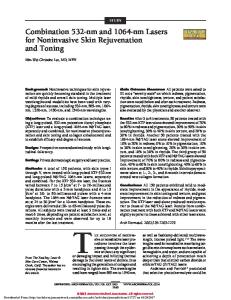
Fig. 3. The laser fiber is positioned adjacent to a third-order chorioallantoic membrane vessel. (A) Before irradiation, the fiber is maintained at working distance of 1 mm (energy 550 mJ, pulse width 30 ms). (B) After two pulses, the third-order vessel is ablated without rupture (arrow). Note the minimal surrounding thermal impact. (C) After fiber removal, there is no recannulation of the vessel and it remains ablated without rupture. Arrows indicate the coagulated and sealed vessel ends. (D) In a different egg, rupture of a third-order vessel is shown with extravasation of blood. Rupture occurred after four pulses (working distance 1 mm, energy 550 mJ, pulse width 10 ms).
were used to study the effects of pulsed KTP laser energy because they most closely resemble typical vessels treated in phonatory mucosa. A 532-nm pulsed KTP laser (Aura i with StarPulse; Laserscope Inc., San Jose, CA) was fitted with a fused silica, 0.2 numeric aperture 600-m fiber, which was attached to a threeaxis micromanipulator (Fine Science Tools, Inc.) enabling precise control of the fiber-to-tissue distance. The fiber was positioned using an operating microscope that provided magnification and recording capabilities. All personnel observed laser safety precautions during the experiments. Laser energy settings tested were 471 mJ, 510 mJ, and 550 mJ. At each energy setting, the fiberto-tissue working distance to the vessel being treated was tested at 1 and 3 mm. For each working distance, pulse width was also varied (10, 15, and 30 ms). In all cases, a pulse rate of two pulses per second was used. The exposure spot diameter at 1 mm and 3 mm from the fiber was approximately 1.1 mm and 2.1 mm, respectively. The range of pulse fluence achieved by varying both laser pulse energy and distance from the fiber was 13 to 58 J/cm2. Thermal relaxation time (cooling) of vessels in the size range tested occurs in approximately 10 ms or less,1,2 such that the 500-ms time between laser pulses delivered at two pulses per second allows nearly complete cooling of the target vessels between pulses. The laser pulses were delivered until the vessel was either “ablated” or until it ruptured (Fig. 3). We defined ablation by creation of a sealed bloodless segment.
RESULTS Segments of 135 third-order vessels from 23 eggs were tested. The energy was applied until there was vessel ablation or vessel wall rupture, which was easily observed Laryngoscope 117: February 2007
222
as blood extravasation. Figure 3 shows a typical sequence of events that occurred with the application of pulsed KTP laser energy. When the laser energy was applied, several characteristic changes in the blood vessel morphology were noted. Initially, the vessel wall constricted followed by either ablation or rupture with subsequent pulses. The number of pulses needed to achieve a particular effect varied depending on the various settings of energy, pulse width, and fiber-to-tissue distance. In general, more pulses were required for a vessel effect with lower energy settings and increased working distances. Minimal perivascular thermal effect within the albumen was noted when the vessel treatment end point (ablation or rupture) was seen. Photothermal trauma to the albumen was noted as white coagulum when further laser irradiation was administered beyond the vascular treatment objective. The following portion of the collected data set illustrates the effect of various laser settings on third-order vessel segments. When laser energy and working distance were held constant (550 mJ and 1 mm, respectively), increasing the pulse width led to fewer vessel ruptures. Twenty percent of vessels ruptured at 10 ms (2 of 10), 13% ruptured at 15 ms (2 of 15), and 10% ruptured at 30 ms (1 of 10) (Fig. 4). At the same constant laser energy (550 mJ) but at a longer working distance of 3 mm, increasing the pulse width eventually led to complete ablation (coagulation) with no vessel rupture. The same pattern of fewer vessel ruptures with increased pulse width was observed Broadhurst et al.: Pulsed KTP Laser Vessel Ablation
Fig. 4. Graph showing the effect of pulse width on vessel rupture rate at 1 mm. Note that increasing the pulse width decreased the percentage of vessels ruptured.
with fewer ruptures overall at the longer working distance. Fourteen percent of vessels ruptured at 10 ms (1 of 7), and no ruptures occurred at either 15 ms (0 of 5) or 30 ms (0 of 7) (Fig. 5). Increasing the working distance also led to a reduced incidence of rupture. With pulse width (10 ms) and energy (550 mJ) held constant, increasing working distance from 1 to 3 mm decreased rupture rates from 20% (2 of 10) to 14% (one of seven). Rupture rates decreased to 0% (0 of 5) from 13% (2 of 15) at a longer pulse width of 15 ms when the working distance was increased from 1 to 3 mm, and the rupture rate remained 0% (0 of 7) when the pulse width was increased further to 30 ms. When the working distance and pulse width were held constant (1 mm and 10 ms, respectively), increasing the total energy delivered led to more vessel ruptures (Fig. 6). Seven percent of vessels ruptured at an energy setting of 471 mJ (one of 14), 18% of vessels ruptured at 510 mJ (2 of 11), and 20% of vessels ruptured at 550 mJ (2 of 10).
Fig. 5. Graph showing the effect of pulse width on vessel rupture rate at 3 mm. Note that at this longer working distance, no rupture occurred at pulse widths of 15 and 30 ms.
Laryngoscope 117: February 2007
Fig. 6. Graph showing the effect of increasing total energy delivered on rupture rate at constant working distance and pulse width. Note that increasing the total energy delivered causes more vessel rupture. *These data points represent rupture rates for the pulsed dye laser at an equivalent energy and working distance.18
DISCUSSION This is the first study examining damage to target microvasculature using a pulsed KTP laser under conditions similar to laryngologic use, i.e., propagation of low repetition rate pulses from an optical fiber tip. From previous studies of both PDLs and pulsed KTP lasers similar to those in our study,1,9 it is known that 1) shorter pulses tend to cause hemorrhages and 2) vessel response depends on pulse fluence (energy delivered per unit area). Light emerging from the tip of a fiber diverges such that a combination of pulse energy and fiber-to-tissue distance determines the fluence in laryngologic use. One goal of this study was to determine whether vessel ablation without hemorrhage was indeed more easily achieved with longer pulses provided by the KTP laser and this is what was found. Selectivity for vessel damage (thermal confinement) also depends on pulse width.1,2,9 Heat flow in tissue depends on the square root of time.1 In this study, a range of KTP laser pulse widths from 10 to 30 ms was studied corresponding to heat flow over a distance of 0.1 to 0.2 mm. Thus, even the longest pulses tested allow thermal confinement primarily to the target vessels. It has become clear that photoangiolysis of the microcirculation of the superficial lamina propria is an effective strategy to treat a number of lesions of the phonatory mucosa, including papillomatosis,10 –14 dysplasia,5,8,15,16 and microvascular angiomata.4,7,17 The effectiveness of this approach in treating epithelial diseases in the operating room allowed for the creation of a new office-based paradigm by using a 585-nm PDL5 and subsequently a 532-nm pulsed KTP laser8 through the operating channel of a flexible laryngoscope. Although the 585-nm PDL was the initial laser to be used for office-based laser laryngeal surgery, this laser tended to cause frequent vessel wall disruption and visible extravasation of blood as a result of its extremely short pulse width.5 This clinical observation was also noted in a prior basic investigation using the CAM model.18 Vessel Broadhurst et al.: Pulsed KTP Laser Vessel Ablation
223
wall rupture and ecchymosis was an expected and acceptable occurrence in dermal applications because the minor soft tissue bleeding did not result in obvious scar and palpable diminished pliability. However, unlike skin, blood extravasation can theoretically be detrimental when treating phonatory epithelium, which must remain extremely supple to allow for entrained oscillation at a wide range of frequencies. More obvious is the fact that extralumenal blood located on the surface of the epithelial disease preferentially absorbs laser energy more so than the intralesional or sublesional microcirculation. Although this bleeding is generally mild, in an office setting it is often aspirated into the trachea, which causes coughing and potentially more bleeding. Taken together, the therapeutic time window provided by the local topical anesthesia is diminished. When blood extravasates into the delicate superficial lamina propria, there is indiscriminate absorbance of the 585-nm irradiation into the blood-stained, and otherwise normal, SLP. Although our experience is that the PDL is generally a safe laser, we have already encountered patients from another institution who have permanently lost vocal fold pliability from injudicious use of this laser. Review of that surgery revealed that repeated application of laser energy in tissues with marked subepithelial bleeding resulted in photothermal trauma of the SLP. Our clinical observations of the broadly available angiolytic lasers for laryngology reveal that the performance of the 532-nm pulsed KTP laser is superior to the 585-nm PDL.6 – 8 The enhanced microcirculatory hemostasis is easily observed, which the surgeon perceives as more predictable laser tissue interactions and response to treatment. We typically use the pulsed KTP laser between approximately 450 mJ to 550 mJ, a 15-ms pulse width, and a fiber-to-tissue distance of 2 to 4 mm. Within these clinical parameters, effective photoangiolysis without vessel wall rupture was easy to achieve in this investigation. It was rare for third-order vessels to rupture when the pulsed KTP laser was used at 3-mm fiber-to-tissue distance (Fig. 5) and even unlikely at 1 mm (Fig. 4), 10-ms pulse width, or at higher energy (550 mJ) (Fig. 6). This wide zone of safety is extremely useful when performing procedures in an office-based setting where there is a moving microcirculatory target. The results here with the pulsed KTP laser are dramatically different from the experience with the CAM model using the PDL (Fig. 6), which led to frequent blood extravasation at similar energy and working distance.18 The asterisk data points on the graph depict vessel rupture rates that are much greater for the PDL as compared with the pulsed KTP laser at equivalent energy and working distance. The ease and effectiveness by which the pulsed KTP laser (compared with the 585-nm PDL) achieves selective photoangiolysis is because the 532-nm wavelength is more strongly absorbed by oxyhemoglobin and because the surgeon can calibrate a longer pulse-width (15–20 ms) based on observations of soft tissue interactions. We believe that the frequent vessel wall disruption while using the 585-nm PDL was the result of the extremely short pulse width (approximately .450 ms). Essentially, the blood often heats too quickly and not uniformly within the vessel lumen, which Laryngoscope 117: February 2007
224
disrupts the vessel wall before complete intravascular coagulation. Because of this, there is a narrow zone of optimal energy delivery to achieve complete intravascular coagulation. In part this can be compensated for with experience and acquired skill by adjusting fiber-to-tissue distances as well as the energy settings for each pulse. This investigation further demonstrates that the chick CAM is an excellent model to simulate the superficial lamina propria of the phonatory mucosa and especially the microcirculation.18 Like with other phonosurgical procedures, a surgical model/simulator19 will be a valuable hands-on educational platform to familiarize surgeons with this emerging laser technology. We are currently investigating more extreme settings using the pulsed KTP laser by extending pulse width ranges. Increasing pulse width will likely effectively photocoagulate small vessels but will result in extravascular thermal trauma, which is detrimental to phonatory mucosa. This is illustrated by increasing the pulse width until it simulates the KTP in a continuous-wave mode, which is known to vaporize epithelium. Decreasing pulse width will lead to early vessel wall rupture resulting from rapid energy delivery (like the PDL). Given these variations, we hope to determine how extremes in pulse width ranges can be modulated by adjusting fiber-to-tissue distance and increasing energy output to better define the clinical limitations of the 532-nm pulsed KTP laser as an angiolytic instrument.
CONCLUSIONS 1. The settings for the pulsed KTP laser that most effectively produced vessel ablation without rupture were with pulse widths of 15 or 30 ms and a fiberto-tissue distance of 3 mm. 2. The parameters stated previously represent zones of effectiveness and are interdependent with energy output. Therefore, surgeons should familiarize themselves with this new technology potentially using the CAM as an educational model. 3. In general terms, selective photoangiolysis without vessel wall rupture is optimized with longer pulse widths, increased fiber-to-tissue distance, and lower energy. 4. Although longer pulse width parameters (i.e., ⬎30 ms) may be effective at ablation without vessel wall disruption and extravasation of blood, the pulse width setting that results in extravascular thermal trauma was not determined. This is planned for a future investigation. 5. Clinically, the 532-nm pulsed KTP laser is more effective than the 585-nm PDL in selective photoangiolysis and substantially easier to use when treating laryngeal pathology. The investigation here coupled to prior work using the CAM model substantiates these clinical observations. Acknowledgments The authors thank Steven Adlard, Pamelia Cutulle, RN, and Harold “Sonny” Cruz for assistance with equipment handling. Broadhurst et al.: Pulsed KTP Laser Vessel Ablation
The authors received the KTP laser on loan from Laserscope Inc.
BIBLIOGRAPHY 1. Anderson R, Parrish J. Selective photothermolysis: precise microsurgery by selective absorption of pulsed radiation. Science 1983;220:524 –527. 2. Anderson RR, Parrish JA. Microvasculature can be selectively damaged using lasers: a basic theory and experimental evidence in human skin. Lasers Surg Med 1981;1:263–276. 3. Anderson RR, Jaenicke KF, Parrish JA. Mechanisms of selective vascular changes caused by dye lasers. Lasers Surg Med 1983;3:211–215. 4. Zeitels SM, Healy GB. Laryngology and phonosurgery. N Engl J Med 2003;349:882– 892. 5. Zeitels SM, Franco RA Jr, Dailey SH, Burns JA, Hillman RE, Anderson RR. Office-based treatment of glottal dysplasia and papillomatosis with the 585-nm pulsed dye laser and local anesthesia. Ann Otol Rhinol Laryngol 2004;113: 265–276. 6. Zeitels SM, Burns JA. Laser applications in laryngology: past, present, and future. Otolaryngol Clin North Am 2006; 39:159 –172. 7. Zeitels SM, Akst L, Burns JA, Hillman RE, Broadhurst MS, Anderson RR. Pulsed angiolytic laser treatment of ectasias and varices in singers. Ann Otol Rhinol Laryngol 2006;115: 571–580. 8. Zeitels SM, Akst L, Burns JA, Hillman RE, Broadhurst MS, Anderson RR. Office based 532nm pulsed-KTP laser treatment of glottal papillomatosis and dysplasia. Ann Otol Rhinol Laryngol 2006;115:686 – 689. 9. Suthamjariya K, Farinelli WA, Wooseok K, Anderson R. Mechanisms of microvascular response to laser pulses. J Investig Dermatol 2004;122:518 –525.
Laryngoscope 117: February 2007
10. Bower CM, Flock S, Waner M. Flash pump dye laser treatment of laryngeal papillomas. Ann Otol Rhinol Laryngol 1998;107:1001–1005. 11. Zeitels SM. Papillomatosis. In: Atlas of Phonomicrosurgery and Other Endolaryngeal Procedures for Benign and Malignant Disease. San Diego: Singular, 2001:119 –131. 12. Franco RA, Zeitels SM, Farinelli WA, Anderson RR. 585-NM pulsed dye laser treatment of glottal papillomatosis. Ann Otol Rhinol Laryngol 2002;111:486 – 492. 13. McMillan K, Shapshay SM, McGilligan JA. A 585-nanometer pulsed dye laser treatment of laryngeal papillomas; preliminary report. Laryngoscope 1998;108:968 –972. 14. Valdez TA, McMillan K, Shapshay SM. A new laser treatment for vocal cord papillomas—585 nanometer pulsed dye laser. Otolaryngol Head Neck Surg 2001;124:421– 425. 15. Zeitels SM. Vocal fold atypia/dysplasia and carcinoma. In: Atlas of Phonomicrosurgery and Other Endolaryngeal Procedures for Benign and Malignant Disease. San Diego: Singular, 2001:177–218. 16. Franco RA, Zeitels SM, Farinelli WA, Faquin W, Anderson RR. 585-nm pulsed dye laser treatment of glottal dysplasia. Ann Otol Rhinol Laryngol 2003;112:751–758. 17. Zeitels SM, Hillman RE, Desloge RB, Mauri M, Doyle PB. Phonomicrosurgery in singers & performing artists: treatment outcomes, management theories, & future directions. Ann Otol Rhinol Laryngol 2002;111(suppl 190):21– 40. 18. Broadhurst MS, Kobler JB, Akst L, et al. Effects of 585nm pulsed-dye laser settings on vessel ablation in the avian chorioallantoic membrane: implications for vocal-fold mucosal photoangiolysis. Annual meeting of the American Society of Lasers in Medicine & Surgery, 2006. 19. Dailey SH, Kobler JB, Zeitels SM. A laryngeal dissection station: educational paradigms in phonosurgery. Laryngoscope 2004;114:878 – 882.
Broadhurst et al.: Pulsed KTP Laser Vessel Ablation
225